5082
Analysis of 31P chemical-shift signatures of high-energy phosphates in glioma patients measured at 7 Tesla1Division of Medical Physics in Radiology, German Cancer Research Center (DKFZ), Heidelberg, Germany, 2Faculty of Physics and Astronomy, Heidelberg University, Heidelberg, Germany, 3International Max Plank Research School for Quantum Dynamics in Physics, Chemistry, and Biology (IMPRS-QD)), Max Plank for Nucelar Physics (MPIK), Heidelberg, Germany, 4Division of Radiology, German Cancer Research Center (DKFZ), Heidelberg, Germany, 5Faculty of Medicine, Heidelberg University, Heidelberg, Germany, 6Division of Neuroradiology, University Hospital Bonn, Bonn, Germany
Synopsis
Keywords: Non-Proton, Spectroscopy, Phosphorus, 31P, pH, magnesium, glioma
Motivation: The detailed characterization of tumor micro-environment can be supported by 31P MRSI enabling the non-invasive determination of pH and magnesium ion content (Mg).
Goal(s): The aim of this study was to investigate whether a discrimination between different glioma micro-environments might be possible based on the differences in 31P chemical-shift signatures.
Approach: For this purpose, 31P MRSI datasets from 12 patients with glioma acquired at 7T were analyzed and used to estimate the underlying pH and Mg values.
Results: For the analyzed cohort, different trends in 31P chemical-shift signatures, as well as in pH and Mg values were observed for different tumor subtypes.
Impact: The analysis of 31P chemical-shift signatures in glioma patients could potentially be used for stratifying tumor subtypes, and might help in the characterization of tumor microenvironments, e.g. by determining potential biomarkers such as pH and the magnesium ion content.
Introduction
31P magnetic resonance spectroscopic imaging (MRSI) is a valuable tool to non-invasively characterize tumor microenvironment in vivo, i.e. via pH values or the magnesium ion content (Mg)1-4. Recently, we observed that intracellular pH (pHi) (as determined via the chemical-shift difference of inorganic phosphate (Pi) and phosphocreatine (PCr) resonances) is strongly correlated to proliferation markers, but lacked the ability to discriminate for diverse mutation status5. However, adding complementary information using more chemical-shifts, e.g. those of adenosine-5’-triphosphate (ATP) peaks which can be used to determine Mg, could potentially enable such an improved stratification.In this study, the differences in 31P chemical-shift signatures between tumor and healthy tissue were investigated in a cohort of 12 patients with glioma. Using an advanced version of a recently proposed look-up algorithm for the determination of pH and Mg under different conditions6,7, an initial attempt was taken to link the observed chemical-shift signatures to individual characteristics of different tumor microenvironments.
Methods
31P MRSI datasets of 12 patients with glioma acquired at B0 = 7 T were used retrospectively for the analysis of chemical-shift signatures observed in tumor and healthy tissue. The detailed measurement and evaluation protocols can be found in [5].Mean values of the quantified chemical-shifts of Pi, γ-, α- and β-ATP across regions-of-interest (ROIs) covering (i) the whole tumor volume (WHT) and (ii) healthy white matter on the contralateral side (WM) were calculated for each patient separately. These mean chemical-shifts across the WHT and WM ROIs were compared between groups of patients with different histopathology: glioblastoma, IDH-wildtype, WHO grade IV (GBM; n = 7), astrocytoma, IDH-mutant, WHO grade II / III (Astro; n = 4), pleomorphic glioma, IDH-wildtype, WHO grade II (Pleo; n = 1).
Moreover, for each patient separately, the mean values of the chemical-shifts of Pi and ATP were fed into an advanced look-up algorithm as recently proposed6, but with novel look-up elements7, in order to obtain estimated values for pH and Mg.
Results
Figure 1 illustrates local differences in quantified chemical-shifts of the Pi and ATP resonances (relative to PCr) between tumor and healthy tissue for a representative patient with glioblastoma.For all patients, the resonances of Pi as well as of γ- and β-ATP are clearly shifted downfield in tumor tissue compared to healthy tissue (with stronger shifts in the GBM group than in the Astro group; cf. Fig 2). The α-ATP resonance only shows minor changes with the highest variation in the group Astro (Fig. 2). Note, that the absence of strong variations in the chemical-shift of α-ATP relative to PCr justifies referencing all input chemical-shifts to α-ATP as required for the advanced look-up algorithm proposed in [7].
The values determined for pH and Mg are generally elevated in the WHT ROI compared to the WM ROI (Fig. 3A). While for the GBM group pH and Mg are increased at nearly constant levels for the estimated ionic strength, the Astro group showed an increased pH but a variable Mg trend with decreased values for the estimated ionic strengths (Fig. 3B).
Discussion
The differing trends of δPi and δβ in the individual groups (i.e. higher δPi in the GBM group than in the Astro group, but no clear difference in δβ between the groups GBM and Astro, Fig. 2) might indicate a different underlying microenvironment in the tumor subtypes. The trends in the estimated values for pH / Mg / Ion (Fig. 3) lead to the hypothesis, that the chemical-shift alterations are driven not only by changes in pH, but presumably also by changes in the magnesium ion content (both the Pi and ATP resonances shift downfield with increasing Mg). The stronger variability in determined Mg values for the group Astro (Fig. 3) might also be related partly to trends towards a decreased ionic strength (Mg2+ also contributes to the ionic strength).Consistent with the conventional pH estimation (by means of the modified Henderson-Hasselbalch equation8), lower-grade tumors showed a lower pH increase compared to healthy white matter, which might be related to the Ki-67 score and should be investigated further. However, the estimated pH values determined by the advanced look-up algorithm are systematically shifted by approximately -0.2 pH compared to the pH calculated using the modified Henderson Hasselbalch equation8, as discussed in [7].
Conclusion
The results of this study indicate that the 31P chemical-shift signatures obtained from Pi and ATP resonances in vivo could potentially be used to differentiate glioma subtypes. By using these chemical-shift signatures for the determination of pH and Mg, potential differences in the specific tumor microenvironments might be quantifiable.Acknowledgements
References
- Korzowski A, Weinfurtner N, Mueller S, et al. Volumetric mapping of intra- and extracellular pH in the human brain using 31P MRSI at 7T. Magn Reson Med. 2020; 84: 1707– 1723. https://doi.org/10.1002/mrm.28255
- Taylor JS, Vigneron DB, Murphy-Boesch J, et al. Free magnesium levels in normal human brain and brain tumors: 31P chemical-shift imaging measurements at 1.5 T. Proc Natl Acad Sci U S A. 1991;88(15):6810-6814. doi:10.1073/pnas.88.15.6810
- Wenger KJ, Hattingen E, Franz K, et al. Intracellular pH measured by 31P-MR-spectroscopy might predict site of progression in recurrent glioblastoma under antiangiogenic therapy. J Magn Reson Imaging. 2017;46(4):1200-1208.
- Klomp DWJ, van de Bank BL, Raaijmakers A, et al. 31P MRSI and 1H MRS at 7 T: initial results in human breast cancer. NMR Biomed. 2011;24(10):1337-1342.
- Paech D, Weckesser N, Franke VL et al. Whole-brain Intracellular pH Mapping of Gliomas using High-resolution 31P MR Spectroscopic Imaging at 7 T. Radiology: Imaging Cancer 2023. In Press.
- Franke VL. In vivo determination of pH and magnesium ion concentration by means of 31P MRSI: A multi-parametric look-up approach - heiDOK. Published online 2023. Accessed May 25, 2023. https://archiv.ub.uni-heidelberg.de/volltextserver/32909/
- Seng B, Franke VL, Platek J, et al. Advancement of a novel 31P MRS based approach for the in vivo determination of pH and magnesium ion content under different chemical conditions. Submitted Abstract ISMRM 2024. Abstract #5626.
- de Graaf RA. In Vivo NMR Spectroscopy: Principles and Techniques: 2nd Edition.; 2007. doi: 10.1002/9780470512968.
Figures
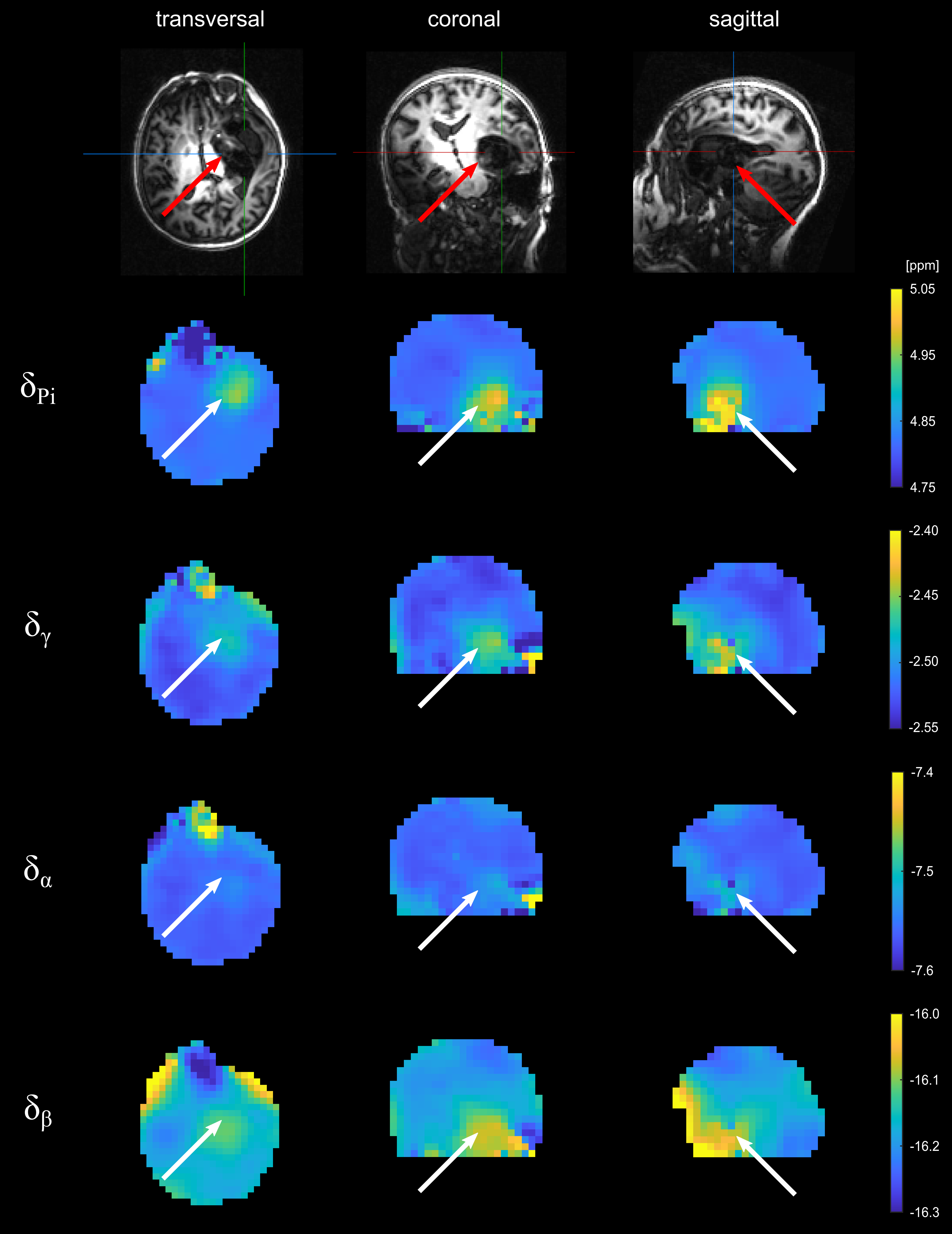
Figure 1: Volumetric maps of the quantified chemical-shifts of inorganic phosphate (Pi) and ATP (γ-, α- and β-ATP) relative to phosphocreatine of one representative patient with glioblastoma. The 31P MRSI datasets were acquired at B0 = 7T with an isotropic, nominal spatial resolution of (12.5 mm)³ and spatially zero-filled with a factor of 2. The position of the tumor is indicated by arrows.
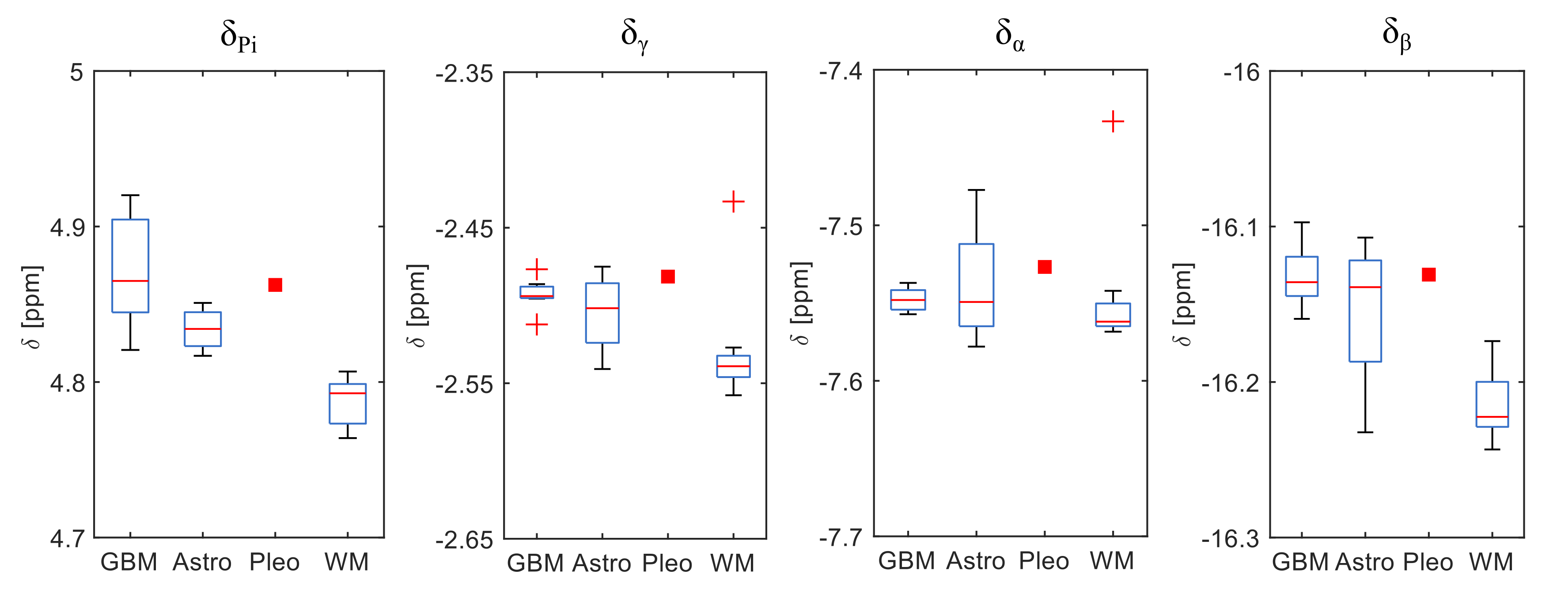
Figure 2: Comparison of the mean chemical-shifts of Pi and ATP (relative to PCr) averaged across the respective whole tumor ROI and the contralateral white matter ROI (WM). The patients were grouped based on their histopathology: glioblastoma, IDH-wildtype, WHO grade IV (GBM, n = 7), astrocytoma, IDH-mutant, WHO grade II/III (Astro, n = 4), pleomorphic glioma, IDH-wildtype, WHO grade II (Pleo, n = 1). The red bars in the boxplots show the median values across all patients of the respective group. The WM boxplot includes the mean chemical-shifts across all 12 patients.
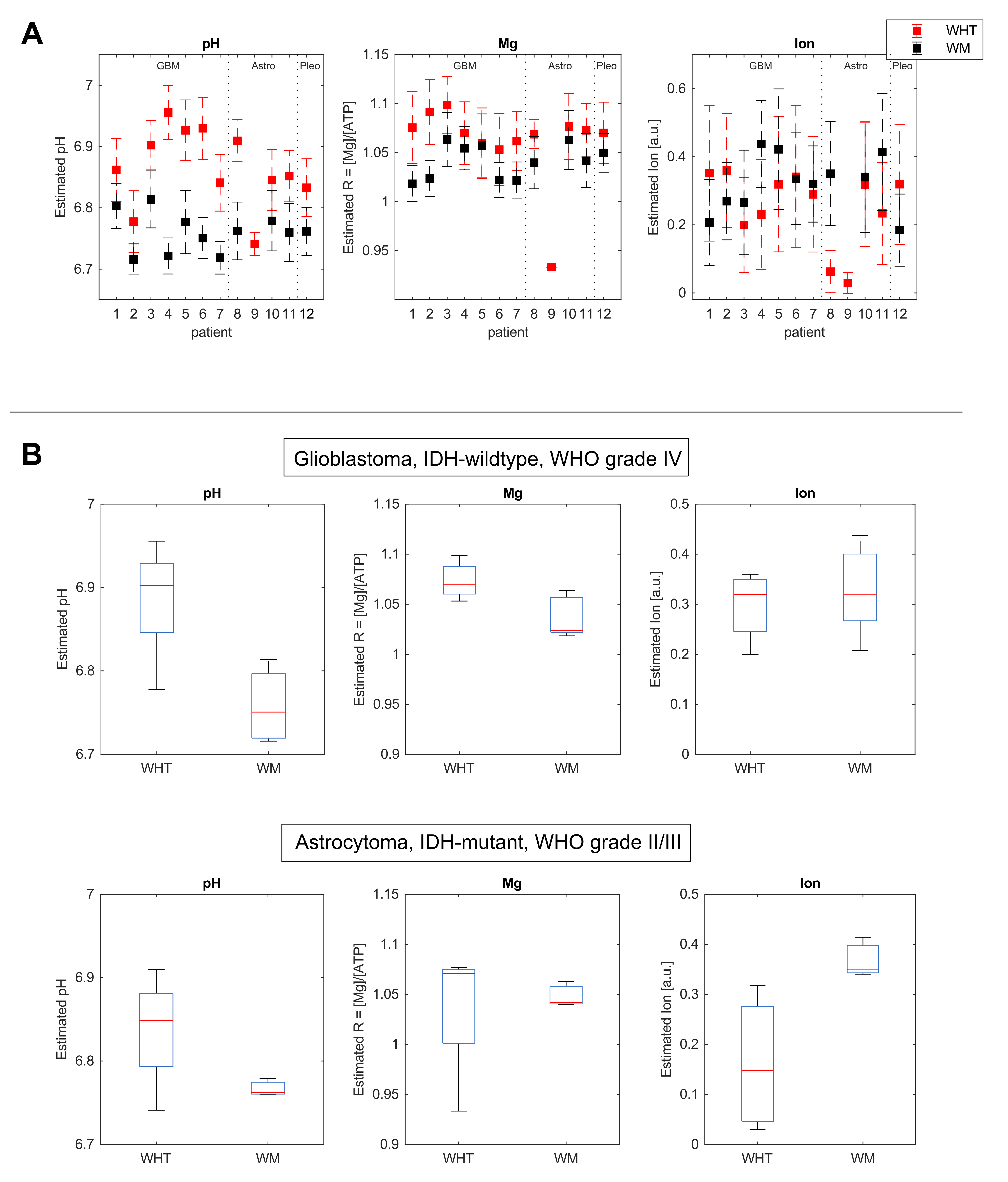
Figure 3: Values for pH, magnesium ion content (R = [Mgtot]/[ATPtot]) and ionic strength (Ion) estimated by the advanced look-up algorithm based on the mean chemical-shifts across the whole tumor (WHT) and the contralateral white matter (WM) ROI. Panel A: Estimated values for each patient separately. For patient 9, no values could be determined for the WM ROI by the look-up algorithm. Panel B: Boxplots showing the median values across all patients of the respective groups: glioblastoma, IDH-wildtype, WHO grade IV (top), and astrocytoma, IDH-mutant, WHO grade II / III (bottom).