5080
In Vivo Imaging of Tumor Metabolic Heterogeneity Using [1-13C]Pyruvate-d3 Hyperpolarized By Reversible Exchange With Parahydrogen1Division of Medical Physics, Department of Diagnostic and Interventional Radiology, Faculty of Medicine, University of Freiburg, University Medical Center Freiburg, Freiburg 79106, Germany, 2German Cancer Consortium (DKTK), partner site Freiburg, German Cancer Research Center (DKFZ), Heidelberg 69120, Germany, 3Department of Nuclear Medicine, School of Medicine, Klinikum rechts der Isar, Technical University of Munich, Munich 81675, Germany, 4Institute of Molecular Medicine and Cell Research, Faculty of Medicine, University of Freiburg, Freiburg 79104, Germany, 5NVision Imaging Technologies GmbH, Ulm 89081, Germany, 6Department of Chemistry, Integrative Biosciences (Ibio), Karmanos Cancer Institute (KCI), Wayne State University, Detroit 48202, MI, United States, 7Sektion Biomedizinsche Bildgebung, Molecular Imaging North Competence Center MOINCC, Klinik für Radiologie und Neuroradiologie, University Hospital Schleswig-Holstein, Kiel University, Kiel, Germany, 8Werner Siemens Imaging Center, Department of Preclinical Imaging and Radiopharmacy, Eberhard Karls University Tübingen, Tübingen 72076, Germany, 9German Cancer Consortium (DKTK), partner site Tübingen, German Cancer Research Center (DKFZ), Heidelberg 69120, Germany, 10German Cancer Consortium (DKTK), partner site Munich, German Cancer Research Center (DKFZ), Heidelberg 69120, Germany
Synopsis
Keywords: Hyperpolarized MR (Non-Gas), Contrast Agent
Motivation: Hyperpolarized [1-13C]pyruvate MRI is promising for studying cancer metabolism and assessing early therapy response but requires high-throughput and less complex hyperpolarization techniques for wide availability.
Goal(s): Demonstrating metabolic imaging in a genetic mouse model of metastasizing breast cancer (MMTV-PyMT) using purified [1-13C]-pyruvate-d3 hyperpolarized via parahydrogen-based Signal Amplification By Reversible Exchange (SABRE).
Approach: Our rapid (6 min) and efficient hyperpolarization method yielded highly-polarized (>10%) [1-13C]-pyruvate-d3 in safe aqueous solutions.
Results: Administered to two PyMT-mice, 13C chemical shift imaging detected the injected pyruvate and metabolized [1-13C]-lactate. Analysis revealed elevated lactate levels in tumors compared to healthy breast tissue, highlighting tumor compartments with distinct metabolic profiles.
Impact: We showcase our rapid, cost-effective SABRE hyperpolarization approach, yielding safe, highly-polarized pyruvate for inaugural cancer metabolic investigations. This innovation expands high-throughput preclinical HP-MRI research, enabling comprehensive exploration of tumor biology, metabolic processes, and therapeutic responses in cancer.
Introduction
Hyperpolarized (HP) [1-13C]pyruvate MRI has shown great promise for studying cancer metabolism and assessing early therapy response 1–3. However, a high-throughput, low foot-print and less complex hyperpolarization technology is needed to make this promising technique widely available. Recently, we introduced a fast (6 min) and cost-effective method using Signal Amplification By Reversible Exchange (SABRE) 4 and Spin-Lock Induced Crossing (SLIC-SABRE )5 at microtesla fields (Fig. 1) 6 and a rapid purification to produce safe HP solutions of aqueous pyruvate 7. We successfully demonstrated in vivo HP metabolic MRI in healthy mice 7. Here, we report the first application of this promising high-throughput hyperpolarization technique to investigate a genetic model for proliferating breast cancer in mice (MMTV-PyMT). Particularly, for studying the PyMT model, pyruvate appears as a promising agent as these luminal type of cancers do overproduce lactate under anaerobic conditions and typically present enhanced levels of LDHA and MCT1 8.Methods
50mM [1-13C]pyruvate-d3 was hyperpolarized in methanol-d4 up to 24.1% using an iridium-based catalyst and reversible exchange with >90% parahydrogen (pH2) in a home-build SLIC-SABRE setup (B0=50µT, B1=1.995µT, average HP 21.2±1.7%, N=8, Fig. 2). The hyperpolarized solution was purified in about 60s by adding 1ml PBS in D2O, evaporating methanol at 100°C and 5mbar, and filtering the solution to remove the precipitated catalyst. After purification, as demonstrated in our recent study, a clean, non-acute toxic, sterile and pH-neutral injection solution of HP [1-13C]pyruvate-d3 was obtained (P~11%, c~30mM, V~250µL, residual 50 mM methanol and 20 μM catalyst) 7. This purified pyruvate solution was administered to breast tumor-bearing mice (~13 weeks, ~24g, bolus up to 5µL/g) within ≈8s and a 2D FID chemical shift imaging (FIDCSI) sequence was applied (FOV: 26x32mm2, matrix: 13x16, axial slice 5/7mm, spectral bandwidth 2500Hz (200 points), TR=85ms, α=12°, Bruker 70/20). T2w RARE MRI was acquired for anatomical reference (FOV: 26x32mm2, matrix: 173x213, slice 1mm, TR=4s, N=9). Pyruvate and lactate images were generated by integrating the area-under the curve of a multi-peak Voigt profile that was fitted to the line-broadened spectra (30Hz) using (a custom) python 3.10 script (based on 8).In the PyMT mouse model of metastasizing breast cancer the oncogene (Polyoma Middle T) is expressed only in the breast epithelium, therefore the (FVB) mice develop breast tumors in all ten mammary glands with palpable tumors at an age of about 8 weeks 9.
Results
Strong 13C CSI signals were detected 10s and 20 s after injection in mouse 1 and 2, respectively (Fig. 4, 5). Mouse 1: The tumor was readily identified on the T2w MRI, showing a hyper- (T2w+) and hypo- (T2w-) intense region. The strongest 13C pyruvate and lactate signals were observed in the heart. Lactate levels were elevated in the tumor compared to adjacent non-cancerous breast tissue. The T2w- region showed less pyruvate and lactate signal than the T2w+ region.Mouse 2: T2w MRI showed a large tumor with a necrotic, a T2w+, a T2w- region, and another smaller tumor. At 20 seconds post-injection, the pyruvate signal had significantly diminished, primarily remaining within the blood vessels. However, similar to mouse 1, robust lactate signals were detected within the T2w- tumor region, while limited lactate was observed in the T2w+ part. No pyruvate or lactate was detected in the necrotic region. In mouse 2, no healthy breast tissue was present for reference; instead, a smaller metabolically active tumor was identified.
Discussion
This fast and cost-effective hyperpolarization technique has proven valuable for preclinical investigations of cancer metabolism. It allowed a clear differentiation between healthy breast tissue and the tumor, with lactate predominantly appearing in the latter. Additionally, CSI images unveiled metabolic heterogeneity within distinct tumor regions, including necrotic areas lacking perfusion and regions with varying metabolic activity, possibly linked to differences in perfusion. We eagerly await the histological results that will provide a deeper understanding of the tissue differences among these tumor compartments.Using the robust CSI sequence, initial metabolic information was obtained from the 13C images; however, they lack spatial and temporal resolution and are highly reliant on precise timing. Hence, our future metabolic MRI studies may benefit from 3D dynamic 13C imaging to improve the interpretation of the findings.
Conclusion
The characterization of tumor biology, focusing on metabolic activity and heterogeneity, holds significant promise for studying cancer, improving therapies, and monitoring early response. Fast, low-cost SABRE hyperpolarization enables high-throughput HP-MRI, thus providing a potent resource for preclinical cancer research. This development extends the accessibility of HP-MRI to a broader biomedical community, offering new avenues for research in the field of oncology.Acknowledgements
Research reported in this publication was supported by the German Federal Ministry of Education and Research (BMBF) in the funding program “Quantum Technologies – from Basic Research to Market” under the project “QuE-MRT” (contract number: 13N16448), the German Cancer Consortium (DKTK), the Research Commission of the University Medical Center Freiburg, the Core Facility AMIRCF (DFG-RIsources N° RI_00052), B.E.S.T. Fluidsysteme GmbH I Swagelok Stuttgart, and the German Research Foundation (DFG #SCHM 3694/1-1, #SCHM 3694/2-1, #SFB1479).
References
1. Wang ZJ, Ohliger MA, Larson PEZ, et al. Hyperpolarized 13C MRI: State of the Art and Future Directions. Radiology. 2019;291(2):273-284. doi:10.1148/radiol.2019182391
2. Sushentsev N, McLean MA, Warren AY, et al. Hyperpolarised 13C-MRI identifies the emergence of a glycolytic cell population within intermediate-risk human prostate cancer. Nat Commun. 2022;13(1):466. doi:10.1038/s41467-022-28069-2
3. Woitek R, McLean MA, Ursprung S, et al. Hyperpolarized Carbon-13 MRI for Early Response Assessment of Neoadjuvant Chemotherapy in Breast Cancer Patients. Cancer Res. 2021;81(23):6004-6017. doi:10.1158/0008-5472.CAN-21-1499
4. Adams RW, Aguilar JA, Atkinson KD, et al. Reversible Interactions with para-Hydrogen Enhance NMR Sensitivity by Polarization Transfer. Science. 2009;323(5922):1708-1711. doi:10.1126/science.1168877
5. Theis T, Truong M, Coffey AM, Chekmenev EY, Warren WS. LIGHT-SABRE enables efficient in-magnet catalytic hyperpolarization. J Magn Reson. 2014;248:23-26. doi:10.1016/j.jmr.2014.09.005
6. Schmidt AB, Eills J, Dagys L, et al. Over 20% Carbon-13 Polarization of Perdeuterated Pyruvate Using Reversible Exchange with Parahydrogen and Spin-Lock Induced Crossing at 50 μT. J Phys Chem Lett. 2023;14(23):5305-5309. doi:10.1021/acs.jpclett.3c00707
7. de Maissin H, Groß PR, Mohiuddin O, et al. In Vivo Metabolic Imaging of [1-13C]Pyruvate-d3 Hyperpolarized By Reversible Exchange With Parahydrogen**. Angew Chem Int Ed. 2023;62(36):e202306654. doi:10.1002/anie.202306654
8. Asghar Butt S, Søgaard LV, Ardenkjaer-Larsen JH, et al. Monitoring mammary tumor progression and effect of tamoxifen treatment in MMTV-PymT using MRI and magnetic resonance spectroscopy with hyperpolarized [1-13C]pyruvate. Magn Reson Med. 2015;73(1):51-58. doi:10.1002/mrm.25095
9. Gottwald W. TUMKRI NVIS PHIPvsDNPInVivoStudy.; 2023. https://github.com/wgottwald/2023_TUMKRI_NVIS_PHIPvsDNPInVivoStudy. Accessed November 7, 2023.
10. Lin EY, Jones JG, Li P, et al. Progression to malignancy in the polyoma middle T oncoprotein mouse breast cancer model provides a reliable model for human diseases. Am J Pathol. 2003;163(5):2113-2126. doi:10.1016/S0002-9440(10)63568-7
Figures
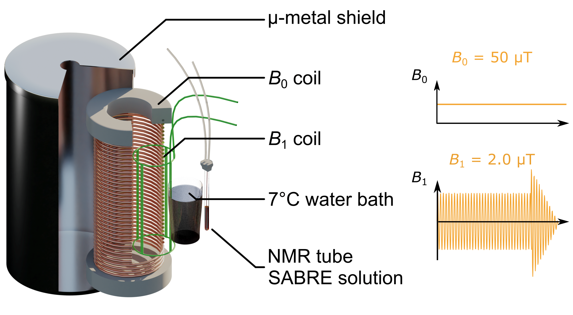
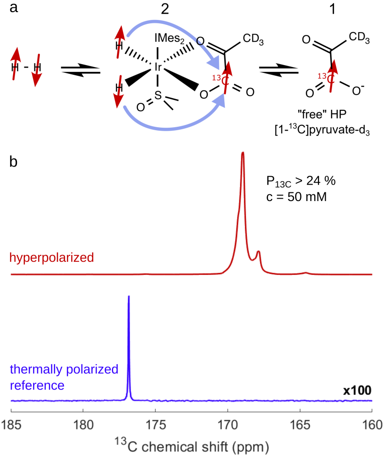
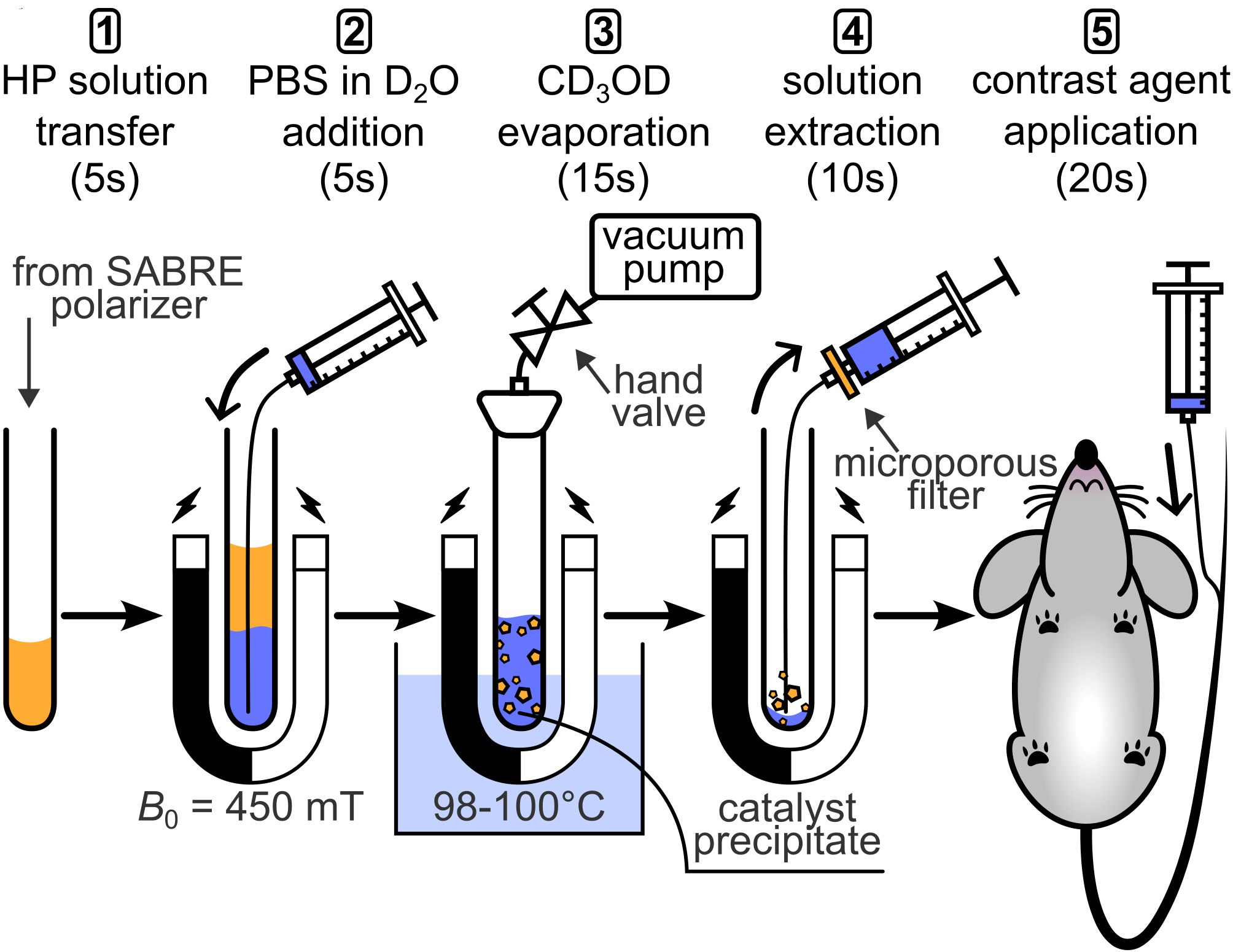
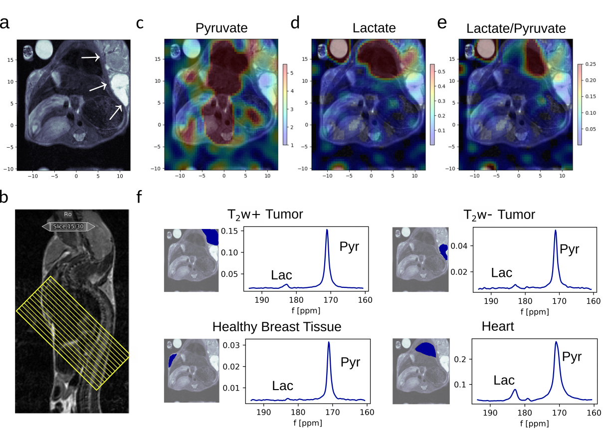
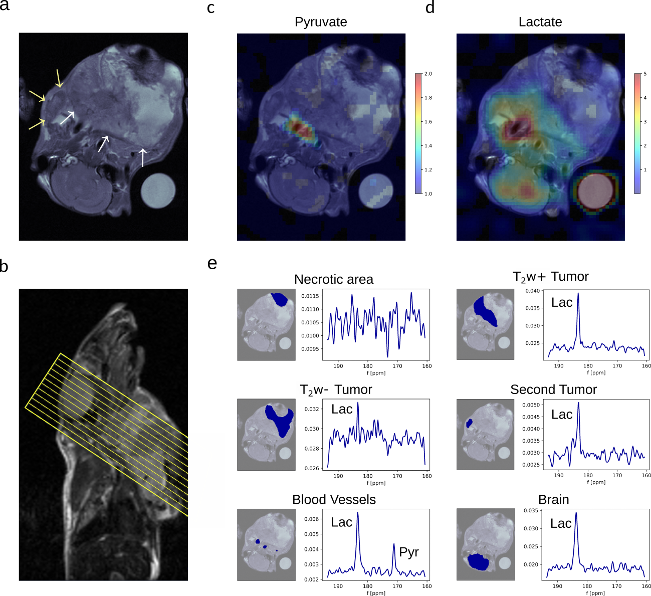
Figure 5. Metabolic imaging of breast cancer, mouse 2. a T2w RARE 1H MRI showing an axial anatomic slice used for metabolic imaging with white and yellow arrows pointing at the tumors. b The slice orientation is indicated by yellow lines in the sagittal image. c,d 4x interpolated 13C MRI of pyruvate (c) and lactate (d) signal superimposed on the T2w MRI (a).e 13C chemical shift spectra of a necrotic tumor compartment, different tumor regions (T2w+ = hyperintense, T2w- = hypointense) blood vessels and brain. The 13C images and spectra were acquired 20s post-injection.