5074
Microstructural Abnormalities of Grey Matter and White Matter in Children with Basic-type Intermittent Exotropia using NODDI Technology1Beijing Tongren Hospital, Beijing Eye Center, Beijing, China, 2Chinese University of Hong Kong (Shenzhen) School of Medicine, Shenzhen Research Institute of Big Data, Hong Kong, China, 3Chinese University of Hong Kong (Shenzhen) School of Data Science, Hong Kong, China, 4Department of Radiology, Beijing Tongren Hospital, Beijing, China, 5MR Research Collaboration, Siemens Healthineers Ltd., Beijing, China, 6Shenzhen Research Institute of Big Data, Hong Kong, China
Synopsis
Keywords: Microstructure, Diffusion/other diffusion imaging techniques, NODDI; intermittent exotropia; microstructural changes; TBSS; binocular function
Motivation: Intermittent exotropia (IXT) is quite common in children. The exact pathogenesis is not clear, which makes it difficult to make early diagnosis and evaluate progression.
Goal(s): To investigate the microstructural changes in vivo in children with IXT and explore the potential neuropathological mechanisms.
Approach: 31 IXT children and 37 controls were enrolled. Brain gray matter (GM) and the white matter (WM) regions extracted from their whole-brain MRI data were compared using the NODDI technology and TBSS method.
Results: Microstructural changes in GM and WM between the two groups were found, located in the visual perception and oculomotor control associated areas.
Impact: The parameters derived from NODDI can demonstrate the microstructural abnormalities in primary, advanced visual center and oculomotor center pathway, which maybe a potential tool in diagnosis and evaluation the IXT patients.
Introduction
Intermittent exotropia (IXT) is one of the most prevalent types of ophthalmopathy in children1,2. The basic type is the most common type3, which can lead to binocular function impairment and stereopsis loss. Several studies have examined altered functional activity and structural abnormalities in children with basic-type IXT. However, little is known about brain microstructural changes to spot early abnormalities in IXT children4. Neurite orientation dispersion and density imaging (NODDI) is a practical technique which allows evaluation of nerve axon and dendrite structure5. It produces 3 parameters: the orientation dispersion index (ODI), the isotropic volume fraction (ISOVF) and the intra-cellular volume fraction (ICVF). Tract-based spatial statistics (TBSS), which combines the strengths of both voxel-wise and tractography-based analyses, is also widely used to investigate neurological disorders6. This study aimed to investigate microstructural changes of gray matter (GM) in children with IXT using NODDI method, as well as alterations of white matter (WM) tracts using TBSS method.Methods
We enrolled 31 IXT children and 37 age-, sex-, handedness-, and education-matched healthy controls (HCs). All participants underwent MRI scanning and detailed ophthalmic examinations. Data were collected using a 3.0 T MRI scanner (Siemens Healthineers, Erlangen, Germany) with a 64 eight-channel head coil. The NODDI parameter values of the subjects' primary and advanced visual center, oculomotor center, and the subcortical white matter fiber are determined based on the ROIs, including ODI, ISOVF and ICVF. Voxel wise statistical analysis of the FA data was carried out using TBSS7. Each subject's aligned ICVF data was then projected onto this skeleton and the resulting data fed into voxel wise cross-subject statistics. All statistical analyses were performed using SPSS, and statistical significance was reported at P <0.05.Results
Differences in NODDI Parameters between GroupsCompared with the HCs, IXT patients had significantly higher ODI values in bilateral precuneus and left superior occipital gyrus (SOG). Lower ODI and ICVF values were shown in bilateral inferior occipital gyrus (IOG), bilateral middle occipital gyrus (MOG), left lingual gyrus (LG), bilateral middle frontal Gyrus (MFG), bilateral Inferior frontal gyrus (IFG), left middle temporal gyrus (MTG). IXT patients also exhibited lower ISOVF values in bilateral cerebellum, bilateral superior frontal gyrus (SFG) and bilateral MFG.
Results from the ROI-restricted TBSS group analysis
ROI-restricted TBSS analysis revealed increased ICVF value in the arcuate fasciculus tracts (AF), superior longitudinal fasciculus (SLF) and inferior longitudinal fasciculus (ILF).
Discussion
Differences in NODDI ParametersCompared with HCs, IXT children had lower ODI and ICVF values in the binocular function associated brain areas, including bilateral IOG, bilateral MOG and the left LG, as well as oculomotor control associated regions, such as the bilateral MFG and the bilateral IFG, which implied a loss of axonal density and an increase of axonal coherence. As a part of ventral visual stream, IOG is correlated with spatial and face-part selectivity8. LG plays an important role in visual memory9. Fei found reduced fALFF values in the left LG and right IOG of IXT children, indicating potential impairment of spatial attention and selectivity in IXT10. Meanwhile, MOG participants in the stereopsis, spatial processing, and so-called “category-selective attention-modulated face/tool processing”11,12. The posterior part of MFG is the motor center for eye movement13. Moreover, the IFG is involved in affective processes, and cognitive control14. These findings may be related to the impairment of visual perception and eye movement control caused by IXT. IXT patients also had significantly higher ODI values in the bilateral precuneus and the left SOG, implying a loss of axonal coherence. The SOG was involved in higher level visual association processes15. The precuneus was critical in visual and vestibular information processing16. A NODDI study of patients with MS suggested that an increased ODI is a sign of active demyelination17. The loss of axonal coherence in the above regions suggested that disruption of axonal coherence precedes axonal loss in GM degeneration of IXT children.
TBSS group analysis
IXT children exhibited increased ICVF value in the AF, SLF and ILF. The AF is the main component of the dorsal language information flow. The SLF is a part of parietal occipital connective fiber related to visual spatial function and metacognitive function. ILF is a large connective bundle connecting the occipital and temporal lobes, playing an important role in visual memory and emotional processing. These findings indicated that the improved neurite density of WM fiber bundles may be related to the compensatory mechanisms, resulting in abnormal WM development in IXT children.
Conclusion
Microstructural changes in brain GM and WM in IXT children may help reveal the pathological mechanism of IXT.Acknowledgements
No acknowledgement found.References
1. Yu CB, Fan DS, Wong VW, et al. Changing patterns of strabismus: a decade of experience in Hong Kong. Br J Ophthalmol. Aug 2002;86(8):854-6.
2. McKean-Cowdin R, Cotter SA, Tarczy-Hornoch K, et al. Prevalence of amblyopia or strabismus in asian and non-Hispanic white preschool children: multi-ethnic pediatric eye disease study. Ophthalmology. Oct 2013;120(10):2117-24.
3. Kim DH, Jung JH, Choi MY, et al. A cross-sectional study of ophthalmologic examination findings in 5385 Koreans presenting with intermittent exotropia. Sci Rep. Jan 24 2023;13(1):1329.
4. Jiao Y, Chen R, Ke X, et al. Predictive models of autism spectrum disorder based on brain regional cortical thickness. Neuroimage. Apr 1 2010;50(2):589-99.
5. Zhang H, Schneider T, Wheeler-Kingshott CA, et al. NODDI: practical in vivo neurite orientation dispersion and density imaging of the human brain. Neuroimage. Jul 16 2012;61(4):1000-16.
6. Govindan RM, Behen ME, Helder E, et al. Altered water diffusivity in cortical association tracts in children with early deprivation identified with Tract-Based Spatial Statistics (TBSS). Cereb Cortex. Mar 2010;20(3):561-9.
7. Smith SM, Jenkinson M, Johansen-Berg H, et al. Tract-based spatial statistics: Voxelwise analysis of multi-subject diffusion data. NeuroImage. 2006;31(4):1487-1505.
8. de Haas B, Sereno MI, Schwarzkopf DS. Inferior Occipital Gyrus Is Organized along Common Gradients of Spatial and Face-Part Selectivity. J Neurosci. Jun 23 2021;41(25):5511-5521.
9. Sulpizio V, Committeri G, Lambrey S, et al. Selective role of lingual/parahippocampal gyrus and retrosplenial complex in spatial memory across viewpoint changes relative to the environmental reference frame. Behav Brain Res. Apr 1 2013;242:62-75.
10. Zhang W, Fei N, Wang Y, et al. Functional changes in fusional vergence-related brain areas and correlation with clinical features in intermittent exotropia using functional magnetic resonance imaging. Hum Brain Mapp. Oct 15 2023;44(15):5002-5012.
11. Zhu PW, Huang X, Ye L, et al. Altered intrinsic functional connectivity of the primary visual cortex in youth patients with comitant exotropia: a resting state fMRI study. Int J Ophthalmol. 2018;11(4):668-673.
12. Fortin A, Ptito A, Faubert J, et al. Cortical areas mediating stereopsis in the human brain: a PET study. Neuroreport. May 7 2002;13(6):895-8.
13. Doron KW, Funk CM, Glickstein M. Fronto-cerebellar circuits and eye movement control: a diffusion imaging tractography study of human cortico-pontine projections. Brain Res. Jan 11 2010;1307:63-71.
14. Kuusinen V, Cesnaite E, Peräkylä J, et al. Orbitofrontal Lesion Alters Brain Dynamics of Emotion-Attention and Emotion-Cognitive Control Interaction in Humans. Frontiers in human neuroscience. 2018;12:437.
15. Uddén J, Snijders TM, Fisher SE, et al. A common variant of the CNTNAP2 gene is associated with structural variation in the left superior occipital gyrus. Brain and language. Sep 2017;172:16-21.
16. Liu LQ, Li QY, Zhang ZH, et al. Altered functional connectivity of primary visual cortex in adults with strabismus and amblyopia: a resting-state fMRI study. J Integr Neurosci. Jan 21 2022;21(1):4.
17. Sacco S, Caverzasi E, Papinutto N, et al. Neurite Orientation Dispersion and Density Imaging for Assessing Acute Inflammation and Lesion Evolution in MS. AJNR Am J Neuroradiol. Dec 2020;41(12):2219-2226.
Figures
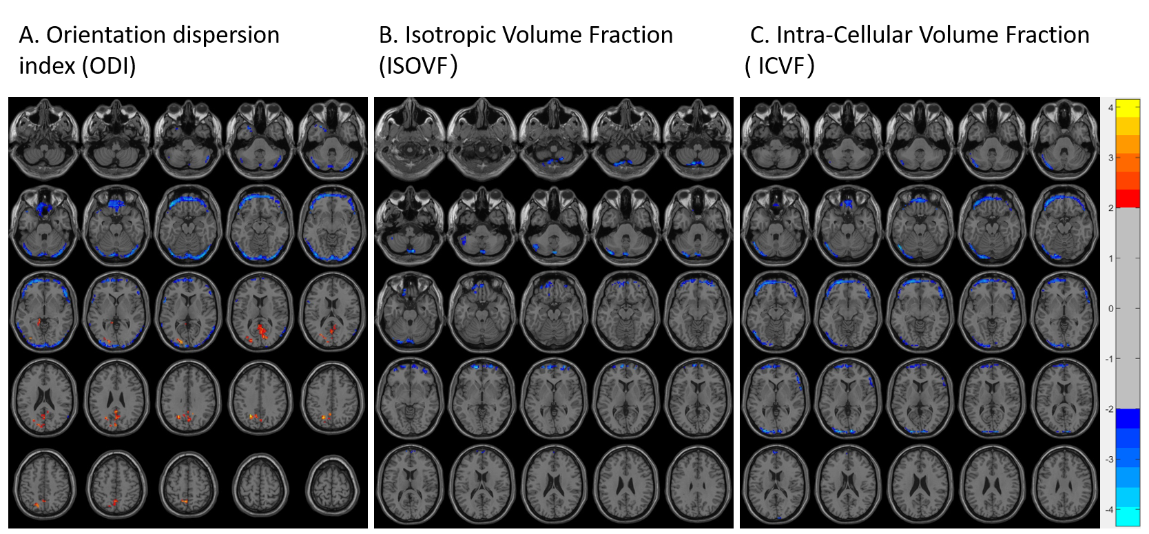
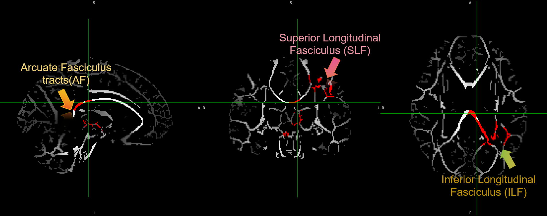
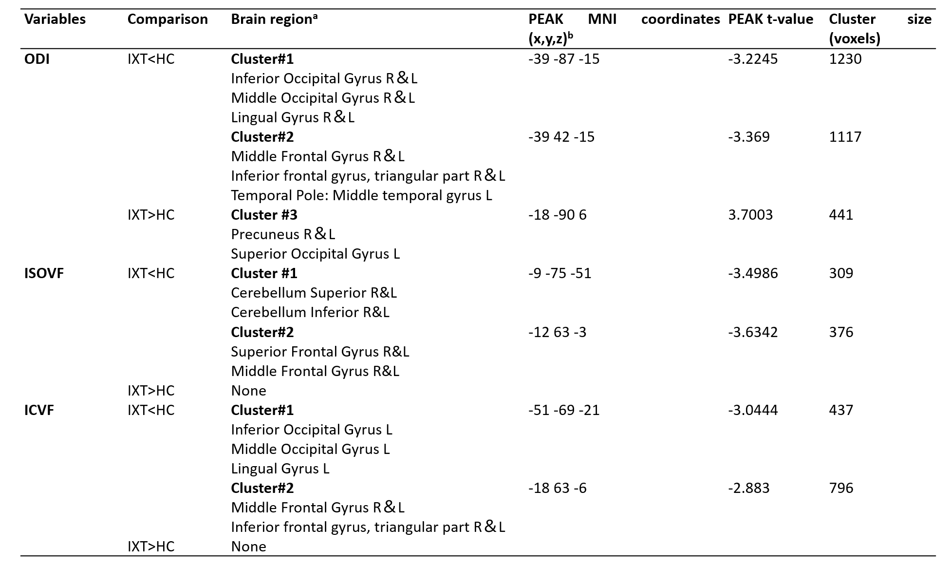
Table1. Significant differences of NODDI values between different brain regions of IXT and HC group.
a L or R refers to the left or right hemisphere. b MNI: Montreal Neurological Institute. x, y, and z reflect coordinates for peak voxel or other local maxima in MNI space. IXT, intermittent exotropia; HC, healthy control; ODI, the orientation dispersion index, ISOVF, the isotropic volume fraction;ICVF, the intra-cellular volume fraction.