5073
Synthetic high-b-value MUSE DWI in preoperative differentiation of high-grade glioma from low-grade glioma1Department of Radiology, Taihe Hospital, Hubei University of Medicine, Shiyan 442000, China, ShiYan, China, 2GE Healthcare, MR Research China, Beijing, TaiWan, China
Synopsis
Keywords: DWI/DTI/DKI, Diffusion/other diffusion imaging techniques
Motivation: High-b-value diffusion weighted imaging (DWI) and apparent diffusion coefficient (ADC) with b value = 3000 s/mm2 are independent predictors of overall survival.
Goal(s): To explore the feasibility of synthetic high-b-value DWI and ADC values in diagnosis of glioma using actually scanned ones as standard reference.
Approach: To assess scanned and synthetic multiple high-b-value MUSE DWI and ADC of 5 patients with low-grade glioma and 3 patients with high-grade glioma.
Results: All synthetic high-b-value DWIs had superior image quality and diagnostic efficacy of glioma to all scanned DWIs. The scanned and synthetic ADC were significantly different between different b values.
Impact: Synthetic high-b-value MUSE DWI can provide better image quality and diagnostic efficiency of grading glioma without consuming extra scan time, encouraging us to promote the utility of setting up synthetic high-b-value MUSE DWI as routine.
Introduction:
Glioma is the most common primary malignant tumors in the brain. It is of great magnificence to accurately distinguish between low-grade gliomas and high-grade gliomas [1]. At present, grading glioma is still based on pathological detection. The routine MRI including diffusion weighted imaging (DWI) before surgery is used to visualize tumor edge, residuals and even fiber structures using diffusion tensor imaging (DTI), for it can reflect water molecules in tissues [2]. Apparent diffusion coefficient (ADC) value is calculated from the DWI images with b value of 0 (low) and 1000 (high) s/mm2 in the preoperative diagnosis and prognosis assessment of brain glioma [3]. A novel Multiplexed Sensitivity Encoding (MUSE) DWI has been used in clinical practices and research on hippocampal sclerosis, endometrial cancer, Crohn's Disease, and pancreas for less geometric distortion and better image quality at high spatial resolution [7-10]. High-b-value DWI imaging has been widely used in clinical applications but is challenging to obtain ones with high signal-to-noise ratio and good image quality. Especially after surgery, distortion around tumor-restationed cavity is often visible to eyes on conventional single-shot echo planar imaging DWI (SS-EPI-DWI). Therefore, we aimed to investigate synthetic multiple high-b-value DWI and ADC values in diagnosis of glioma using actually scanned high-b-value DWI and ADC values as standard reference.Materials and methods:
A total of 8 glioma patients were recruited in this study, and the patients were divided into a high-grade group and a low-grade group according to the World Health Organization Classification of Central Nervous System Tumors published in 2021. All participants underwent an MRI examination with a multiple-b-value DWI protocol (TR/TE = 4500/88.5ms, FOV= 22×18cm2, matrix size = 110×92, slice thickness = 4, b value = 1500, 2000, 2500, 3000, 3500, 4000 s/mm2) and with a two-b-value DWI protocol (TR/TE = 4500/88.5ms, FOV= 22×18cm2, matrix size = 110×92, slice thickness = 4, b value = 0, 1000 s/mm2) to synthesize high-b-value DWI scheme (b = 1500, 2000, 2500, 3000, 3500, and 4000 s/mm2). To avoid bleeding, necrosis, and calcification, three regions of interest (ROI) were mapped at the same maximum cross-section of the tumor, and the signal intensity and corresponding ADC values of the actual and synthetic images were measured.Variance homogeneity and normality were assessed by Levene test and Shapiro-Wilk test, and differences in signal strength (SI) and derived ADC values between the two groups were compared using Wilcoxon test or paired T-test. Independent sample t test or Mann-Whitney U test were used to compare the signal intensity and derived ADC values of synthetic DWI with different b values and actual scanned DWI with different b values, respectively. The diagnostic sensitivity, specificity and AUC of each parameter were calculated through the analysis of subjects' operating characteristics.Results:
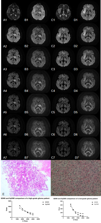
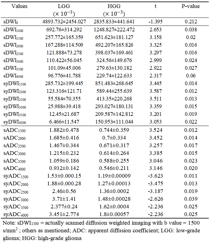
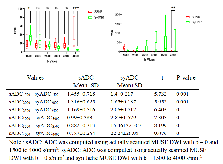
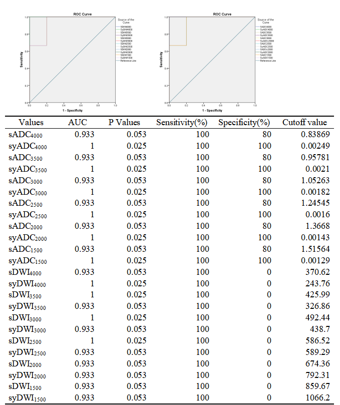
HGG had higher signal intensity of DWI and lower ADC values of all different b values of either actually scanned or synthetic ones than LGG (p<0.001, Figure 1, Table 1). There were significant differences of the SI on DWIs, ADC values, SNR and CNR between actually scanned DWI and synthetic DWI (Table 2 and Table 3). Scanned and synthetic high-b-value DWI and ADC values had excellent diagnostic efficacy on differentiation of HGG and LGG (AUC = 0.933 and AUC = 1, p < 0.05, Figure 2).Discussion
Synthetic high-b-value MUSE DWI showed significant higher SNR and CNR than scanned ones, improving the diagnostic efficiency [11]. Despite significant different of synthetic and scanned ADC values, synthetic ADC values showed better diagnostic performance as a previous study of small field-of-view synthetic high-b-value diffusion-weighted imaging (rFOV-syDWI) in the diagnosis of cervical cancer [12]. In Figure 1, SI of syDWIs with b value over 2000 s/mm2 showed different decreasing tendency, for high-grade glioma has higher cell density and angiogenesis than low-grade glioma. As the synthetic-DWI fitting equation was obtained using only two b values, linear SI decreasing did not appropriately reflect water motion in glioma. Even so, decreasing rate of SI still may distinguish high-grade gliomas than low-grade ones. Additionally, it should be cautious to refer synthetic ADC when b value = 2000 s/mm2 or more. Despite small sample size, synthetic high-b-value MUSE DWI showed the superior diagnostic efficiency to actual scanned ones, encouraging to future attempt in different part of body in particularly without additional scan time.Conclusion
Synthetic high-b-value MUSE DWI generated by using only two scanned MUSE DWI with b value = 0 and 1000 s/mm2 greatly encouraged clinics to set up this function and promoted utility when diagnosis.Acknowledgements
No acknowledgement found.References
[1] Kang XW, Xi YB, Liu TT, et al. Grading of Glioma: combined diagnostic value of amide proton transfer weighted, arterial spin labeling and diffusion weighted magnetic resonance imaging[J].BMC Med Imaging, 2020, 20(1): 50.
[2] Förster A, Brehmer S, Seiz- Rosenhagen M, et al. Heterogeneity of glioblastoma with gliomatosis cerebri growth pattern on diffusion and perfusion MRI[J]. J Neurooncol, 2019, 142(1): 103-9.
[3] Zhang L, Min ZQ, Tang M, et al. The utility of diffusion MRI with quantitative ADC measurements for differentiating high- grade from low-grade cerebral gliomas: evidence from a meta- analysis[J]. J Neurol Sci, 2017, 373: 9-15.
[4] Eyüboğlu İ, Çakir İM, Aslan S, et al. Diagnostic efficacy of apparent diffusion coefficient measurements in differentiation of malignant intra-axial brain tumors[J]. Turk J Med Sci, 2021, 51(1): 256-67.
[5] Cao HM, Xiao X, Hua J, et al. The added value of inflow-based vascular-space-occupancy and diffusion-weighted imaging in preoperative grading of gliomas[J]. Neurodegener Dis, 2020, 20(4): 123-30.
[6] Zeng Q, Ling C, Shi F, et al. Glioma infiltration sign on high b-value diffusion-weighted imaging in gliomas and its prognostic value[J]. J Magn Reson Imaging, 2018: 2018Mar1.
[7] Li J, Bai YC, Wu LH, et al. Synthetic relaxometry combined with MUSE DWI and 3D-pCASL improves detection of hippocampal sclerosis. Eur J Radiol. 2022;157:110571. doi:10.1016/j.ejrad.2022.110571.
[8] Ota T, Tsuboyama T, Onishi H, et al. Diagnostic accuracy of MRI for evaluating myometrial invasion in endometrial cancer: a comparison of MUSE-DWI, rFOV-DWI, and DCE-MRI. Radiol Med. 2023;128(6):629-643. doi:10.1007/s11547-023-01635-4.
[9] Chang HC, Chen G, Chung HW, et al. Multi-shot Diffusion-Weighted MRI With Multiplexed Sensitivity Encoding (MUSE) in the Assessment of Active Inflammation in Crohn's Disease. J Magn Reson Imaging. 2022;55(1):126-137. doi:10.1002/jmri.27801.
[10] Bai Y, Pei Y, Liu WV, et al. MRI: Evaluating the Application of FOCUS-MUSE Diffusion-Weighted Imaging in the Pancreas in Comparison With FOCUS, MUSE, and Single-Shot DWIs. J Magn Reson Imaging. 2023;57(4):1156-1171. doi:10.1002/jmri.28382.
[11] Harder F N, Jung E, Weiss K, et al. Computed high-b-value high-resolution DWI improves solid lesion detection in IPMN of the pancreas[J]. Eur Radiol, 2023,33(10):6892-6901.
[12] Tang Q, Zhou Q, Chen W, et al. A feasibility study of reduced full-of-view synthetic high-b-value diffusion-weighted imaging in uterine tumors[J]. Insights Imaging, 2023,14(1):12.
Figures