5072
Impact of rTMS on the glymphatic system in patients with first-episode depression before and after treatment1Lanzhou University Second Hospital, Lanzhou, China, 2GE HealthCare MR Research, Beijing, China, Beijing, China
Synopsis
Keywords: Diffusion Reconstruction, Tractography & Fibre Modelling, rTMS,first-episode depression
Motivation: The neuropathological mechanisms of repetitive transcranial magnetic stimulation (rTMS) for the treatment of depression are unclear.
Goal(s): The aim of this study was to explore the effects of rTMS on the brain glymphatic system using Diffusion MR Technique in patients with first-episode depression before and after treatment.
Approach: Diffusion tensor image analysis along the perivascular space (DTI-ALPS) as a non-invasive method for evaluating the activity of the glymphatic system in human brain by using diffusion images.
Results: The left ALPS index of patients with first-episode depression is lower than that of healthy people, and it is increased after rTMS treatment.
Impact: This study demonstrated the DTI-ALPS method to assess lymphatic system activity in first-episode depression patients before and after rTMS treatment. The abnormality of ALPS index may provide a potential biomarker for explaining depression, and rTMS can cause changes in ALPS.
Introduction
Repetitive transcranial magnetic stimulation (rTMS), as an effective treatment for depression, can rapidly and powerfully change brain activity, but its neural basis is still unclear1. The glymphatic system is a recently discovered waste drainage system in the brain. This system promotes elimination of soluble proteins, also facilitates the distribution of glucose, lipids, amino acids, and neuromodulators. It has been suggested that glymphatic dysfunction may be exacerbating the depressive and cognitive behavioral phenotypes 2. In addition, depressive disorder is also a risk factor for neurodegenerative diseases3. Diffusion tensor image analysis along the perivascular space (DTI-ALPS) as a non-invasive method for evaluating the activity of the glymphatic system in human brain by using diffusion images4. Therefore, the aim of this study was to apply DTI-ALPS to compare the glymphatic system differences between before and after rTMS treatment in first-episode depression and to explore the relationship between depression and psychiatric scales.Methods
Twenty patients with first-episode depression treated with rTMS (The left dorsolateral prefrontal lobe cortex was stimulated with 10 Hz rTMS for 2 weeks) and Twenty age-, sex-, and educational-matched healthy controls (HCs) were recruited. All subjects underwent the same neuropsychological testing (Montreal Cognitive Assessment (MoCA),sedation-agitation scale (SAS) and Hamilton Depression Scale (HAMD)). Diffusion imaging was acquired by using a 3T MR scanner (SIGNATM Premier; GE Healthcare, Waukesha, WI, USA) with a 48-channel head coil. A whole Brain diffusion data was performed with a multi-slice single-shot spin-echo echo-planar image (SE-EPI, TR = 5705 ms, TE = 68.8 ms, MPG = 120 directions, FOV = 240 mm, matrix = 120 × 120, 78 slices, slice thickness = 2 mm) set in 20 directions at b = 1000 s/mm2, 40 directions at b = 1800 s/mm2, and 60 directions at b = 2500 s/mm2. Diffusion MRI data analysis was performed by using FSL (FSL; www.fmrib.ox.ac.uk/fsl). After calculating the fractional anisotropy (FA) map and co-registering ICBM DTI-81 Atlas, we extracted the periventricular projection (superior and posterior corona radiata) and association fibers (superior longitudinal fasciculus) (Fig. 1). ALPS indexes from projection and association fibers derived from the ICBM DTI-81 Atlas were calculated.Results
There were no group differences in age, sex composition, and years of education. For the neuropsychological testing, there was no significant difference among first-episode depression group, post-treatment group and HC. Depression scores were significantly different among the three groups. (Table 1). The the left ALPS indexes showed significant differences between first-episode depression and HC (P = 0.001). The average ALPS indexes, the left ALPS indexes, and the right ALPS indexes showed no significant differences between post-treatment group and HC (Fig.2). The spearman analysis showed that the left ALPS indexes were negatively correlated with HAMD scores in the first-episode depression group (Fig. 3).Discussion
This study showed that the left ALPS index was lower in the first-episode depression group compared to the HC group. After rTMS treatment, the left ALPS index of the post-treatment group increase. The occurrence of depressive is the result of the synergistic effect of many factors, and the specific etiology is still unclear. It had shown a direct link between the lack of brain neurotransmitters such as serotonin (5-HT) and the development of depression5. The recycling and metabolism of these neurotransmitters depend on the functional integrity of astrocytes. The latest research findings that the glymphatic system also serves as a key player in nutrient distribution, brain energy supply (via glucose delivery), and lipid transport and signaling. The damage of the lymphatic system leads to the abnormal calcium signal transduction of astrocytes, which results in the dysfunction of astrocytes6. Meanwhile, a higher ALPS index represented better glymphatic activity. The left ALPS index was lower in the first-episode depression group, suggesting that the lymphatic function was impaired and neurotransmitter transport was weakened, which may be one of the causes of depression.In addition, we found that ALPS increased after rTMS stimulation of the left dorsolateral prefrontal cortex. We hypothesized that high-frequency rTMS stimulation of the corresponding cortex could increase the metabolic level of adjacent brain regions, increase neuronal excitability, and further transmit through white matter fibers, resulting in changes in projection fibers, association fibers, and subcortical fibers cross fiber diffusivity and thus leads to an increase in the APLS index.
Conclusion
The DTI-APLS method can be used for evaluating the activity of the glymphatic system. A higher ALPS index represented better glymphatic activity. The left ALPS index is decreased in first-episode depression, which may indicate lymphatic system dysfunction. The treatment with rTMS can improve APLS in patients with depression.Acknowledgements
This work was supported by Gansu Province Clinical Research Center for Functional and Molecular Imaging (No. 21JR7RA438) and Gansu Provincial Science and Technology Program Projects(No. 22JR11RA084).References
1. Luo X, Hu Y, Wang R, Zhang M, Zhong X, Zhang B. Individualized rTMS Treatment for Depression using an fMRI-Based Targeting Method. J Vis Exp. 2021 Aug 2;(174). doi: 10.3791/62687. PMID: 34398163.
2. Liang S, Lu Y, Li Z, Li S, Chen B, Zhang M, Chen B, Ji M, Gong W, Xia M, Verkhratsky A, Wu X, Li B. Iron Aggravates the Depressive Phenotype of Stressed Mice by Compromising the Glymphatic System. Neurosci Bull. 2020 Dec;36(12):1542-1546. doi: 10.1007/s12264-020-00539-x. Epub 2020 Jun 24. PMID: 32578069; PMCID: PMC7719143.
3. Miebach L, Wolfsgruber S, Frommann I, Fließbach K, Jessen F, Buckley R, Wagner M. Cognitive Complaints in Memory Clinic Patients and in Depressive Patients: An Interpretative Phenomenological Analysis. Gerontologist. 2019 Mar 14;59(2):290-302. doi: 10.1093/geront/gnx208. PMID: 29325011.
4. Taoka T, Masutani Y, Kawai H, et al. Evaluation of glymphatic system activity with the diffusion MR technique: diffusion tensor image analysis along the perivascular space (DTI-ALPS) in Alzheimer's disease cases. Jpn J Radiol. 2017;35(4):172-178. doi:10.1007/s11604-017-0617-z.
5. Yohn CN, Gergues MM, Samuels BA. The role of 5-HT receptors in depression. Mol Brain. 2017 Jun 24;10(1):28. doi: 10.1186/s13041-017-0306-y. PMID: 28646910; PMCID: PMC5483313.
6. Xuan X, Zhou G, Chen C, Shao A, Zhou Y, Li X, Zhou J. Glymphatic System: Emerging Therapeutic Target for Neurological Diseases. Oxid Med Cell Longev. 2022 Jun 11;2022:6189170. doi: 10.1155/2022/6189170. Retraction in: Oxid Med Cell Longev. 2023 Aug 2;2023:9806739. PMID: 35726332; PMCID: PMC9206554.
Figures
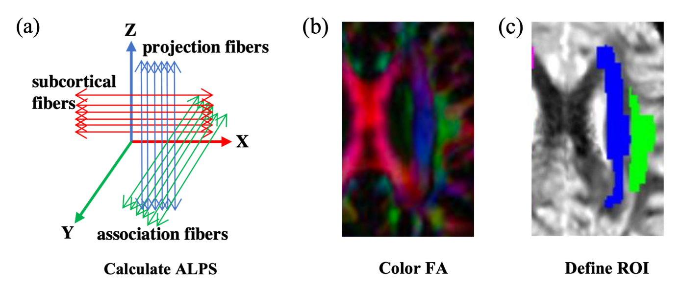
Fig. 1 The concept of the diffusion tensor image analysis along with the perivascular space (DTI-ALPS) method for perivascular diffusion.
Note: (a) Schematic diagram showing the relationship between the direction of the perivascular space (gray cylinder) and the direction of the fibers. (b) The DTI superimposed color display shows the distribution of projection fibers. (c)Using ICBM DTI-81 Atlas to extract the periventricular projection (superior and posterior corona radiata) and association fibers (superior longitudinal fasciculus).
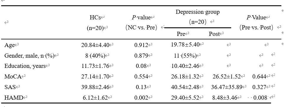
Table 1. The results of Neuropsychological test in normal controls and before and after depression treatment
Note: Data are expressed as mean ± standard deviation. p, differences in values between normal controls and Depression group according to t test and differences in values before and after rTMS therapy according to paired t test.
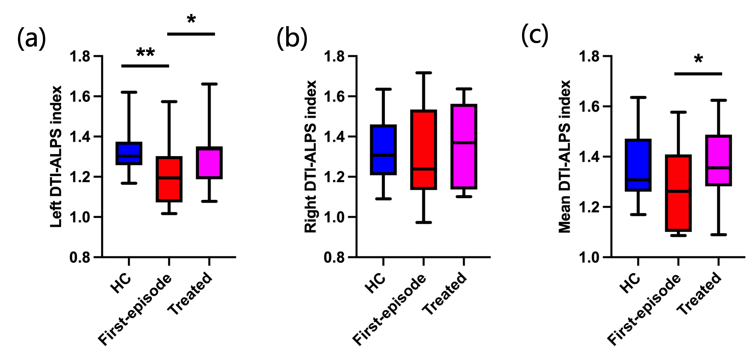
Fig. 2 The results of ALPS index in normal controls and depression before and after treatment .
Note: (a) The left ALPS index:The the left ALPS indexes showed significant differences between first-episode depression and HC (P = 0.001). (b) The right ALPS index,(C) The mean ALPS index:The average ALPS indexes, the left ALPS indexes, and the right ALPS indexes showed no significant differences between post-treatment group and HC
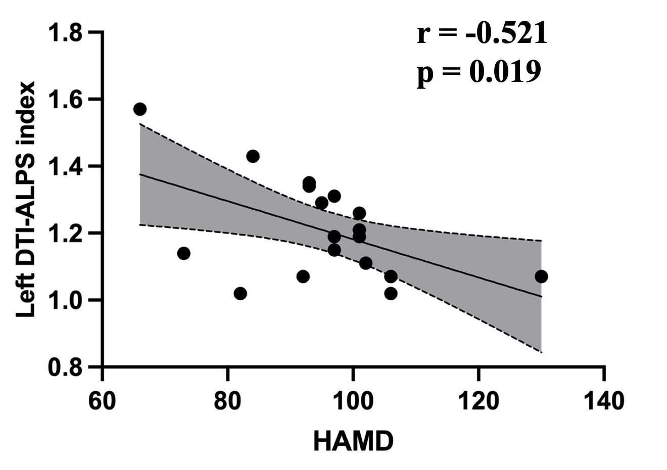
Fig. 3 Correlation analysis between ALPS index and Hamilton Depression Scale score.
Note: The left ALPS indexes were negatively correlated with Hamilton Depression Scale score in the first-episode group (r=-0.521,p=0.019).