5068
Associations between time-dependent diffusion MRI parameters and WHO/ISUP grade of clear cell renal cell carcinoma1Radiology, West China Hospital of Sichuan University, Chengdu, China, 2Department of Clinical, Philips Healthcare, China, Chengdu, China
Synopsis
Keywords: Microstructure, Kidney
Motivation: OGSE-MRI provides more information on kidney cancer prognosis, metastasis possibility, specific survival rate in the future.
Goal(s): To explore whether parameters derived from OGSE can demonstrate microstructure in clear cell renal cell carcinoma
Approach: Patients suspected of ccRCC who underwent diffusion-weighted imaging were employed to quantify the time-dependent diffusion MRI-based microstructural parameters cell diameter (D), intracellular volume fraction (vin), cellularity, and diffusivities (Dex).
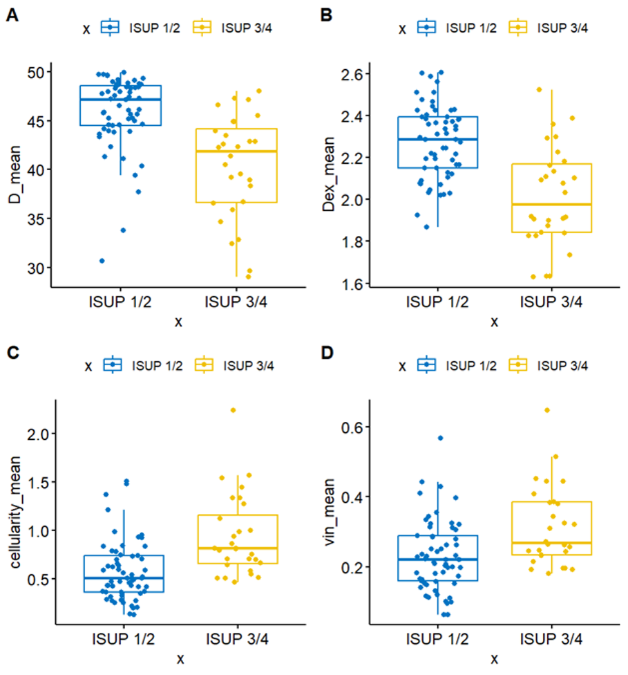
Results: ccRCC with high WHO/ISUP grade show higher cellularity and vin, while lower D and Dex parameters.
Impact: The OGSE MRI parameters correlates with pathologic grades and demonstrates promise for characterizing renal carcinoma.
Introduction
Oscillating gradient diffusion magnetic resonance imaging (MRI) enables elucidation of microstructural characteristics in cancers; however, there are limited data to evaluate its utility in patients with renal carcinoma. This study aimed to investigate the values of time-dependent diffusion MRI parameters in predicting the grade of clear cell renal cell carcinoma (ccRCC).Methods
Patients suspected of ccRCC who underwent diffusion-weighted imaging were prospectively recruited between October 2022 and September 2023. The MR examination was performed on a 3T MR scanner (Elition, Philips). A two-compartment model was employed to quantify the time-dependent diffusion MRI-based microstructural parameters cell diameter (D), intracellular volume fraction (vin), cellularity, and diffusivities (Dex). Finally, the patient's post-operative histological results confirmed the World Health Organization/International Society of Urology Pathology (WHO/ISUP) classification.Results
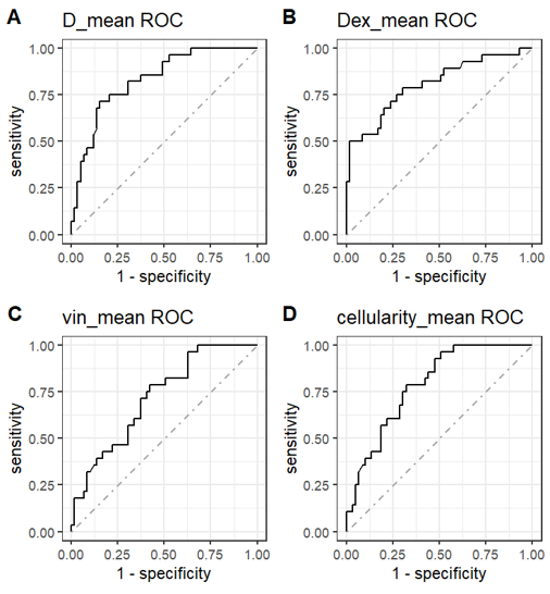
87 patients with ccRCC (mean age: 54 ± 13.2 years) including 3 WHO/ISUP grade I, 56 WHO/ISUP grade II, 17 WHO/ISUP grade III and 11 WHO/ISUP grade IV were finally enrolled. No significant difference was observed between low-grade and high-grade regarding age, sex and surgery method. Compared to high-grade (WHO/ISUP grade III/IV) tumors, low-grade (WHO/ISUP grade I/II) tumors exhibited lower cellularity (0.57 ± 0.04 vs. 0.93 ± 0.08 ;p<0.0001 ) and vin values (0.23 ± 0.01 vs. 0.31± 0.02 ;p<0.0001), while showing relatively higher D (45.86 ± 0.49 vs. 40.37 ±1.00 ;p<0.0001) and Dex values ( 2.28 ± 0.02 vs. 2.01 ± 0.05;p<0.001). For histological classification of low-grade and high-grade ccRCC, the D values showed the highest diagnostic performance (AUC: 0.83, 95% CI: 0.74-0.92), slightly higher than the Dex values (AUC: 0.80, 95%CI: 0.70-0.91), cellularity mean values (AUC: 0.79, 95% CI: 0.69-0.88), and vin mean values (AUC: 0.72, 95% CI: 0.61-0.83).Discussion
Our findings reveal that cellularity and vin obtained from OGSE sequence are strongly connected with ccRCC WHO/ISUP grade, however D and Dex parameters are adversely correlated. This suggests that OGSE sequences can reflect cellular microstructure and provide information regarding CCRCC tissue grading. This is because high-grade ccRCC has larger nucleoli, higher cell density, less extracellular space, faster cell proliferation, and a more complicated architecture, making it more susceptible to diffusion limitation1. ccRCC is the most common renal tumor and is more malignant and aggressive, compared with chromone renal cell carcinoma and papillary renal cell carcinoma2,3. WHO/ISUP classification is one of the critical independent factors suggesting the prognosis of RCC, high-grade ccRCC is more likely to metastasize and usually requires comprehensive management treatments4. Consequently, preoperative ccRCC grading of ccRCC has a substantial value in shaping tumor management strategies and surgical plans. Time-dependent diffusion MRI shortens the diffusion time by substituting the long diffusion sensitizing gradients used in PGSE methods with rapid oscillation gradients5. Therefore, OGSE can be utilized to build biophysical models for characterizing the cellular microstructure, such as cell size, cell volume fraction, and transcytolemmal water exchange. OGSE-DWI has the ability to discriminate cell size in the inflammatory environment which has potential applications in tumorigenesis and metastasis6. Studies have shown that OGSE has been applied to characterize the grading and staging of gliomas, prostate cancer grading, breast tumors, etc., and has been validated by histopathology5,7-8. In a prospective study, Ba et al. verified that OGSE parameters were associated with Her 2 and LN expression in breast cancer and offered information regarding patient prognosis and specific survival rate8. We look forward to OGSE also providing more information on kidney cancer prognosis, metastasis possibility, and specific survival rate in the future.Conclusion
The time-dependent diffusion MRI parameters correlate with pathologic grades and demonstrate promise for characterizing renal carcinoma.Acknowledgements
None.References
- Kinouchi T, Mano M, Matsuoka I, Kodama S, Aoki T, Okamoto M, Yamamura H, Usami M, Takahashi K. Immature tumor angiogenesis in high-grade and high-stage renal cell carcinoma. Urology. 2003 Oct;62(4):765-70.
- Moch H, Cubilla AL, Humphrey PA, Reuter VE, Ulbright TM. The 2016 WHO Classification of Tumours of the Urinary System and Male Genital Organs-Part A: Renal, Penile, and Testicular Tumours. Eur Urol. 2016 Jul;70(1):93-105.
- Finelli A, Cheung DC, Al-Matar A, Evans AJ, Morash CG, Pautler SE, Siemens DR, Tanguay S, Rendon RA, Gleave ME, Drachenberg DE, Chin JL, Fleshner NE, Haider MA, Kachura JR, Sykes J, Jewett MAS. Small Renal Mass Surveillance: Histology-specific Growth Rates in a Biopsy-characterized Cohort. Eur Urol. 2020 Sep;78(3):460-467.
- Abu-Ghanem Y, Powles T, Capitanio U, Beisland C, Järvinen P, Stewart GD, Gudmundsson EO, Lam TB, Marconi L, Fernandéz-Pello S, Nisen H, Meijer RP, Volpe A, Ljungberg B, Klatte T, Dabestani S, Bex A. The Impact of Histological Subtype on the Incidence, Timing, and Patterns of Recurrence in Patients with Renal Cell Carcinoma After Surgery-Results from RECUR Consortium. Eur Urol Oncol. 2021 Jun;4(3):473-482.
- Wu D, Jiang K, Li H, Zhang Z, Ba R, Zhang Y, Hsu YC, Sun Y, Zhang YD. Time-Dependent Diffusion MRI for Quantitative Microstructural Mapping of Prostate Cancer. Radiology. 2022 Jun;303(3):578-587.
- Hoffmann E, Gerwing M, Niland S, Niehoff R, Masthoff M, Geyer C, Wachsmuth L, Wilken E, Höltke C, Heindel WL, Hoerr V, Schinner R, Berger P, Vogl T, Eble JA, Maus B, Helfen A, Wildgruber M, Faber C. Profiling specific cell populations within the inflammatory tumor microenvironment by oscillating-gradient diffusion-weighted MRI. J Immunother Cancer. 2023 Mar;11(3):e006092.
- Zhang H, Liu K, Ba R, Zhang Z, Zhang Y, Chen Y, Gu W, Shen Z, Shu Q, Fu J, Wu D. Histological and molecular classifications of pediatric glioma with time-dependent diffusion MRI-based microstructural mapping. Neuro Oncol. 2023 Jun 2;25(6):1146-1156.
- Ba R, Wang X, Zhang Z, Li Q, Sun Y, Zhang J, Wu D. Diffusion-time dependent diffusion MRI: effect of diffusion-time on microstructural mapping and prediction of prognostic features in breast cancer. Eur Radiol. 2023 Sep;33(9):6226-6237.
Figures