5067
Differentiation of benign and malignant breast lesions using DWI with a fractional-order calculus(FROC) model based on SMS technique1Anqing Municipal Hospital, Anqing, China, 2MR scientific Marketing,Diagnostic Imaging, Siemens Healthineers Ltd., Shanghai, China
Synopsis
Keywords: Diffusion Modeling, Breast
Motivation: This study focus on improving imaging time and image quality while accurately distinguishing between benign and malignant breast lesions.
Goal(s): To assess the diagnostic capabilities of the FROC diffusion model combined with SMS technology for breast lesions.
Approach: Compare two diffusion model, the traditional single-index DWI model and the FROC, and then evaluate their diagnostic performance, image quality, and the consistency of results using SMS technology.
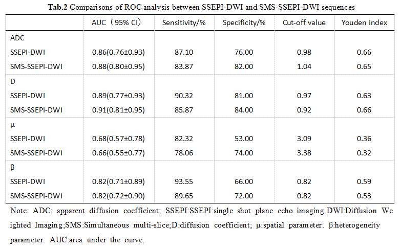
Results: FROC combined with SMS offered a feasible and effective approach for distinguishing benign and malignant breast lesions, while the diagnostic performance of D and β values from FROC was potentially superior to the traditional ADC values.
Impact: The research introduces an innovative approach that combines the FROC-DWI model with SMS technology to enhance the efficiency and diagnostic capabilities of breast imaging, potentially improving patient outcomes and reducing unnecessary invasive procedures.
Introduction
Breast cancer is one of the most common malignancies in women, and magnetic resonance imaging (MRI) plays a significant role in the diagnosis and therapeutic evaluation. The FROC model is more sensitive to microstructural and heterogeneity changes in tumor tissue, providing a better reflection of the complexity and heterogeneity of tissue microstructure. It provides a new set of parameters, including the abnormal diffusion coefficient (D), intravoxel diffusion heterogeneity parameter (β), and spatial parameter (μ).Moreover, the FROC model involves a variety of b-values with inconsistent settings, resulting in relatively long sequence scan times, limiting its clinical applicability. And Simultaneous multi-slice (SMS) can reduce scan time. Therefore, the objective of this study is to explore the diagnostic capabilities of breast FROC diffusion models in conjunction with SMS technology concerning imaging time, image quality, and the derived parameters.Clinical Data
A retrospective collection was conducted on 234 female patients who underwent breast MRI scans at our hospital between January 2021 and December 2022. Exclusion criteria included a history of breast lesion radiotherapy, chemotherapy, or surgery, unconfirmed lesions without pathological proof, poor MRI image quality, lesion size less than 5mm, and inflammatory breast lesions. This project received approval from Ethics Commttee of our hospital and all patients granted the exemption from.MRI Examination
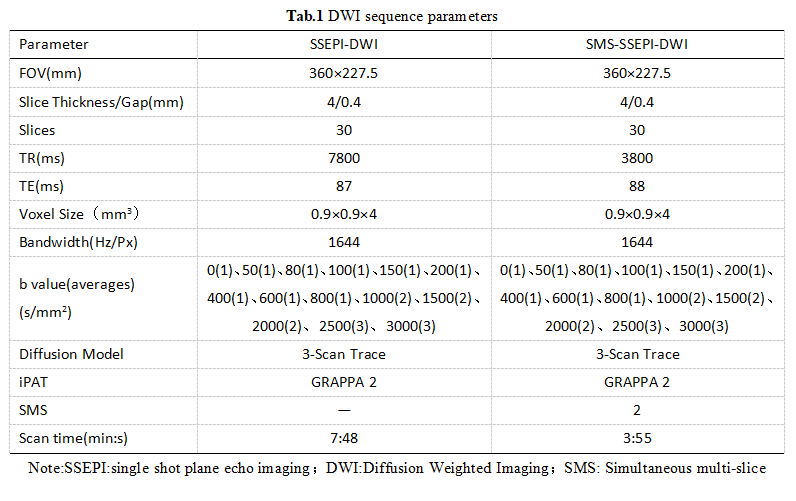
A 3T MRI scanner(MAGNETOM Skyra, Siemens Healthcare) with a dedicated 16-channel phased-array coil for breast MRI scans was used. Two sets of DWI sequences were scanned, one using conventional SSEPI-DWI and the other combining SMS technology (SMS-SSEPI-DWI). (Table 1) Parameters from both the DWI single-exponential model and the FROC model were separately calculated by Body DiffusionLab(BoDiLab).In the region of the lesions, two radiologists with over 5 years of experience in breast MRI diagnosis independently evaluated image artifacts, image sharpness, lesion conspicuity, and overall image quality using a 5-point scale, where (1 point: very poor; 2 points: poor; 3 points: satisfactory; 4 points: good; 5 points: excellent). Both radiologists outlined the images while avoiding cystic, necrotic, and hemorrhagic areas. Statistical AnalysisStatistical analysis was conducted using SPSS 23.0 and MedCalc 20.0 software. Quantitative data were expressed as mean ± standard deviation. Comparisons were made using independent-sample t-tests or Mann-Whitney U tests, and ROC analysis was used to evaluate diagnostic performance. Intraclass correlation coefficients (ICC) were calculated for inter-observer agreement. p<0.05 indicates that the observed difference is statistically significant.Results
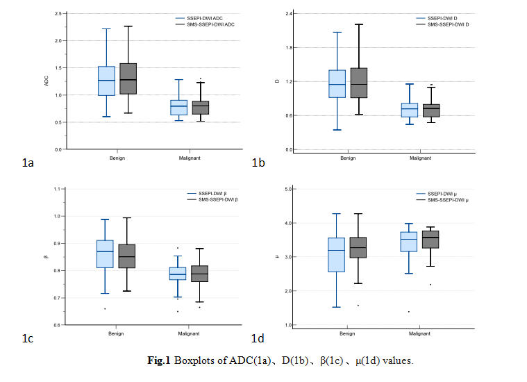
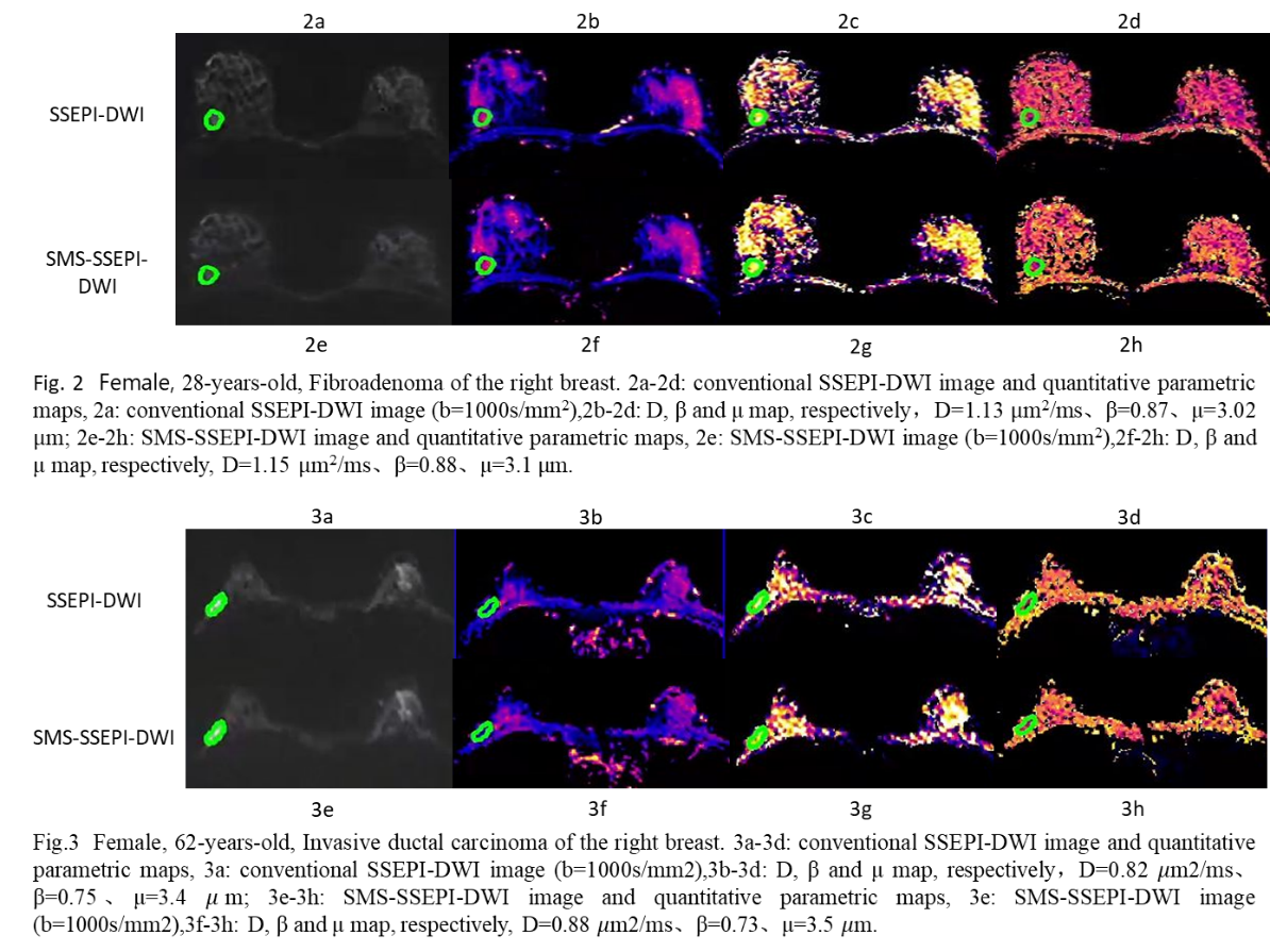
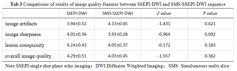
In the study, a total of 124 patients with 141 breast lesions were included. There were 74 left breast lesions and 67 right breast lesions, and the distribution of lesions showed no statistically significant difference (t=2.93, p=0.98). Among these patients, there were 52 cases(age: 43.77±13.11years, range: 25 to 64) of benign breast lesions. And 89 cases(age: 49.13±15.71 years, range: 27 to 71) of malignant breast lesions. The age difference between patients with benign and malignant breast lesions was not statistically significant (t=7.51, p=0.18).Both observers demonstrated good inter-observer agreement for DWI-derived parameters and image quality assessment, with ICC values above 0.75. ICC of SSEPI-DWI was 0.78-0.95, and SMS-SSEPI-DWI was 0.79-0.98. Malignant lesions exhibited significantly lower ADC, D, and β values and a higher μ value compared to benign lesions(p<0.05).D showed the highest AUC value and sensitivity, while β showed the highest specificity.Both DWI sequences showed no statistically significant differences in image quality parameters, including image artifacts, sharpness, lesion conspicuity, and overall image quality.Discussion
This study demonstrates that ADC values and FROC-derived parameters (D, β, μ) obtained from SSEPI-DWI and SMS-SSEPI-DWI sequences can be used to differentiate between benign and malignant breast lesions. These parameters show significant differences between benign and malignant lesions, with D exhibiting the highest AUC and β showing greater sensitivity than ADC. SMS-SSEPI-DWI offers similar diagnostic performance and improved scan time, potentially benefiting future clinical diagnoses and scientific research using multi-b-value DWI for breast lesions.Conclusion
The FROC diffusion imaging model based on non-Gaussian distributions to identify benign and malignant breast lesions is feasible, with D and β showing better diagnostic potential than ADC. SMS-SSEPI-DWI significantly shortens image acquisition time while maintaining comparable image quality and diagnostic performance for derived parameters, which may facilitate clinical breast lesion diagnosis and scientific research using multi-b-value DWI.Acknowledgements
No acknowledgement found.References
[1] XU W, ZHENG B, LI H. Identification of the Benignity and Malignancy of BI-RADS 4 Breast Lesions Based on a Combined Quantitative Model of Dynamic Contrast-Enhanced MRI and Intravoxel Incoherent Motion[J].Tomography,2022,8(6):2676-2686.
[2] SALLAM. H, LENGA L, SOLBACH C,et al. Correlation of background parenchymal enhancement on
breast MRI with breast cancer[J]. Clin Radiol,2023,78(9):e654-e659.
[3] YUAN C, JIN F, GUO X, et al. Correlation Analysis of Breast Cancer DWI Combined with DCE-MRI Imaging Features with Molecular Subtypes and Prognostic Factors[J]. J Med Syst,2019,43(4):83.
Figures