5065
How to best match voxels – evaluating different sequential co-registration strategies for ultra-high b-value DWI in multiparametric breast MRI1Radiology, University hospital Erlangen, Erlangen, Germany, 2Department of Artificial Intelligence in Biomedical Engineering (AIBE), Friedrich-Alexander-University Erlangen-Nuernberg, Erlangen, Germany, 3Chair of Medical Informatics, Friedrich-Alexander-University Erlangen-Nuernberg, Erlangen, Germany
Synopsis
Keywords: DWI/DTI/DKI, Breast
Motivation: Breast MRI increasingly includes ultra-high b-value DWI, but technical and patient-related challenges can cause mismatches to morphologic sequences. Co-registration strategies are needed to reliably apply AI-algorithms to the data.
Goal(s): The study examines how different co-registration orders for ultra-high b-value acquisitions in multiparametric breast MRI impact co-localization to morphologic sequences, both quantitatively and qualitatively.
Approach: This IRB-approved retrospective study of 144 multiparametric breast MRI exams with ultra-high b-value DWI, assessed different co-registration algorithms using ANTs library methods quantitatively and qualitatively.
Results: The sequential arrangement of contrasts used during co-registration significantly affects cross-sequence congruence of lesions in multiparametric breast MRI incorporating ultra-high b-value DWI.
Impact: The study highlights the relevance of considering different sequential arrangements used during co-registration in ultra-high b-value DWI.
INTRODUCTION
Breast magnetic resonance imaging (MRI) with dynamic contrast-enhanced (DCE) acquisitions is the most sensitive imaging modality to detect suspicious breast lesions1. Integrating diffusion-weighted imaging (DWI) can add to MRI protocols, offering complementary value in lesion detection and characterization. Ultra-high b-value DWI (at around b=1500s/mm²) is increasingly investigated in specific settings, e.g. early detection of cancer. However, technical challenges such as distortion artifacts and fat-saturation issues, along with lengthy examination times, can impact image quality and pose challenges to DWI.With the increasing use of artificial intelligence (AI) algorithms in breast MRI, e.g. for lesion detection, characterization or for deriving virtual enhancement images from unenhanced breast MRI acquisitions2, technical cross-sequence congruence of the acquisitions become increasingly relevant. This is important since the commonly relatively small lesions in breast MRI might miss overlapping voxels in the acquisition matrix already with small movements or artifacts and can thus impede performance of AI algorithms (Figure 1).
This study explores different approaches of sequential arrangements during the co-registration, aiming to decipher the optimal order and to increase conformity of voxel-wise tissue co-locations in between ultra-high b-value DWI and the morphologic MRI sequences.
METHODS
Data: This IRB-approved retrospective study included n=144 female patients (mean age: 52 ± 12 years) examined in clinical routine in a university hospital. A 3T MRI (Siemens MAGNETOM Vida and Skyra Fit) was used to acquire the routine multiparametric breast MRI protocol, which includes T1-weighted (one before and five after GBCA-injection), T2-weighted fat saturated and a multi b-value DWI (DWI_50: b=50s/mm2, DWI_750: b=750s/mm2, DWI_1500: b=1500s/mm2).Algorithms: We used the Ants library, a medical image registration and segmentation toolkit, for co-registration of the independent test dataset3. Using the 'SyN' registration method, which integrates symmetric normalization (affine + deformable transformation) with mutual information as the optimization metric, all sequences were co-registered. The following n=4 different successions of co-registration were applied A: DWI_1500 to T1w; B: DWI_1500 to DWI_750 to T1w; C: DWI_1500 to DWI_50 to T1w, D: DWI_1500 to DWI_750 to DWI_50 to T2w_fs to T1w (Figure 2).
Experiments: A qualitative assessment was performed on n=50 randomly selected images, each with lesion segmentation provided by a medical reviewer. A 5-point Likert scale was used to evaluate overall image quality and lesion congruency in between the ultra-high b-value DWI and T1w enhanced subtraction sequences for the different co-registration scenarios. Means and standard deviations were calculated and frequency counts of each rating were analyzed. Statistical analysis was performed using the chi-squared test for comparison4. In addition, a quantitative evaluation was performed on all n=144 subjects by calculating signal intensity (SI) in segmented lesions in the DWI_1500 images after co-registration as compared to the original DWI_1500 acquisition.
RESULTS
Despite the chi-squared test lacking statistical significance, both mean and frequency counts indicated enhanced lesion co-localization in the DWI_1500 images and the T1w enhanced subtraction images after co-registration, particularly with the methods B and C. Notably, qualitative evaluation revealed a substantial increase in lesion congruency for DWI_1500 images when using these methods, with mean scores improving from 3.44±1.17 to 3.51±0.75 and 3.51±0.81, respectively (Figure 3). The image quality remained consistent for all images post co-registration (Figure 4).This matched the quantitative analysis, demonstrating significant advancements in SI when looking at the segmentation of lesions, with method B displaying the best performance (Figure 4). Specifically, it exhibited the least frequent occurrence of the lowest (B: 5) and the most frequent highest (B: 22) and second highest (B: 41) SI in lesion segmentation. These findings underscore the effectiveness of co-registration method B in improving qualitative and quantitative aspects of lesion localization in DWI_1500 images.
DISCUSSION
Multiparametric breast MRI increasingly incorporates (ultra-high b-value) DWI acquisitions complementing the morphologic sequences during image analysis. Using the data for advanced image processing tasks, such as lesion detection, lesion characterization and/or deriving virtual contrast enhanced image series demands for a high level of congruency of the position of findings in between the acquired sequences. As DWI, especially high b-value acquisitions, can be influenced by motion and distortion, this is of special relevance. Non-matching of the commonly small lesions, with screening detected lesion demonstrating an average of 15mm, might impede the performance and generalizability of AI algorithms for breast MRI.CONCLUSION
This study demonstrates the relevance of carefully considering co-registrations` sequential arrangements as a potential influencing factor for co-registration success and the consecutive performance e.g. of AI algorithms trained and deployed in multiparametric breast MRI when including ultra-high b-value DWI. Future research might further improve the approaches, e.g., by combining global and focal co-registration approaches with regards to individual findings.Acknowledgements
No acknowledgement found.References
1. Turnbull L. Dynamic contrast-enhanced MRI in the diagnosis and management of breast cancer. NMR Biomed. 2009;22(1):28-39.
2. Chen C, Raymond C, Speier W, Jin X, Cloughesy T, Enzmann D, et al. Synthesizing MR image contrast enhancement using 3D high-resolution ConvNets. IEEE Trans Biomed Eng. 2022;70(2):401-412.
3. Avants B, Tustison N, Song G. Advanced normalization tools (ANTS). Insight J. 2009;2(365):1-35.
4. Tallarida R, Murray R. Chi-square test. In: Tallarida R, Murray R, eds. Manual of Pharmacologic Calculations: With Computer Programs. 1987:140-142.Figures
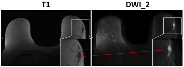
Figure 1: Demonstration of the challenge of lesion displacement within a single examination in different sequences, here as an example between morphologic T1-weighted (T1) and Diffusion Weighted Imaging with b=1500s/mm2 (DWI_1500) sequences showing a lesion in the left breast.
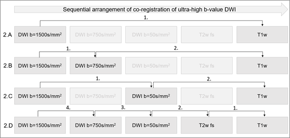
Figure 2: Varied sequential arrangements of co-registration of Diffusion Weighted Imaging with b=1500s/mm2 (DWI_1500) to T1-weighted (T1), incorporating direct co-registration and step-wise co-registration using additional sequences from multiparametric breast MRI, such as T2 weighted images and DWI with b=1500s/mm2 and =1500s/mm2.
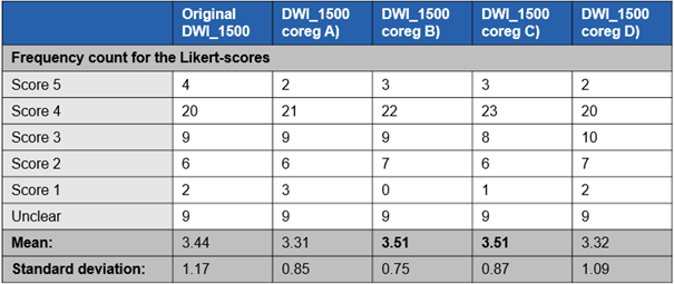
Figure 3: Comparing visually the original Diffusion Weighted Images with b=1500s/mm2 (DWI_1500) and DWI_1500 after applying different co-registration methods, while looking at the 5-point Likert scale ratings from one reader for lesion congruency across 50 test images. Displayed are the frequency counts, mean score and standard deviation of the 5-point Likert scale ratings.
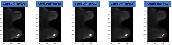
Figure 4: Example of better lesion congruency after applying co-registration to Diffusion Weighted Images with b=1500s/mm2 (DWI_1500), showing consistent image quality across the images. Compared are the original DWI_1500 on the left with different co-registered DWI_1500 on the right. The lesion location is marked with a red square.
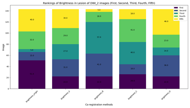
Figure 5: Quantitative evaluation of signal intensities in lesion segmentations for n=144 images, comparing the original Diffusion Weighted Images with b=1500s/mm2 (DWI_1500) and DWI_1500 after applying different co-registration methods. The Figure shows, how often a specific method showed the highest signal intensity in a lesion, hinting to where the segmentation fits best the lesion position.