5056
“Black Bone” MRI: a potential alternative to X-ray with three-dimensional reconstruction for the diagnosis of Legg-Calvé-Perthes disease1Hunan Children's Hospital, Changsha, China, 2MR Collaboration, Siemens Healthineers Ltd, Wuhan, China
Synopsis
Keywords: Bone, Bone
Motivation: Femoral head necrosis can be clearly displayed by Black Bone MRI.
Goal(s): This study aimed to investigate the diagnostic performance of MR imaging with Black Bone sequence in the lateral pillar classification and femoral head extrusion index of Legg-Calvé-Perthes disease (LCPD) in children.
Approach: The study included thirty-eight children with LCPD who underwent both X-ray and MR scans. The diagnostic performance of Black Bone MRI was assessed in comparison to X-ray.
Results: The results showed that Black Bone had no significant difference in measuring the femoral head extrusion index compared to X-ray. For Herring classification, Black Bone MRI performed very well.
Impact: Black Bone MRI can be used as a conventional sequence for the assessment of LCPD in children, allowing patients to avoid ionizing radiation.
Introduction
Legg-Calvé-Perthes disease (LCPD) is a pediatric hip disorder characterized by ischemic injury to the growing femoral head, potentially leading to early-onset osteoarthritis [1]. Clinical investigation of LCPD involves various imaging techniques such as X-rays, arthrography, tomodensitometry, and magnetic resonance imaging (MRI). For long-term follow-up, radiographs are usually sufficient. The modified lateral pillar classification and femoral head extrusion index, measured by pelvic X-ray, are well-known radiologic risk factors closely associated with poor outcomes [2-5]. However, the routine use of X-rays in pediatric patients is limited due to their increased sensitivity to ionizing radiation. MRI, as a non-invasive and radiation-free method, not only shows the shape of the femoral head cartilage but also provides information about soft tissue changes within the joint, including the synovial membrane, round ligament, and glenoid lip. MRI has become an established tool for early diagnosis of LCPD [6-7]. Nevertheless, traditional MRI has limited ability to predict the clinical outcome in the earlier stages of ischemic injury before severe tissue damage occurs. Therefore, there is a need for X-ray visualization of bone structure provided by MR imaging.Previous studies have demonstrated that perfusion weighted imaging and T1ρ, T2 mapping can estimate LCPD in its early stages [8,9]. However, their widespread adoption is hindered by the relatively high hardware and software requirements. Recently, Black Bone MR images have been introduced as a new sequence for evaluating bone abnormalities, such as skull fractures, orofacial deformity, and craniosynostosis [10-12]. This sequence utilizes TEs and TRs, as well as an optimal flip angle, to minimize soft-tissue contrast and enhance the bone-soft-tissue boundary. Importantly, it requires minimal hardware and software resources. Building on the diagnostic quality of the black bone sequence, our aim is to investigate whether Black Bone MR images can serve as an alternative to X-rays for lateral pillar classification and femoral head extrusion index measurement in LCPD.Methods
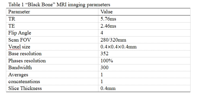
Thirty-eight children (23 boys, mean age, 4.34 years; range, 3.1-11.2years) with LCPD who underwent MRI including “Black Bone” imaging on a 3T system (MAGNETOM Prisma, Siemens Healthcare, Erlangen, Germany), with a short TE, TR, and low flip angle 3D volume acquisition (Table 1). X-ray data and Black Bone MRI were used to determine the severity of LCPD using Herring classification (Herring A, B, and C) and the femoral head extrusion index on both trouble side and control side. According Modified Waldenström classification, which is based on plain radiographic assessment for the staging of LCPD, we reduced to two categories by combining categories Ia and Ib into group 1, categories IIa, IIb, IIIa, IIIb and IV into group 2. Two independent observers, blinded to the others measurement, randomly selected unilateral cases. Inter-reader intraclass correlations (ICC) or Kappa test, paired-sample t test, and receiver operating characteristic (ROC) curve were used to assess the agreement between the two readers, the difference between the MRI and X-ray and between control and trouble sides, and the performance in lateral pillar classification, respectively. P<0.05 was considered statistically significant.Results
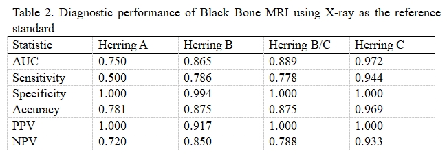
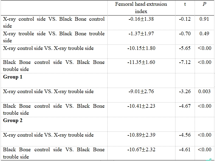
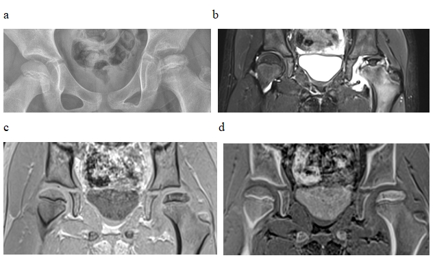
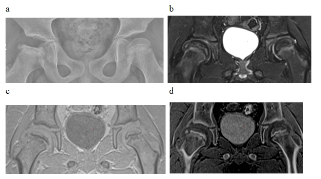
There were high inter-reader agreement for Black Bone MRI in lateral pillar classification (=0.809) and in the femoral head extrusion index (trouble side: ICC=0.988; control side: ICC=0.994). For Herring classification, Black Bone MRI had high diagnostic performance (all AUC0.85) (Table 2). For femoral head extrusion index, there were no significant difference between X-ray dates and Black Bone MRI (p>0.05). In group 1, group 2, and all cases, the differences were statistically significant between the trouble side and control side both Black Bone MRI and X-ray (p<0.05). Figure 1 and Figure 2 shows the patterns of Black Bone MRI and X-ray for femoral head necrosis.Conclusions
Black Bone sequence shows promise as a nonionizing alternative to X-ray for the assessment of LCPD in children.Acknowledgements
The authors thanks ZHT of the MR Scientific Marketing, Siemens Healthineers for data analysis.References
References1. Kim, HK. Pathophysiology and new strategies for the treatment of Legg-Calvé-Perthes disease. J BONE JOINT SURG AM. 2012; 94 (7): 659-69. doi: 10.2106/JBJS.J.01834.2. Herring, JA, Kim, HT, Browne, R. Legg-Calve-Perthes disease. Part I: Classification of radiographs with use of the modified lateral pillar and Stulberg classifications. J BONE JOINT SURG AM. 2004; 86 (10): 2103-20. PMID: 15466719.3. Park, MS, Chung, CY, Lee, KM, et al. Reliability and stability of three common classifications for Legg-Calvé-Perthes disease. CLIN ORTHOP RELAT R. 2012; 470 (9): 2376-82. doi: 10.1007/s11999-012-2314-7.4. Liggieri, AC, Tamanaha, MJ, Abechain, JJ, et al. Intra and interobserver concordance between the different classifications used in Legg-Calvé-Perthes disease. Rev Bras Ortop. 2015; 50 (6): 680-5. doi: 10.1016/j.rboe.2015.09.010.5. Alter, A. Epiphyseal extrusion as a prognostic index in Legg-Calve'-Perthes disease J PEDIATR SURG. 1982; 17 (2): 217-218. doi: 10.1016/s0022-3468(82)80298-4.6. Kim, HK, Kaste, S, Dempsey, M, et al. A comparison of non-contrast and contrast-enhanced MRI in the initial stage of Legg-Calvé-Perthes disease. PEDIATR RADIOL. 2013; 43 (9): 1166-73. doi: 10.1007/s00247-013-2664-7.7. Lahdes-Vasama, T, Lamminen, A, Merikanto, J, et al. The value of MRI in early Perthes' disease: an MRI study with a 2-year follow-up. PEDIATR RADIOL. 1997; 27 (6): 517-22. doi: 10.1007/s0024700501718. Chong David Y,Schrader Tim,Laine Jennifer C,et al.Reliability and Validity of Visual Estimation of Femoral Head Hypoperfusion on Perfusion MRI in Legg-Calve-Perthes Disease.JOURNAL OF PEDIATRIC ORTHOPAEDICS.2021;41 (9):e780-e786.doi:10.1097/BPO.00000000000019459. Armstrong A R,Bhave S,Buko E O,et al.Quantitative T2 and T1ρ mapping are sensitive to ischemic injury to the epiphyseal cartilage in an in vivo piglet model of Legg-Calvé-Perthes disease.OSTEOARTHRITIS AND CARTILAGE.2022;30 (9):1244-1253.doi:10.1016/j.joca.2022.05.009.10. Dremmen, MHG, Wagner, MW, Bosemani, T, et al. Does the Addition of a "Black Bone" Sequence to a Fast Multisequence Trauma MR Protocol Allow MRI to Replace CT after Traumatic Brain Injury in Children? AM J NEURORADIOL. 2017; 38 (11): 2187-2192. doi: 10.3174/ajnr.A5405.11. Kupka, MJ, Aguet, J, Wagner, MM, et al. Preliminary experience with black bone magnetic resonance imaging for morphometry of the mandible and visualisation of the facial skeleton. PEDIATR RADIOL. 2022; 52 (5): 951-958. doi: 10.1007/s00247-021-05257-8.12. Eley, KA, Watt-Smith, SR, Sheerin, F, et al. "Black Bone" MRI: a potential alternative to CT with three-dimensional reconstruction of the craniofacial skeleton in the diagnosis of craniosynostosis. EUR RADIOL. 2014; 24 (10): 2417-26. doi: 10.1007/s00330-014-3286-7.Figures