5048
Muscle Kinetics on Diffusion-weighted Imaging during Plantar Flexion for Assessing Sex Differences in Age-related Muscle Quality1First affiliated hospital of SYSU, Guangzhou, China, 2Philips Healthcare, Guangzhou, China
Synopsis
Keywords: Muscle, Aging
Motivation: The exercise-tailored diffusion-weighted imaging (DWI) protocol has never been implemented to assess sex differences in age-related muscle quality in the healthy population.
Goal(s): To investigate the feasibility of muscle kinetics on DWI during exercise for assessing sex differences in age-related muscle quality.
Approach: This study recruited 87 healthy participants with 174 legs imaged and assessed muscle kinetics on DWI during exercise for assessing sex differences in age-related muscle quality.
Results: Males and females showed different age-related pattern with muscle kinetics on DWI during plantar flexion.
Impact: An exercise-tailored MR protocol can be implemented for assessing the muscle quality.
Introduction
Sex is one of the most common influential factors for skeletal muscles in the healthy population. It has multi-dimensional effects on muscle structure, composition, physiology, and power generation [1-3], which constitute the so-called muscle quality that describes muscle’s ability to function [4]. By consensus on sarcopenia, magnetic resonance imaging (MRI) is recommended as one of the imaging modalities to measure muscle quality. However, MR measurements of muscle quality have not yet been thoroughly developed, standardized, or appreciated [5, 6]. Diffusion-weighted imaging (DWI) allows for the noninvasive quantification of water movement in vivo. The effects of diffusion and perfusion are quantified together as the apparent diffusion coefficient (ADC), with diffusion having a bigger contribution [7]. No study has ever implemented an exercise-tailored DWI protocol to investigate the feasibility of muscle kinetics on DWI during exercise for assessing sex differences in age-related muscle quality. Therefore, we aimed to develop one set of muscle kinetics on DWI during exercise and preliminarily explore its feasibility for assessing sex differences in age-related muscle quality.Methods
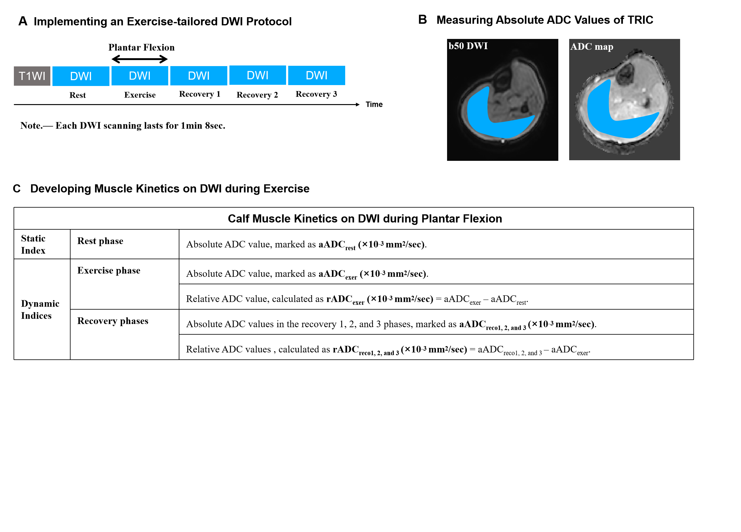
This prospective study recruited 87 healthy participants (20-59 years) from November 2021 to May 2023. The leg dominance was determined by asking the participant to kick a ball, where the kicking leg was defined as the dominant leg and the supporting leg as the non-dominant leg [8]. A plantar flexion-tailored DWI protocol was sequentially implemented with one rest phase, one exercise phase, and recovery 1, 2, and 3 phases. Each phase corresponds to one DWI scanning. The absolute apparent diffusion coefficient (aADC, ×10-3 mm2/sec) of triceps surae (TRIC) was measured in the ADC maps by two radiologists to acquire aADC in the rest phase (aADCrest), aADC in the exercise phase (aADCexer), and aADC in recovery phases (aADCreco1, 2, and 3). Based on aADC, muscle kinetics on DWI was developed by additionally collecting relative ADC (rADC, ×10-3 mm2/sec): rADC in the exercise phase (rADCexer) as aADCexer –aADCrest; rADC in the recovery phases (rADCreco1, 2, and 3) as aADCreco1, 2, and 3 –aADCexer. The independent or paired t-test was performed to compare differences.Results
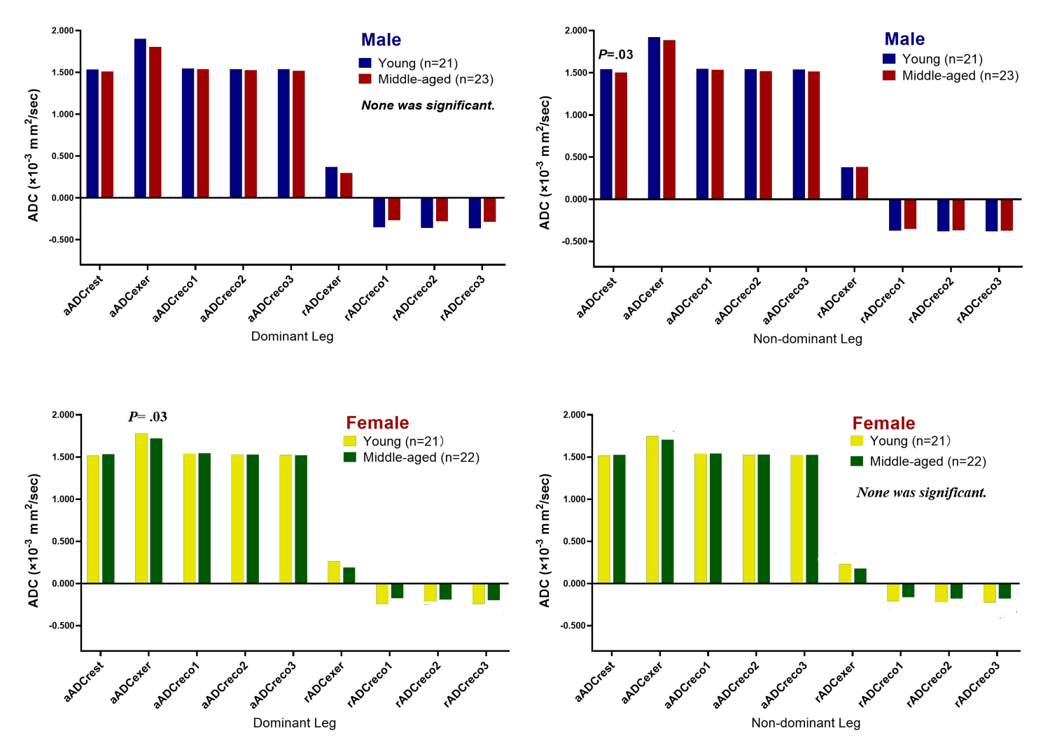
Two legs were imaged in all 87 participants ( age range/mean, 23-58/41±10 years; 44 males; 41 young participants<40 years). Males showed no significant differences of muscle kinetics on DWI with age in the dominant leg and had significantly lower aADCrest in the middle-aged than in the young participants in the non-dominant leg (1.503±0.049 vs. 1.543±0.067×10⁻³ mm2/sec, p=.03). In comparison, females showed significantly lower magnitudes of rADCexer in the middle-aged than in the young participants in the dominant leg (0.187±0.103 vs. 0.267±0.127×10⁻³ mm2/sec, p=.03) and showed no significant differences of muscle kinetics on DWI with age in the non-dominant leg.Discussion
One set of muscle kinetics on DWI during exercise comprised both static and dynamic indices, where the static indices reflect the structural and compositional information of skeletal muscles, and the dynamic indices reflect the dynamic kinetics associated with muscle contraction and relaxation. Physiologically, both diffusion and perfusion may contribute positively to the increased ADC values during exercise. According to a study that used a plantar flexion-tailored intravoxel incoherent motion (IVIM) imaging protocol, true diffusion coefficient (D), perfusion fraction (f), and pseudo-diffusion (D*) were all elevated in the gastrocnemius and soleus muscles during exercise [9]. Functionally, a higher ADC value in the exercise phase may indicate a higher muscle contracting strength, as it has been proved that the ADC value would increase more profoundly in response to a higher exercise intensity [10]. Males and females may show different changing patterns of skeletal muscle with age.Conclusion
A plantar flexion-tailored DWI protocol allowed for the simultaneous assessments of both static and dynamic muscle qualities, which could be used to assess the sex differences in age-related muscle quality.Acknowledgements
None was declared.References
1. Haizlip, K.M., B.C. Harrison, and L.A. Leinwand, Sex-based differences in skeletal muscle kinetics and fiber-type composition. Physiology (Bethesda), 2015. 30(1): p. 30-9.2.
2. Larsson, L., et al., Sarcopenia: Aging-Related Loss of Muscle Mass and Function. Physiol Rev, 2019. 99(1): p. 427-511.3.
3.Suetta, C., et al., The Copenhagen Sarcopenia Study: lean mass, strength, power, and physical function in a Danish cohort aged 20-93 years. J Cachexia Sarcopenia Muscle, 2019. 10(6): p. 1316-1329.4.
4. Fragala, M.S., A.M. Kenny, and G.A. Kuchel, Muscle quality in aging: a multi-dimensional approach to muscle functioning with applications for treatment. Sports Med, 2015. 45(5): p. 641-58.5.
5. Chen, L.K., et al., Asian Working Group for Sarcopenia: 2019 Consensus Update on Sarcopenia Diagnosis and Treatment. J Am Med Dir Assoc, 2020. 21(3): p. 300-307.e2.6.
6. Cruz-Jentoft, A.J., et al., Sarcopenia: revised European consensus on definition and diagnosis. Age Ageing, 2019. 48(1): p. 16-31.7.
7. Le Bihan, D., et al., Separation of diffusion and perfusion in intravoxel incoherent motion MR imaging. Radiology, 1988. 168(2): p. 497-505.
8. Paillard, T. and F. Noé, Does monopedal postural balance differ between the dominant leg and the non-dominant leg? A review. Hum Mov Sci, 2020. 74: p. 102686.
9. Mastropietro, A., et al., Triggered intravoxel incoherent motion MRI for the assessment of calf muscle perfusion during isometric intermittent exercise. NMR Biomed, 2018. 31(6): p. e3922.
10. Yanagisawa, O. and T. Kurihara, Intramuscular water movement during and after isometric muscle contraction: evaluation at different exercise intensities. Clin Physiol Funct Imaging, 2016. 36(5): p. 368-75.
Figures