5047
DTI-MRI as a non-invasive assessment of extracellular matrix remodeling in lumbar paravertebral muscles of rats with sarcopenia1The First Affiliated Hospital of Kunming Medical University, kunming, China
Synopsis
Keywords: Muscle, Aging
Motivation: The evaluation of extracellular matrix levels in skeletal muscle often requires tissue biopsy, which is difficult for patients to accept in clinical practice
Goal(s): We evaluated the extracellular matrix level of skeletal muscle using non-invasive magnetic resonance imaging technology and conducted pathological verification using skeletal muscle samples from rats with sarcopenia
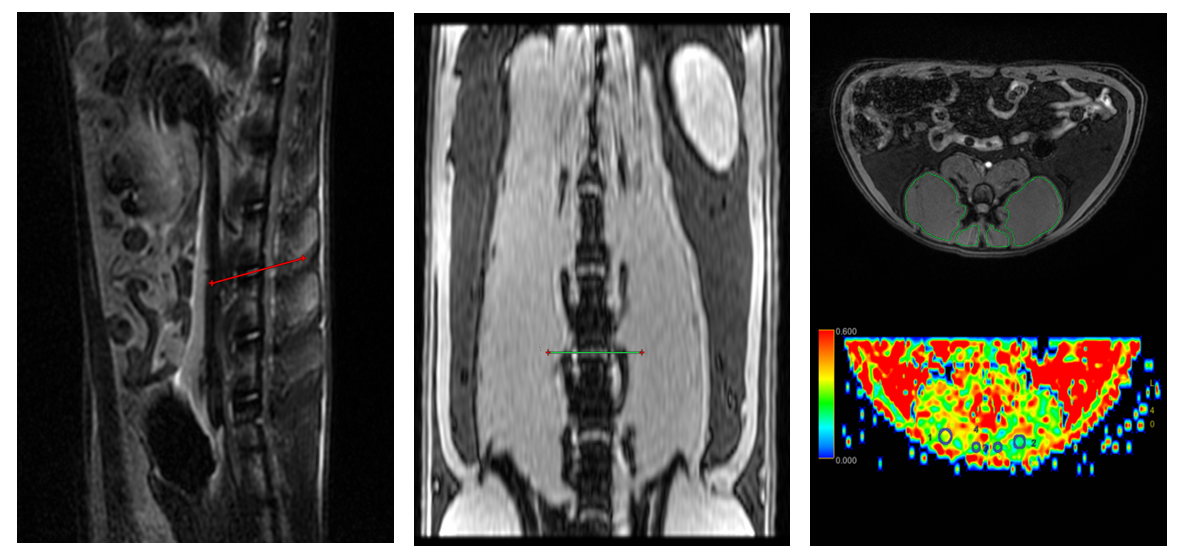
Approach: We constructed a classic sarcopenic rat model , scanned the lumbar paravertebral muscles with a 3.0T MRI-based DTI sequence, and validated through histological experiments
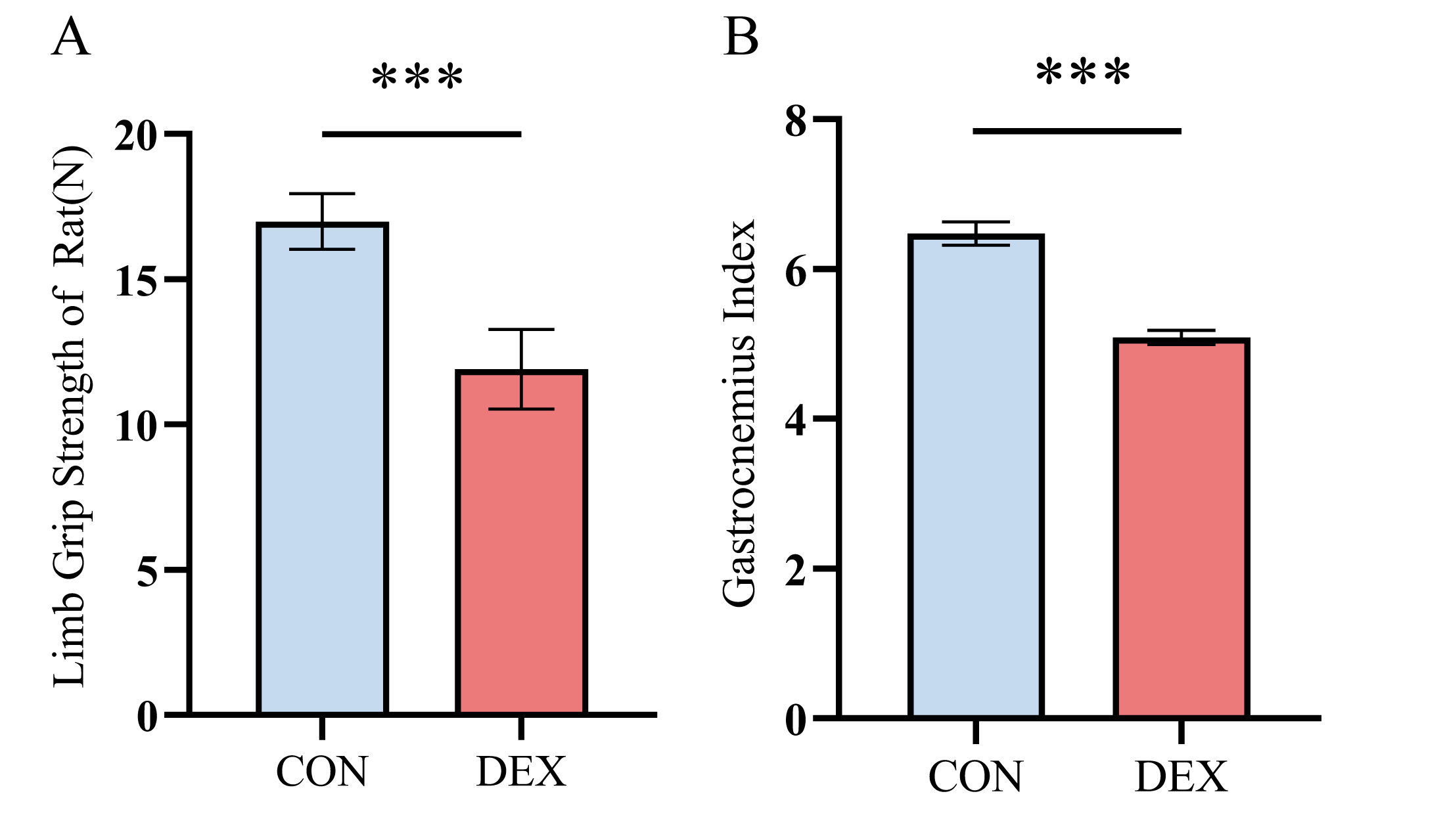
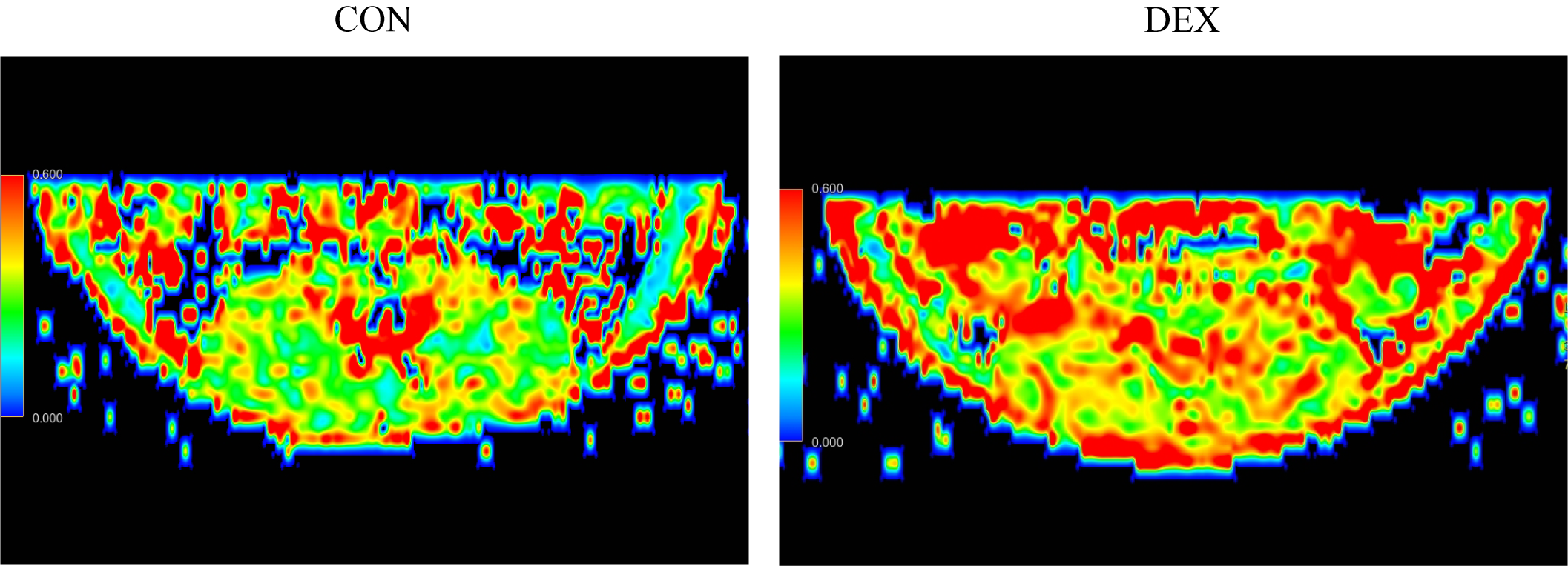
Results: FA and CSA of sarcopenic rats were significantly reduced compared to the normal control group, and there was a strong correlation between them and pathological related parameters
Impact: Our study provides a non-invasive tool for evaluating the extracellular matrix levels of skeletal muscles in sarcopenia
Abstract
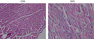
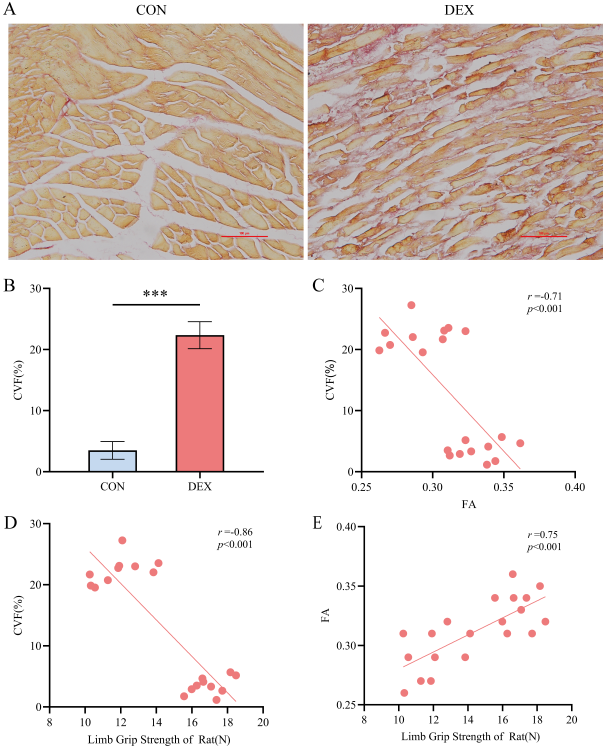
Introduction: Extracellular matrix remodeling in skeletal muscle is one of the important reasons leading to the development of sarcopenia. The aim of this study was to evaluate alterations in extracellular matrix remodeling in lumbar paravertebral muscles of sarcopenic rats by diffusion-tensor magnetic resonance imaging((DT-MRI). Methods: Twenty 6-month-old female Sprague Dawley rats were randomly divided into dexamethasone (DEX) group and normal saline control group. Rats in both groups were subjected to 3.0T magnetic resonance imaging scanning included Mensa, T2WI, and DT-MRI sequences. Observe the changes in muscle fibers and extracellular matrix of the erector spinal muscle using hematoxylin eosin and Sirius red staining. The expressions of chollagen I, III and fibronectin in erector spinae were detected by western blot. Pearson correlation analysis was used to evaluate the correlation strength between MRI quantitative parameters and corresponding histopathology markers. Results: The cross-sectional area and fractional anisotropy values of erector spinae in DEX group rats were significantly decreased compared with those in the control group (p < 0.05). Hematoxylin eosin staining showed atrophy and disordered arrangement of muscle fibers in the DEX group; Sirius red staining showed a significant increase in collagen volume fraction in the DEX group. The results of western blot showed that the expression of collagen I, collagen III, and fibronectin was significantly increased in the dexamethasone group (p < 0.001 for all). The correlation coefficients between fractional anisotropy values and collagen volume fraction, collagen I, collagen III, fibronectin were -0.71, -0.94, -0.85, -0.88, respectively (p < 0.05 for all). Discussion:In our study, compared with the control group, the CSA value of the erector spinalis muscle in the DEX group rats was significantly reduced, while the CSA value of the multifidus muscle did not show significant changes. From the perspective of muscle fiber types, this is because the erector spinalis muscle is mainly composed of type II muscle fibers, while the multifidus muscle is mainly composed of type I muscle fibers[1]. In our results, the skeletal muscles of dexamethasone group rats showed an increase in interstitial fibrous connective tissue, with significant increases in Collagen I and Collagen III contents. Mahdy et al.'s research suggests that the accumulation of fibrous tissue increases the stiffness of skeletal muscles, limits their extension and contraction, and reduces the body's motor ability[2]. Conclusion: The fractional anisotropy value strongly correlates with pathological collagen volume fraction, collagen I, collagen III, and fibronectin, indicating that DT-MRI can non-invasive evaluate the changes of extracellular matrix remodeling in the erector spinal muscle of sarcopenia, providing a potential imaging biomarker for diagnosis of sarcopenia.Acknowledgements
Not ApplicableReferences
[1] AGTEN A, STEVENS S, VERBRUGGHE J, et al. The lumbar multifidus is characterised by larger type I muscle fibres compared to the erector spinae [J]. Anatomy & cell biology, 2020, 53(2): 143-50. [2] MAHDY M A A. Skeletal muscle fibrosis: an overview [J]. Cell and tissue research, 2019, 375(3): 575-88.Figures