5046
A multipurpose Adolescent Idiopathic Scoliosis specific short MRI protocol: feasibility study in volunteers.1Department of Radiology, Image Sciences Institute, UMC Utrecht, Utrecht, Netherlands, 2Department of Orthopaedics, UMC Utrecht, Utrecht, Netherlands, 3Department of Radiology, Division Image and oncology, UMC Utrecht, Utrecht, Netherlands, 4MRIguidance B.V., Utrecht, Netherlands, 5Department of Biomedical Engineering, Eindhoven University of Technology, Eindhoven, Netherlands
Synopsis
Keywords: Skeletal, Skeletal, Scoliosis, MSK, Spine, sCT, synthetic CT
Motivation: To date, there is no description of an optimal MRI protocol for detection of early changes in the scoliotic spine.
Goal(s): The goal is to assess the feasibility of the multi-purpose short MRI protocol based on the MR data of adult volunteers.
Approach: Based on the checklist of relevant landmarks, assessment of the volunteer MR data and identification of the landmarks was performed by 3 readers.
Results: We have demonstrated that the proposed MRI protocol, covering almost the entire spine in 14 min, is efficient for detection of relevant landmarks for: scoliosis assessment, screening for neural axis abnormalities, and surgical planning and navigation.
Impact: A short MRI protocol is developed for accurate assessment of spinal deformity in AIS patients, facilitating the treatment management, monitoring and surgical planning, and simplifying the clinical workflow.
Introduction
Adolescent idiopathic scoliosis (AIS) is a 3D spinal deformity affecting 2-3% of the population1,2. If the deformity progresses, it can heavily reduce the patient's quality of life. Therefore, when a certain Cobb angle is reached, corrective surgery is indicated. To date, there is no description of an optimal MRI protocol for detection of early changes in the scoliotic spine. The UMCU standard non-contrast MR protocol for AIS diagnostics and evaluation includes a radiograph followed by a whole spinal cord clinical MRI protocol, consisting of multiple acquisitions repeated usually in 3 stations, with a total scan duration of about 50-70 min. It is difficult for a patient, especially an adolescent, to tolerate such a long examination.In this work, we present a short 14-min multi-purpose MRI protocol, consisting of 3D T2w TSE scans of thoraco-lumbar and cervical spine, and of 3D BoneMRI scan of thoracolumbar spine, used to generate synthetic CTs. The protocol was developed specifically for AIS patients, with the goal to fulfill multiple purposes:
- detection of early patho-anatomical changes;
- screening for neural axis abnormalities;
- surgical planning/ spinal navigation surgery.
The purpose of this study was to assess the feasibility of the multi-purpose MRI protocol based on the MR data of adult volunteers.
Methods
18 adult volunteers (average age 26, range 21-37 y.o.) participated in this study and written informed consent was signed by all participants. The study was approved by the local IRB. The volunteers were scanned using a clinical 1.5T MR scanner (Philips Healthcare, Best, NL) using the base head coil and the built-in posterior coil. MR images of the thoracolumbar spine were acquired using the protocol described in Table 1. Note, 3D Sag T2w TSE C-spine MR images were acquired in 5 volunteers out of 18.Synthetic CT images were generated using BoneMRI V1.5 (MRIguidance BV, Utrecht, The Netherlands).
The checklist of all relevant landmarks for detection of early stages of scoliosis, screening for neural axis abnormalities, surgical planning, spinal navigation surgery was prepared, see Figure 1, and based on it, the assessment of the volunteer MR data was performed using Vquest software3 by 3 independent readers: a musculoskeletal radiologist, a spine surgeon, and an orthopaedic resident.
The data analysis was performed in Matlab R2002b.
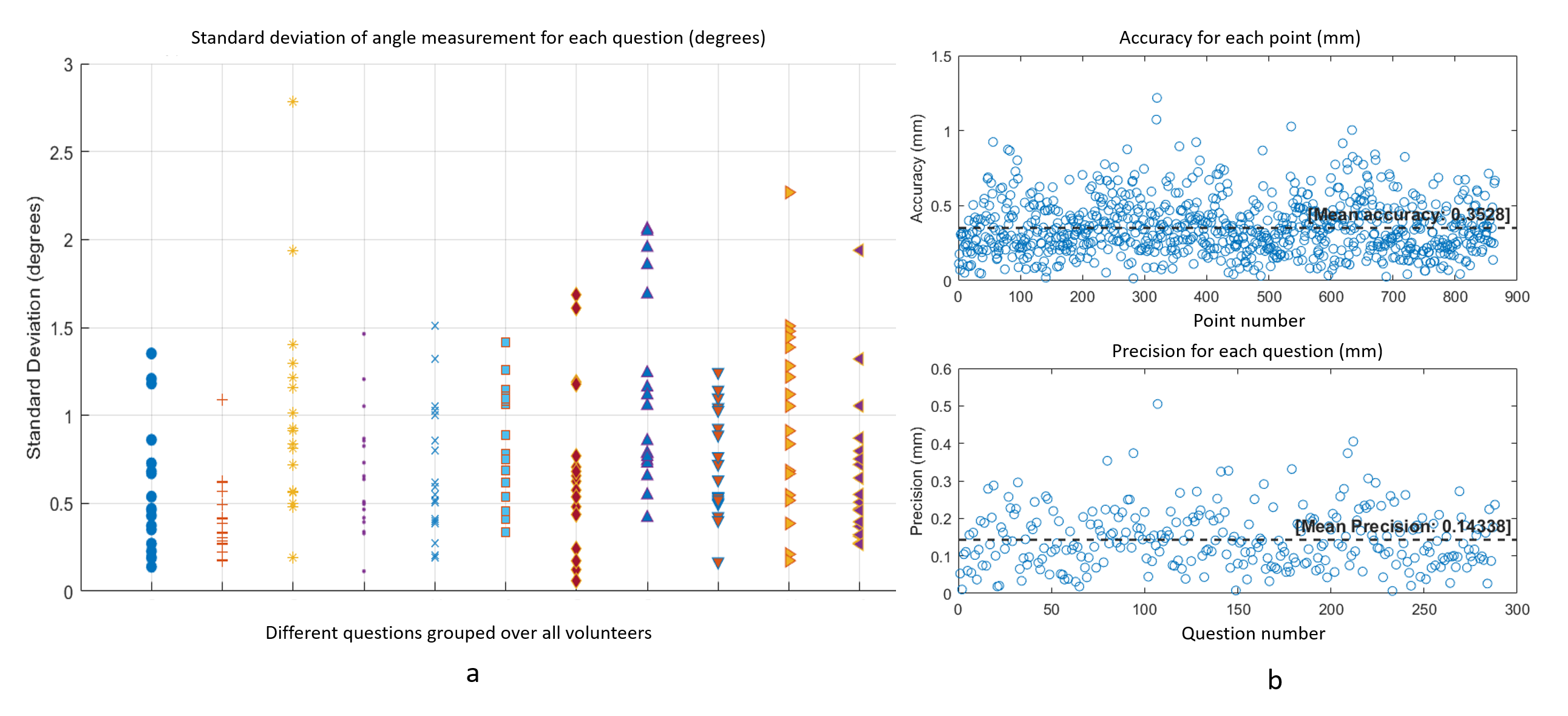
The precision in the line positioning was defined as standard deviation between the angles, formed by the lines placed by readers and reference lines, and was calculated for each question.
The landmark assessment by the readers was analyzed for their accuracy and precision. The accuracy was calculated as the average distance from each reader’s identified point to the mean point of all readers. To describe the consistency, the precision was calculated as standard deviation of the points (the square root of the variance of the distances from each reader's point to the mean point for each landmark).
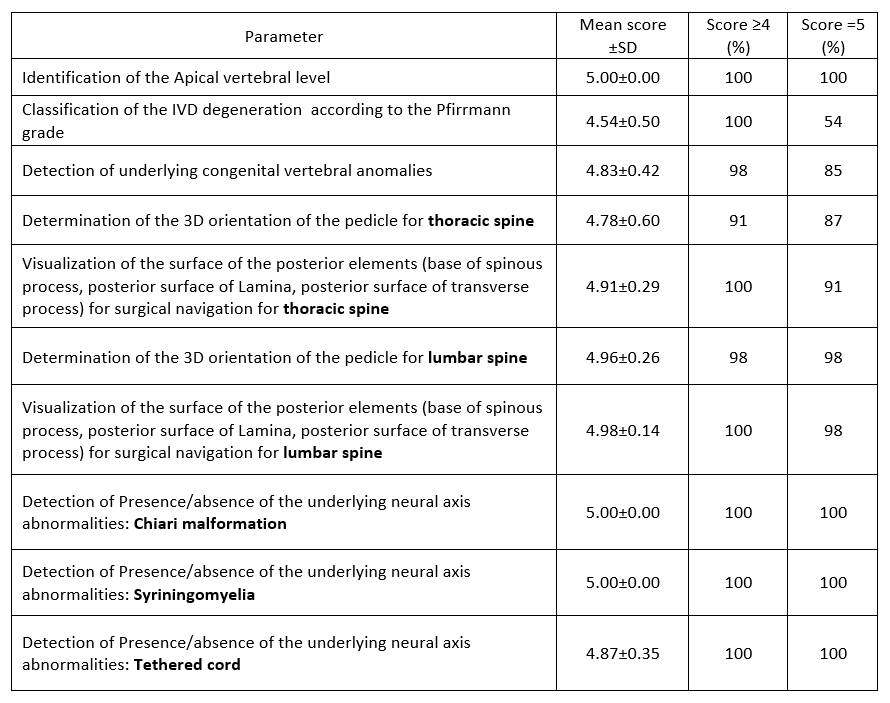
For the Likert-based multiple-choice questions, the mean score among the readers was calculated for each question, as well as the percentage with scores ≥ 4 and score = 5.
Results
An example of MR images acquired in one volunteer is presented in Figure 2.The results of the assessment of line positioning questions are presented in Figure 3(a) for all volunteers and different questions. The majority of angles were under 1.5 degrees variation, indicating high consistency between the readers, and only about 5% of answers had a higher variation of 1.5-2.8 degrees, which still indicated a good agreement between the readers measurements.
The results of the assessment of the dot positioning are presented in Figure 3(b). The mean accuracy of 0.35 mm and the mean precision of 0.14 mm were calculated. Both values were high, which suggested that the readers agreed on where the point is located, and their answers were very similar to each other, clustered together at the mean.
The mean scores among the readers over different parameters are reported in Table 2. The majority of the answers (> 91%) had a score ≥ 4, which indicated moderate to high confidence of the readers in identifying all relevant parameters in the images.
Discussion and Conclusions
We have demonstrated that the proposed MRI protocol, covering almost the entire spine in 14 min, is efficient for evaluation of AIS: for detection of early patho-anatomical changes in a scoliotic spine, for screening for neural axis abnormalities, and for surgical planning and navigation, facilitating the treatment management, monitoring and surgical planning, and simplifying the clinical workflow.Future studies should explore its application in a broader clinical context, including its effectiveness in patients with moderate to severe scoliosis, or in patients with braces.
Acknowledgements
This research was financially supported by the European Research Council (Grant no: 101020004).References
1. S. L. Weinstein, “The Natural History of Adolescent Idiopathic Scoliosis.,” J Pediatr Orthop, vol. 39, no. Issue 6, Supplement 1 Suppl 1, pp. S44–S46, Jul. 2019, doi: 10.1097/BPO.0000000000001350.
2. S. L. Weinstein, L. A. Dolan, J. C. Y. Cheng, A. Danielsson, and J. A. Morcuende, “Adolescent idiopathic scoliosis.,” Lancet, vol. 371, no. 9623, pp. 1527–37, May 2008, doi: 10.1016/S0140-6736(08)60658-3.
3. https://vquest.nl/
Figures
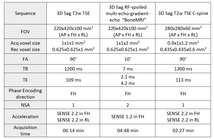
Table 1. The protocol parameter settings for the sequences included in the protocol.
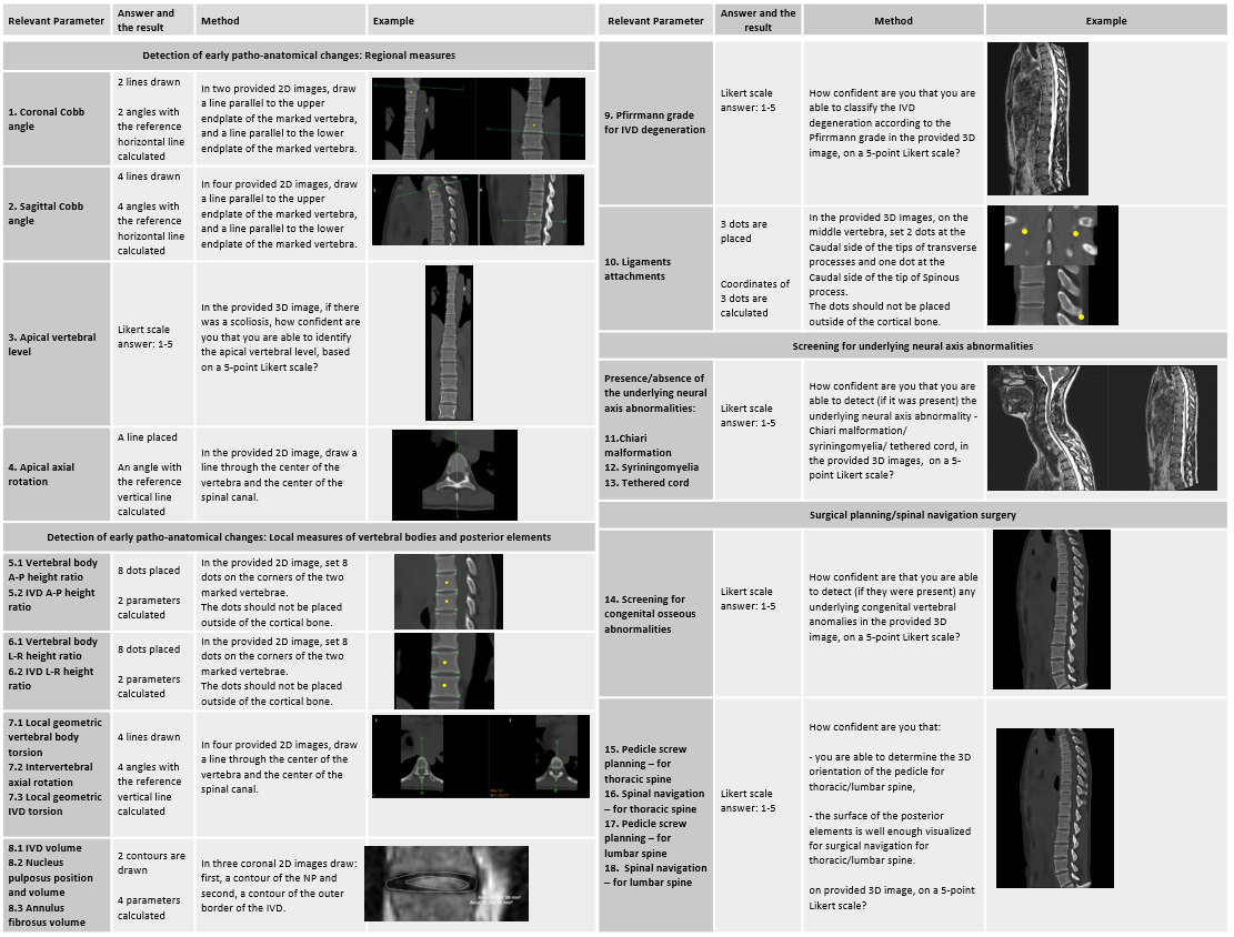
Figure 1. The checklist of all relevant landmarks with the parameters, readers’ answers, methods, and the examples.
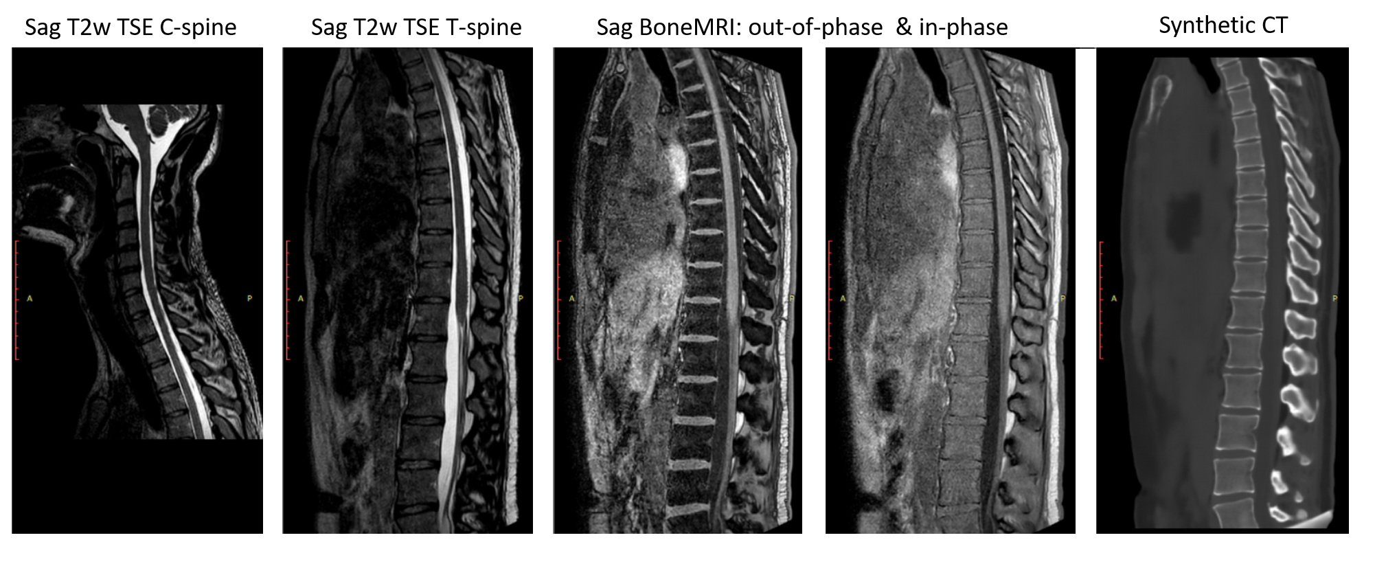
Figure 2. MR images acquired in one volunteer: 3D Sag T2w TSE of C-spine and T-spine, BoneMRI out-of-phase and in-phase images and the reconstructed synthetic CT images.