5045
Use An Automated Pipeline to Quantify Erector Spinae Muscle Asymmetry and Correlate It with Quality of Life in Adult Degenerative Scoliosis1Shanghai Key Laboratory of Magnetic Resonance, East China Normal University, ShangHai, China, 2Institute of Research and Clinical Innovations, Neusoft Medical Systems Co., Ltd., Beijing, China, 3MR Research Collaboration Team, Siemens Healthineers Ltd., ShangHai, China, 4Department of Radiology, The First Affiliated Hospital, Army Medical University, Chongqing, China
Synopsis
Keywords: Muscle, Data Analysis
Motivation: To explore the influence of erector spinae muscle asymmetry to adult degenerative scoliosis (ADS) on patients' quality of life.
Goal(s): To develop a pipeline for automated quantification of the normalized FCSA difference index (CDI) and left-right fat Infiltration asymmetry ratio (LFIAR) from MRI images for assessment of ADS.
Approach: We used nnU-Net for precise anatomical segmentation on T2 images. Then, CDI and LFIAR were calculated and correlated to ADS scales.
Results: The automated model adeptly delineated paravertebral muscle groups, which were used to calculate CDI and LFIAR. Noteworthy correlations between CDI, LFIAR, and ADS scales were found among male subjects.
Impact: This research streamlines clinical workflows, imparts valuable insights into the severity of ADS, and equips clinicians with crucial data for informed clinical decision-making.
Introduction
Adult degenerative scoliosis (ADS) refers to gradual spinal deformations in adults due to degenerative changes, often accompanied by lower back pain and gait abnormalities, significantly impacting quality of life. Erector spinae muscles (ES) are integral components of paravertebral muscles and serve as dynamic stabilizers for the lumbar spine. Dysfunction in these muscles may contribute to lumbar spine conditions [1].Previous studies have indicated the association of fat infiltration and muscle atrophy within the paravertebral muscles with lower back pain (LBP) and spinal deformities [2-5] . The normalized FCSA difference index (CDI) represents the side-to-side ratio of functional muscle area (FCSA), while left-right fat Infiltration asymmetry ratio (LFIAR) represents the side-to-side ratio of fat infiltration[6]. These metrics, which can be calculated from the area of fat and muscle images within the paravertebral muscles on T2-weighted images, can be used to accurately assess muscle status and the asymmetry of fat infiltration between two sides, and offers a deeper understanding of the relationship between ADS, pain, and quality of life.
Manual delineation of paravertebral muscles in MRI is time-consuming and requires expertise. Therefore, we developed a pipeline that can automatically segment intervertebral discs and paravertebral muscle groups, and calculate the associated CDI and LFIAR. Then CDI and LFIAR were correlated to age and scales related to quality of life of ADS patient. We thought the proposed pipeline can make CDI and LFIAR more accessible to physicians and lead to better understandings of the condition of ADS patients and improved clinical decisions.
Methods
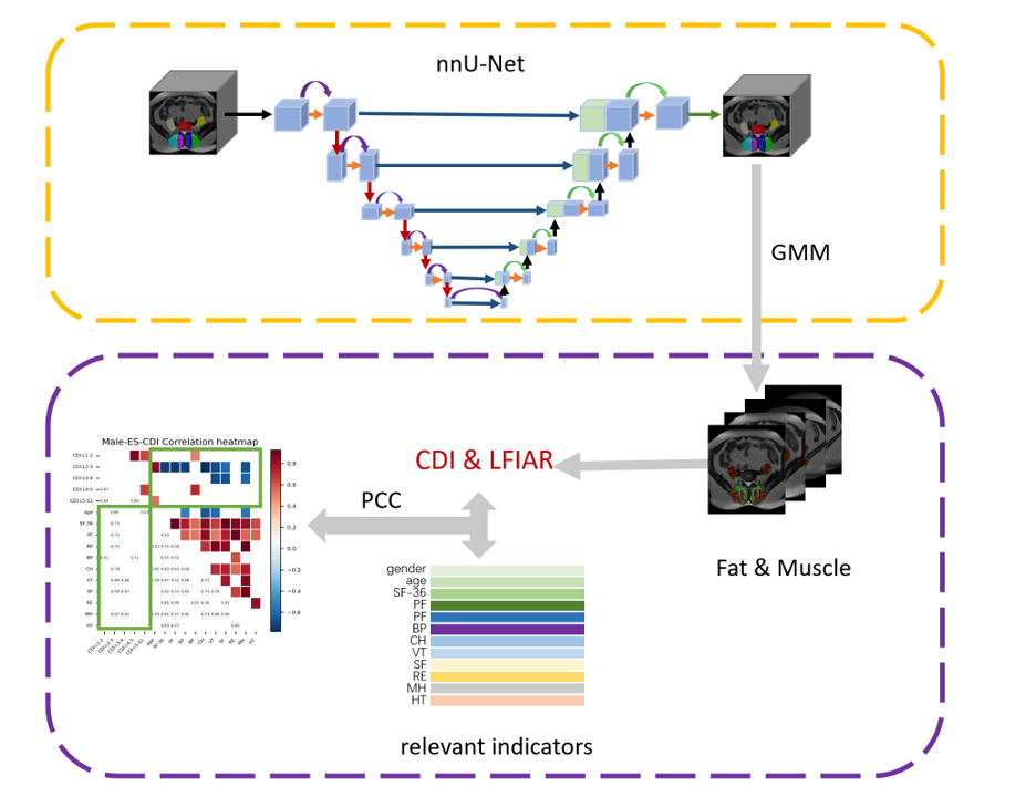
This study obtained T2-weighted data of 66 ADS patients from the First Affiliated Hospital of Army Medical University. Sixty patients completed scales related to the quality of life affected by ADS, which included SF-36, Physical Function (PF), Role Physics (RP), Bodily Pain (BP), Comprehensive Health (CH), Vitality (VT), Social Function (SF), Role Emotion (RE), Mental Health (MH), and Health Transition (HT). Two radiologists with eight years of experience delineated paravertebral muscles, intervertebral discs, and facet joints as regions of interest (ROI). The intersection of their ROIs was used for this study.The workflow was shown in Figure 1. Patients were split into a training (N=46) and a testing cohort (N=20) and five-fold cross-validation was utilized to train the nnUNet-based segmentation model. A Gaussian mixture model was employed to automatically separate fat from muscles in the segmentation results before CDI and LFIAR were calculated for the corresponding paravertebral muscles of each intervertebral disc. Pearson correlation coefficient (PCC) was used to study the correlation analysis between CDI/LFIAR and ADS scales.
Results
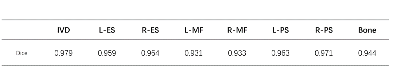
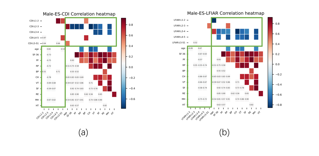
The performance of the segmentation model is presented in Table 1. The model achieved excellent segmentation of paravertebral muscles (including Left Erector Spinae, L-ES; Right Erector Spinae, R-ES; Left Multifidus, L-MF; Right Multifidus, R-MF; Left Psoas, L-PS; Right Psoas, R-PS), bone, and intervertebral discs (IVD), with Dice coefficients ranging from 0.931 to 0.979.From Figure 2a, for males, CDI of ES was positively correlated with age and BP (PCC=0.71), and negatively correlated with SF-36, PF, RP, CH, VT, SF, MH (PCCs, in the range of [-0.78, -0.59]).
LFIAR for ES showed a negative correlation with SF-36, PF, RP, CH, VT, SF, MH (PCCs, in range of [-0.86, -0.55]), as shown in Figure 2b.
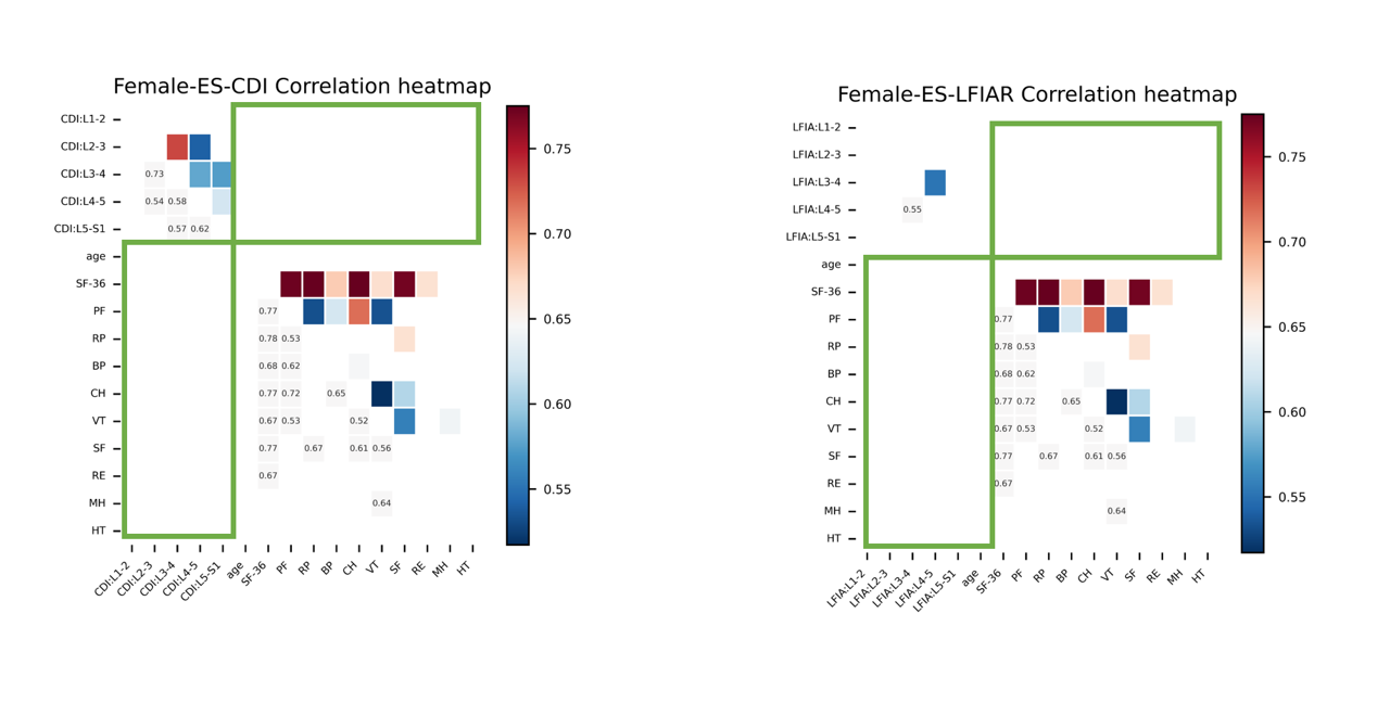
From Figure 3, it can be seen that the correlation between CDI/LFIAR and the ADS scales among female subjects was not as significant as those found in male subjects (|PCC|<0.5).
Discussion
In our study, the segmentation model effectively delineated the paravertebral muscles, achieving high Dice coefficient. Automatically calculated metrics such as CDI and LFIAR characterized the asymmetry of the patient's spine, reducing the workload of clinicians. Notably, stratified correlation analysis revealed gender differences of the influence of CDI and LFIAR to the quality of life. For males, with increasing age, higher CDI values of ES muscle were observed, indicating increased asymmetry in ES muscle and exacerbated pain, leading to a greater impact on daily life. Higher LFIAR correlates with increased severity of fat infiltration asymmetry and more pain, also affecting daily life. For females, CDI and LFIAR of ES muscle demonstrated lower correlations with ADS scales, suggesting a higher tolerance to pain in females.Limitations of our study include a limited sample size, which may affect result stability. The pipeline should be validated in future with larger datasets.
Conclusion
We demonstrated the feasibility of a fully automated pipeline to automatically segment paravertebral muscles, intervertebral discs, and facet joints and compute CDI and LFIAR. The proposed pipeline can make CDI/LFIAR based analysis more accessible to clinicians studying ADS.Acknowledgements
No acknowledgement found.References
[1] Vives MJ. The paraspinal muscles and their role in the maintenance of global spinal alignment. Another wrinkle in an already complex problem. Spine J. 2016;16(4):459-461.
[2] Kim WJ, Shin HM, Lee JS, et al. Sarcopenia and Back Muscle Degeneration as Risk Factors for Degenerative Adult Spinal Deformity with Sagittal Imbalance and Degenerative Spinal Disease: A Comparative Study. World Neurosurg. 2021;148:e547-e555.
[3] Sun XY, Kong C, Lu SB, et al. The Parallelogram Effect of Degenerative Structures Around the Apical Vertebra in Patients with Adult Degenerative Scoliosis: The Influence of Asymmetric Degeneration and Diagonal Degeneration on the Severity of Deformity. Med Sci Monit. 2019;25:3435-3445. Published 2019 May 9.
[4] 4 Yeung KH, Man GCW, Shi L, et al. Magnetic Resonance Imaging-Based Morphological Change of Paraspinal Muscles in Girls With Adolescent Idiopathic Scoliosis. Spine (Phila Pa 1976). 2019;44(19):1356-1363.
[5] Federau C, Kroismayr D, Dyer L, Farshad M, Pfirrmann C. Demonstration of asymmetric muscle perfusion of the back after exercise in patients with adolescent idiopathic scoliosis using intravoxel incoherent motion (IVIM) MRI. NMR Biomed. 2020;33(3):e4194.
[6] Xie D, Zhang J, Ding W, et al. Abnormal change of paravertebral muscle in adult degenerative scoliosis and its association with bony structural parameters. Eur Spine J. 2019;28(7):1626-1637.
Figures