5044
MRI metrics can help explain the loss of functional strength in aging1Radiology, Stanford University, Stanford, CA, United States, 2Bioengineering, Stanford University, Stanford, CA, United States, 3Bernard and Irene Schwartz Center for Biomedical Imaging, Department of Radiology, New York University, New York, NY, United States
Synopsis
Keywords: Muscle, Aging
Motivation: There is a need to investigate the relationship between age-related muscle atrophy and functional strength.
Goal(s): To explore the relationship between volumes of individual knee extensor muscles relative to total volume of the quadriceps as measured by MRI and sit to stand performance.
Approach: Participants performed a sit to stand test while being recorded by a markerless motion capture system and underwent an MRI exam that included a Dixon scan, which was used to calculate muscle volumes in the quadriceps.
Results: There were correlations between age and vastus intermedius and vastus lateralis volume. Rectus femoris volume was correlated with sit to stand performance.
Impact: We utilized a novel experimental design using smartphone-based markerless motion capture and MRI metrics to study the relationship between functional strength and age-related muscle atrophy. This work could inform design of physical therapy programs for the elderly.
Introduction
Functional strength is known to decline with age. The sit to stand test is a test of functional strength used to assess the ability of aging individuals to perform activities of daily living. The quadriceps, as the main knee extensors, are a key muscle for completing this task. It has been extensively reported that 5-repetition sit to stand (5XSTS) time increases with age1, but the changes in relative volume of knee extensor muscles with age and their connection to 5XSTS times has not been explored. We used MRI to measure muscle volumes in the quadriceps of healthy volunteers ages 30-80 who also performed a 5XSTS test while being recorded by a smartphone-based markerless motion capture system. We hypothesized that increasing 5XSTS time associated with aging could be related to changes in the relative volumes of the knee extensor muscles.Methods
Subjects: 22 subjects were included in the study (age 57.0 ± 15.4 years, (30-79) 11 female, 11 male)MRI: All scans were acquired at 3T (GE Healthcare) using an Aircoil. Two Dixon scans were used to cover the whole thigh. Imaging parameters were: matrix size=256x256x42, FOV=450x450x252mm, TR=13.4ms, flip angle=5deg. Each of the muscles in the quadriceps- the rectus femoris (RF), vastus lateralis (VL), vastus intermedius (VI), and vastus medialis (VM)- was manually segmented, and volumes were calculated from the segmentations. The proportion of the total volume of the quadriceps that each muscle in the quadriceps occupies was calculated as $$$Volume_{Muscle}/Volume_{Total}$$$.
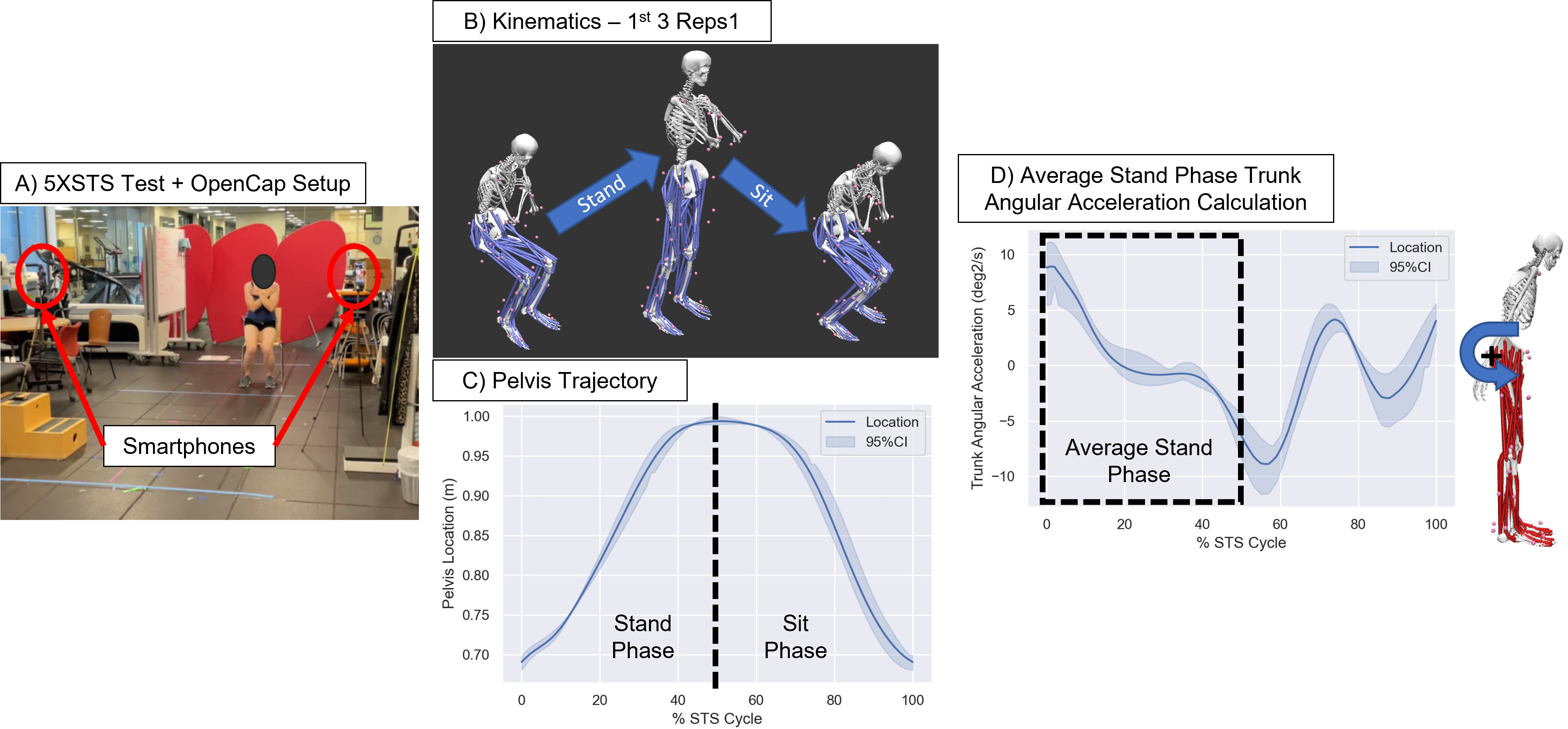
Functional testing (Figure 1): Each participant performed a 5 repetition sit-to-stand test, where the participant rises from a chair then sits back down 5 times, while being recorded by OpenCap2, a freely available open-source markerless motion capture package that calculates kinematics from smartphone videos. Average trunk angular acceleration, a parameter associated with compensation due to age-related strength loss, in the stand phase of the sit-to-stand motion was calculated by averaging trunk angular acceleration from the beginning of the STS cycle to the time point when the pelvis reached the highest point on its trajectory for the first three available STS repetitions for each participant. Kinetics were calculated for the first three available STS repetitions using the OpenCap Processing pipeline3, which generates a muscle-driven dynamic simulation to estimate ground reaction forces, joint moments, and muscle activations, and is validated against gold standard lab-based measurements.
Statistics: Correlations between muscle volume fractions and stand phase trunk acceleration and total 5XSTS time were determined using linear regression. Relationships between total 5XSTS time, stand phase trunk acceleration, and muscle volume fractions and age were also determined using linear regression. A p-value of p<0.05 was considered significant.
Results
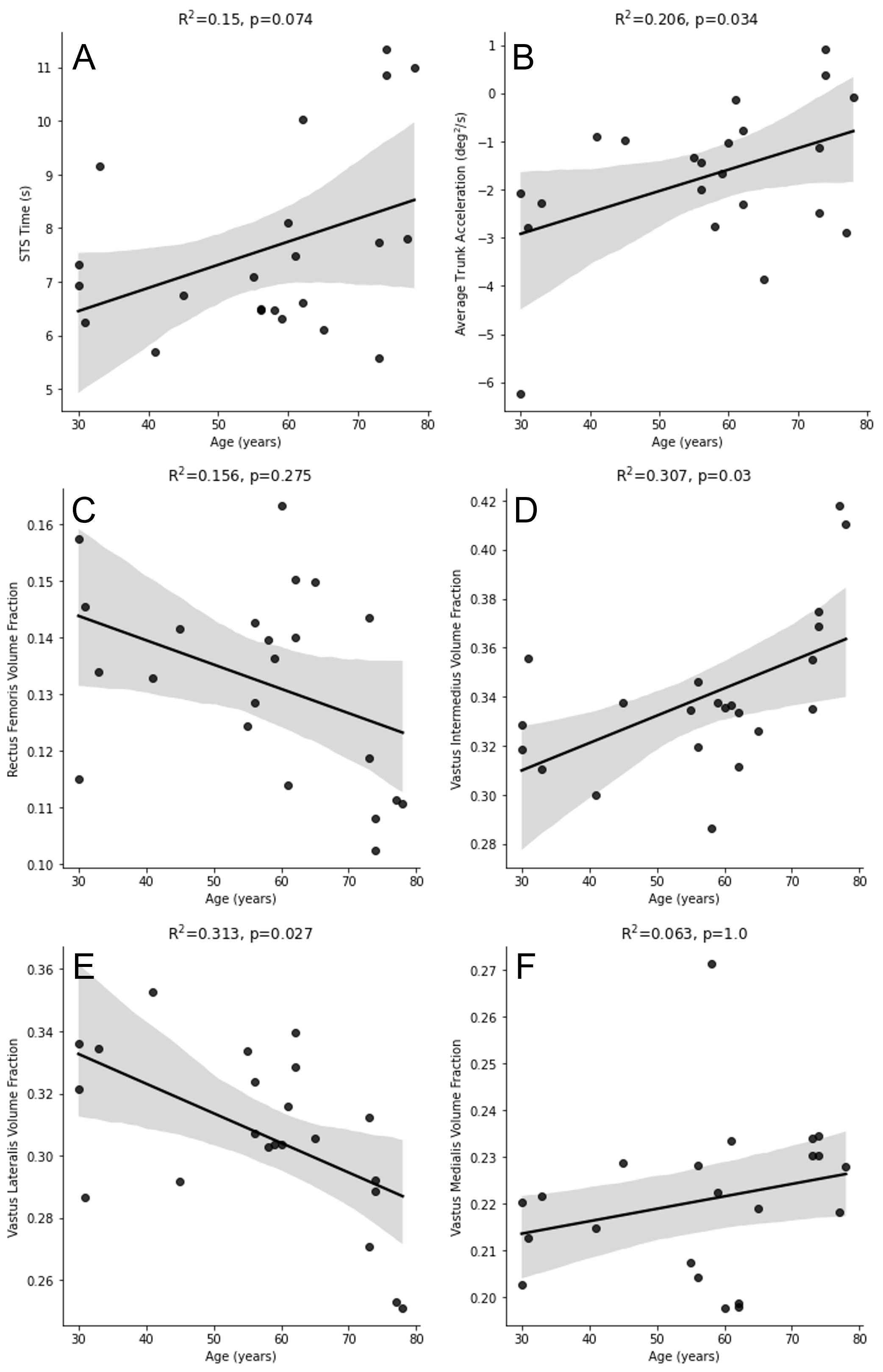
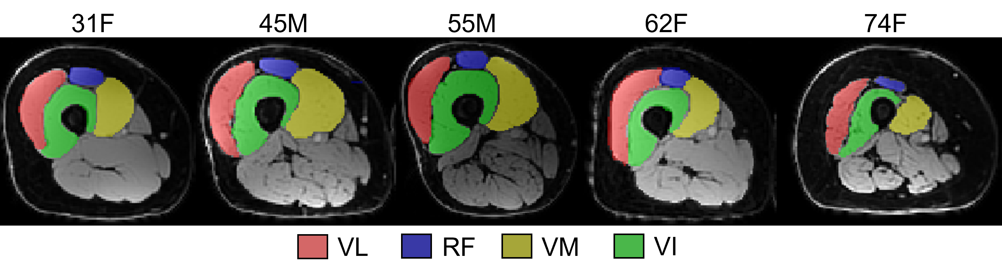
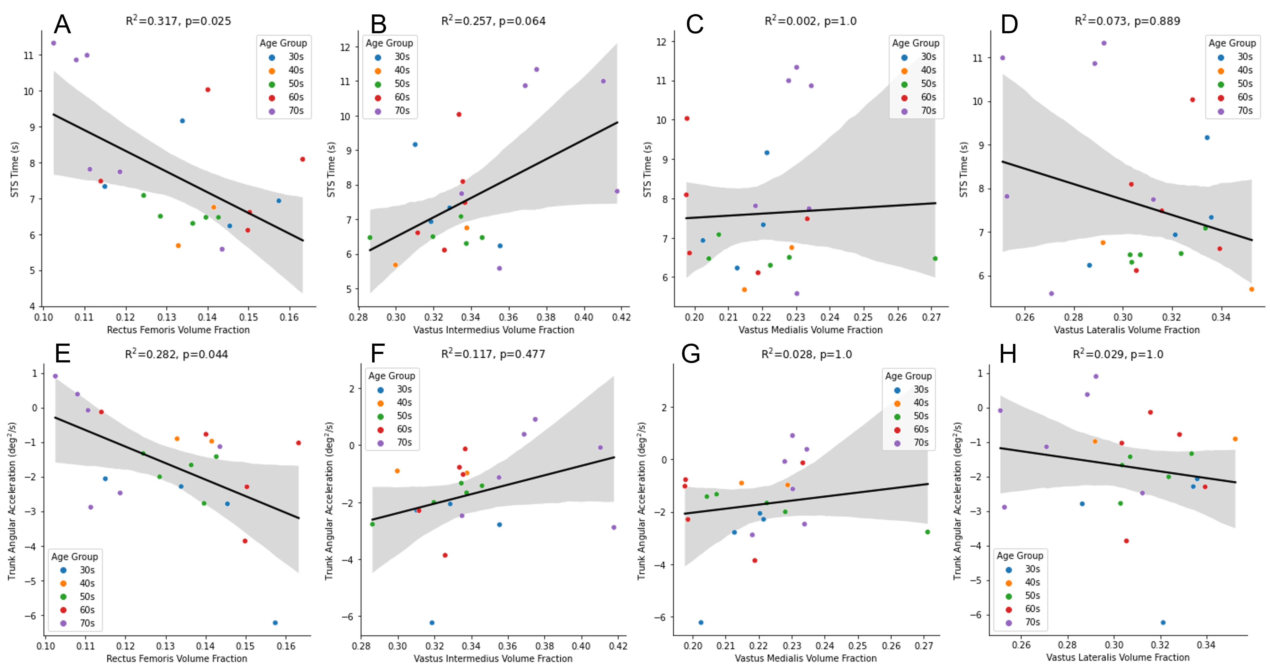
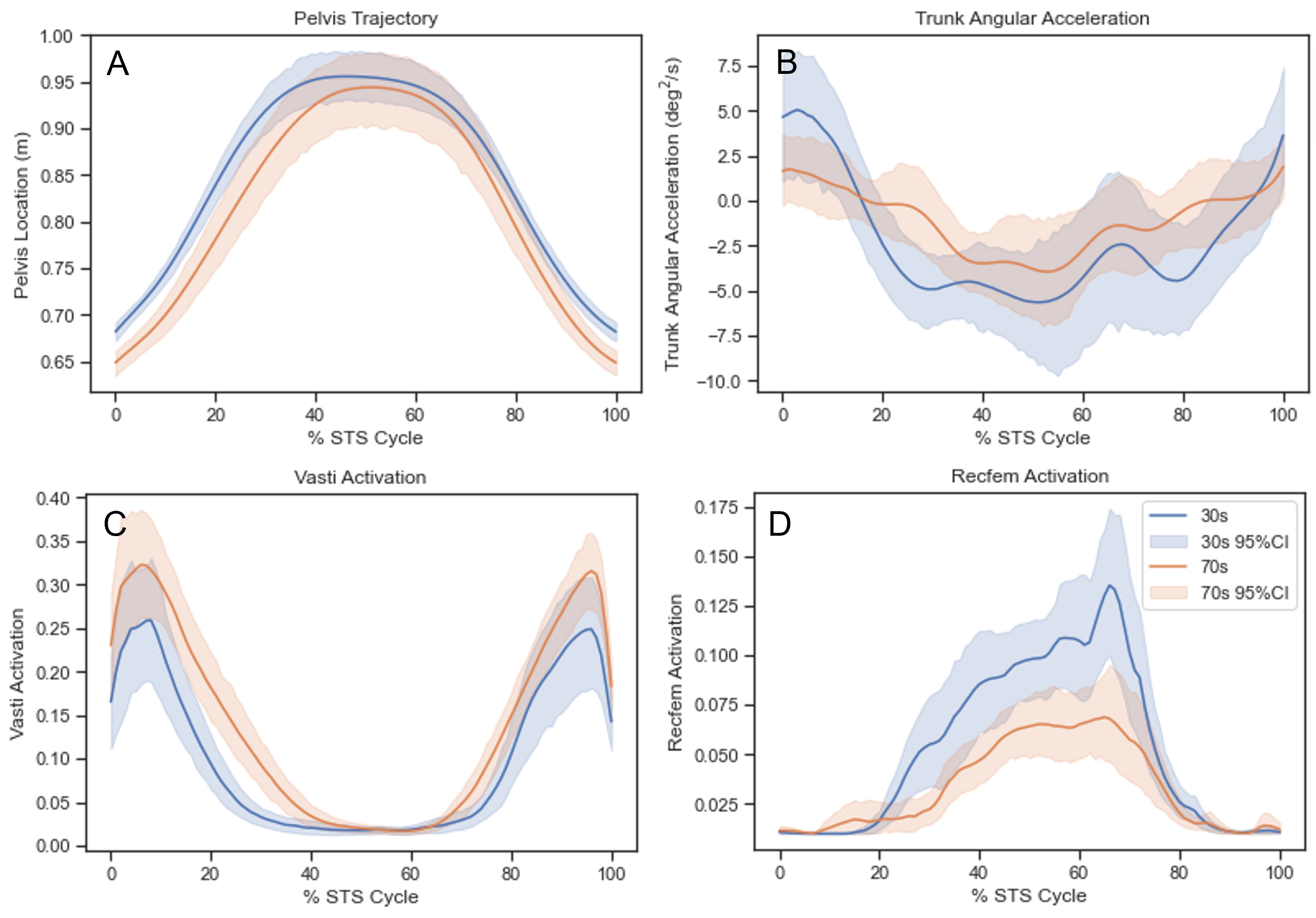
We did not observe a significant correlation between age and 5XSTS time in this dataset (Figure 2a). There was a significant positive correlation between trunk angular acceleration and age (Figure 2b). Relative volumes of the knee extensor muscles changed with age. VI volume fraction was positively correlated with age, while VL volume fraction was negatively correlated with age (Figure 2c-f). These volume changes are visually apparent in MRI images of the quadriceps (Figure 3). RF volume fraction was negatively correlated with 5XSTS time and trunk angular acceleration (Figure 4a&e). No other correlations between muscle volume fractions and 5XSTS time or trunk angular acceleration were significant (Figure 4). Muscle activation patterns show that the vasti are activated at the beginning and the end of the STS cycle, and the RF is activated in the middle of the cycle (Figure 5). On average, RF activation in the youngest group of participants was greater than in the oldest group of participants, but the differences were not significant.Discussion
Both 5XSTS times and trunk acceleration were correlated with RF volume fraction. The RF is a biarticular muscle and thought to be used for upper body balance and coordination4, while the monoarticular vasti may play a greater role in force generation. While there were not significant differences in activation patterns between age groups, reduced relative volume of the RF could be causing compensatory changes in trunk motion in older participants, who had higher (lower magnitude) average trunk angular acceleration. While in previous studies, 5XSTS time increased with age, that was not the case in this population due to lower STS times than reported in previous literature in more elderly participants.Conclusion
This study shows that muscle volume changes with age are not proportional and may affect activities of daily living, such as rising from a chair. These findings increase understanding of functional strength and could help better design rehabilitation or physical therapy programs for the elderly.Acknowledgements
This work was supported by NIH 1K99AG07173501, NIH EB002524, NIH AR062068, GE Healthcare, and the Wu Tsai Human Performance Alliance at Stanford University.References
- Boswell M, Kidzinski L, Hicks J, et al. Smartphone videos of the sit-to-stand test predict osteoarthritis and health outcomes in a nationwide study. npj Digital Medicine. 6.1 (2023): 32.
- Uhlrich S, Falisse A, Kidzinski L, et al. OpenCap: 3D human movement dynamics from smartphone videos. PLOS Computational Biology. 19.10 (2023): e1011462.
- OpenCap Processing pipeline. https://github.com/stanfordnmbl/opencap-processing
- Schumacher C, Berry A, Lemus D, et al. Biarticular muscles are most responsive to upper-body pitch perturbations in human standing. Scientific reports 9.1 (2019): 14492.
Figures