5040
Efficient and Complete Fat Suppression in 2-point Dixon MRI for Improved Qualitative and Quantitative Assessment of Lumbar Plexus1Radiology, UT Southwestern Medical Center, Dallas, TX, United States, 2Advanced Imaging Research Center, UT Southwestern Medical Center, Dallas, TX, United States
Synopsis
Keywords: Other Musculoskeletal, Fat, Fat Suppression, Dixon
Motivation: The use of 2-point Dixon is prevalent for fat/water separation in MRI, but suffers from incomplete fat suppression, leading to residual fat signal in the water image. We have previously developed a “Darkfat” post-processing technique that can reduce this residual signal.
Goal(s): This retrospective study aims to evaluate Darkfat processing in clinical lumbar plexus images.
Approach: Retrospective 2-point T2-weighted lumbar images from 30 patients were processed using Darkfat and evaluated through qualitative and quantitative assessment.
Results: Water-only images from Darkfat processing had significantly better fat suppression (P<0.0001), and subjectively better image quality (P<0.005) compared to original water images from 2-point T2-weighted Dixon acquisition.
Impact: Darkfat image processing significantly improves fat suppression in clinical 2-point T2-weighted lumbar images enabling enhanced anatomical visualization. This could potentially improve pathological visualization.
Introduction
Fat suppression in MR imaging is important for providing appropriate tissue contrast and improving anatomical visualization. Common fat suppression techniques include fat signal saturation, inversion recovery sequences, and opposed-phase imaging1. Opposed-phase imaging, also known as the Dixon method, uses differences in the resonant frequencies of fat and water protons to suppress fat signal. Dixon methods are robust to magnetic field inhomogeneities and provide higher signal to noise ratio (SNR) compared to other fat suppression methods2. However, the commonly used 2-point Dixon only suppresses the MR signal from the main methylene lipid peak while leaving remaining fat signal in the water image3. The residual fat signal can be pre-calculated from known values, which does not account for changes in subject or acquisitions. Previous methods developed a post-processing algorithm that can address this issue from 2-point Dixon methods4. The purpose of this retrospective study was to evaluate this post-processing algorithm, hereby referred to as “Darkfat”, in clinically relevant lumbar plexus images acquired within the past year using 2-point T2-weighted Dixon acquisition.Theory
The Darkfat post-processing method reduces the residual fat signal present in water images acquired from 2-point Dixon scans, leading to improved anatomical visualization. Water-only and fat-only images from conventional Dixon processing are required for the Darkfat processing. Regions in the fat-only image that are comprised of >70% fat signal are identified. The voxels in this region are used to find the average value of the fat signal fraction that remains in the water-only.$$$Fat\ Signal\ Fraction=\ \left(\frac{Original\ Water\ Image}{Original\ Fat\ Image}\right)_{Identified\ Fat\ Voxels}$$$ (Eq. 1)
The fat signal fraction measured through Eq. 1 is spatially invariant for a given set of echo times and is independent of T1 or T2 weighting. This single fraction value is then used to attenuate the residual fat signal in the original water image through Eq. 2.
$$$Darkfat\ Water\ Image=\ Original\ Water\ Image\ - (Fat\ Signal\ Fraction*\ Original\ Fat\ Image)$$$ (Eq. 2)
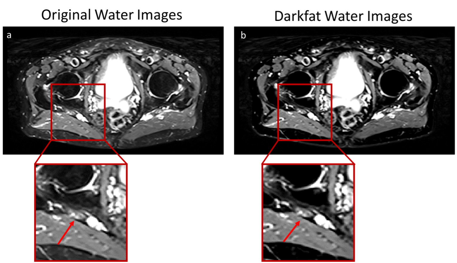
The result of Eq. 2 gives a Darkfat water-only image without the residual fat signal unaccounted for by 2-point Dixon. Darkfat post-processing for water images is performed slice-by-slice independently of imaging sequences (Fig. 1).
Methods
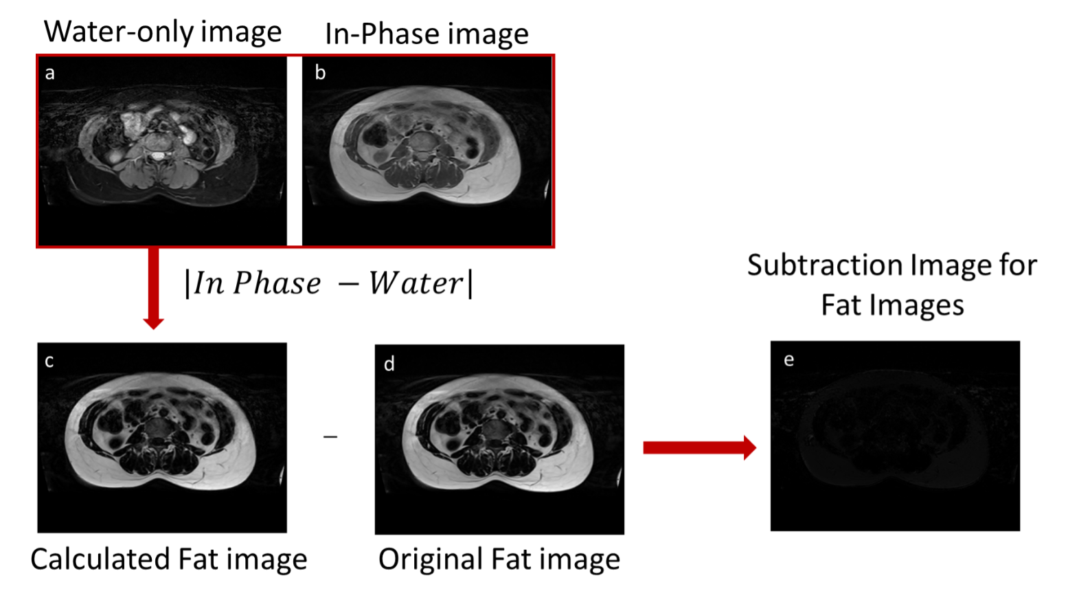
Imaging Subjects: Thirty patients (mean age = 52±14) who had lumbar plexus imaging were compiled under a retrospective IRB. These patients were scanned either on a 3T Ingenia MR (Philips Healthcare) or a 3T Vida MR (Siemens Healthineers) from March-October 2023. The imaging protocol was a 2D T2-weighted turbo spin echo (TSE) 2-point Dixon sequence with the following imaging parameters: resolution = 1x1x4 mm3, TR = 3000-5500 ms, TE = 55-60 ms. Approximate scan time = 3 min.Post-Processing Pipeline: Dixon images were exported and anonymized from the clinical database, but included only water, in-phase, and opposed-phase images. Fat images were generated offline using the exported in-phase and water images. The generated fat images were validated through image difference with original fat images in image sets where the fat image was available (Fig. 2). The water-only and fat-only images were then used to generate the Darkfat images using Matlab (Mathworks) (Fig. 1).
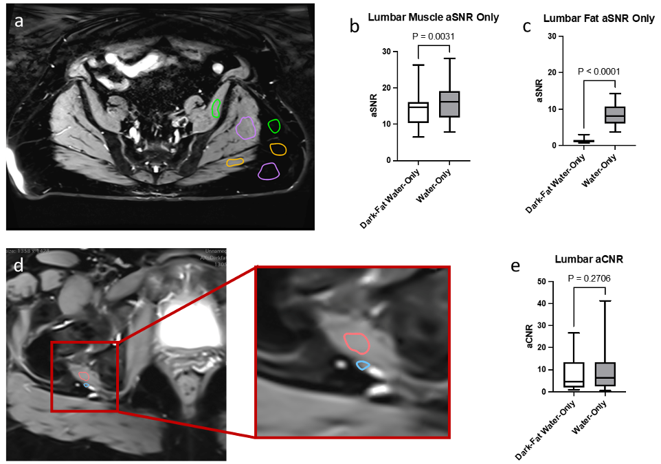
Image Analysis: The apparent SNR (aSNR) and apparent contrast to noise ratio (aCNR) were measured in the same ROIs for both Darkfat processed and conventional Dixon water-only images and compared using paired t-test analysis. The aSNR was calculated by finding the ratio between the signal mean and the signal standard deviation in each ROI. The aCNR was measured by finding the difference in aSNR between the adjacent ROIs (Fig. 5).
An experienced musculoskeletal radiologist with 24 years of experience (A.C.) analyzed the images qualitatively. The images were scored on a scale of 1-5, with 5 being most preferred for the following metrics: overall image quality, anatomical visualization, fat suppression, and appearance of motion artifacts. Images were scored simultaneously but blinded. These scores were analyzed within each subject using paired t-test analysis.
Results
The Darkfat water images had noticeably reduced subcutaneous and visceral fat compared with the original water images from 2-point Dixon (Fig. 3). Overall, Darkfat images were preferred qualitatively in terms of image quality and fat suppression, along with significantly higher scores for anatomical visualization (Fig. 4). The aSNR also showed a clear reduction in the subcutaneous fat while maintaining tissue signal (Fig. 5).Discussion and Conclusion
We have shown that Darkfat processed water images have improved fat suppression and image quality through qualitative and quantitative analyses in clinical lumbar plexus images. This retrospective study provides evidence to support the use of Darkfat processing for 2-point Dixon imaging to improve image quality and subsequently fat-fraction quantification.Acknowledgements
This work was partly supported by Cancer Prevention and Research Institute of Texas (CPRIT) grant RP190049 and NIH/NCI R01CA283663.References
1. Delfaut, E. M. et al. Fat suppression in MR imaging: techniques and pitfalls. Radiographics 19, 373-382, doi:10.1148/radiographics.19.2.g99mr03373 (1999).
2. Dixon, W. T. Simple proton spectroscopic imaging. Radiology 153, 189-194, doi:10.1148/radiology.153.1.6089263 (1984).
3. Yu, H. et al. Multiecho water-fat separation and simultaneous R2* estimation with multifrequency fat spectrum modeling. Magn Reson Med 60, 1122-1134, doi:10.1002/mrm.21737 (2008).
4. Madhuranthakam AJ, Y. H., Alsop DC. System and method for fat suppression in chemical shift based water-fat separated MR imaging. USA patent (2013).
Figures