5038
Feasibility of echo asymmetry and least squares estimation (IDEAL-IQ) in differentiation of atypical spinal hemangiomas from metastases1Peking University Third Hospital, Beijing, China
Synopsis
Keywords: Skeletal, Skeletal
Motivation: It is common for patients with primary tumors to find abnormal signals in the spine, which may be initially reported as “metastases cannot be rule out”. For these cases, differential diagnosis has a significant impact on patient staging and management, which may result in misdiagnosis or additional imaging, ultimately biopsy, and unnecessary costs.
Goal(s): To investigate chemical shift-encoded sequence (IDEAL-IQ) for differential diagnosis of spinal tumors.
Approach: Single-center prospective study.
Results: This work highlights the benefits of adding proton density fat fraction (PDFF) maps yield from IDEAL-IQ to improve diagnostic accuracy of spinal metastases and hemangiomas without contrast agents.
Impact: IDEAL-IQ provides reliable quantification of spinal tumor bone marrow infiltration pattern in clinical settings, which is comparable to the quantitative parameters of DCE-MRI. It may be a valuable tool for providing information for early differential diagnosis in a non-invasive way.
INTRODUCTION
Spinal hemangiomas and metastases are the most common benign and malignant tumors in spine, respectively[1]. Usually, atypical/aggressive vertebral hemangiomas are challenging since they have an atypical radiological appearance and can mimic primary bony malignancies or metastases[2]. It is common for patients with primary tumors to find abnormal signals in the spine, which may be initially reported as “metastases cannot be rule out”. For these cases, differential diagnosis has a significant impact on patient staging and management, which may result in misdiagnosis or additional imaging, ultimately biopsy, and unnecessary costs[3]. As there is considerable overlap in radiologic features of spinal hemangioma and metastases on conventional magnetic resonance imaging, the differential diagnosis still remains challenging[4].PURPOSE
To evaluate the role of chemical shift-encoded sequence (IDEAL-IQ) parameters in differentiation of atypical spinal hemangiomas from metastases.METHODS
- We prospectively enrolled 48 consecutive patients with pathological evidence of metastatic/hemangioma of the spine from July 2022
to March 2023. IDEAL-IQ was performed at 3-T MRI in sagittal position. Our institutional ethics committee approved this study and written informed consent was waived (M2022403).
- The inclusion criteria for spinal metastases were cancer patients with definite pathological diagnosis of metastatic tumor by puncture. The inclusion criteria for spinal hemangioma were as follows: (1) Patients with a clear pathological diagnosis of hemangioma (including typical and atypical hemangioma) by biopsy; (2) Lesions accidentally discovered by enhanced MRI for reasons other than spinal tumors (including spinal demyelination, foraminal schwannoma, prolapsed intervertebral discs, etc.), no tumor history, patients under 40 years of age, no change at 1 year follow-up, and enrolled after evaluation by two senior radiologists consistent with imaging findings (iso-/hypointensity on T1 and hyperintensity on STIR). The exclusion criteria as follows: (1) Age < 18 years; (2) Lesions shorter diameter less than 1 cm; (3) Diffuse multiple metastases, normal vertebral body measurements were not available; (4) Artifact interferes with quantitative measurement.
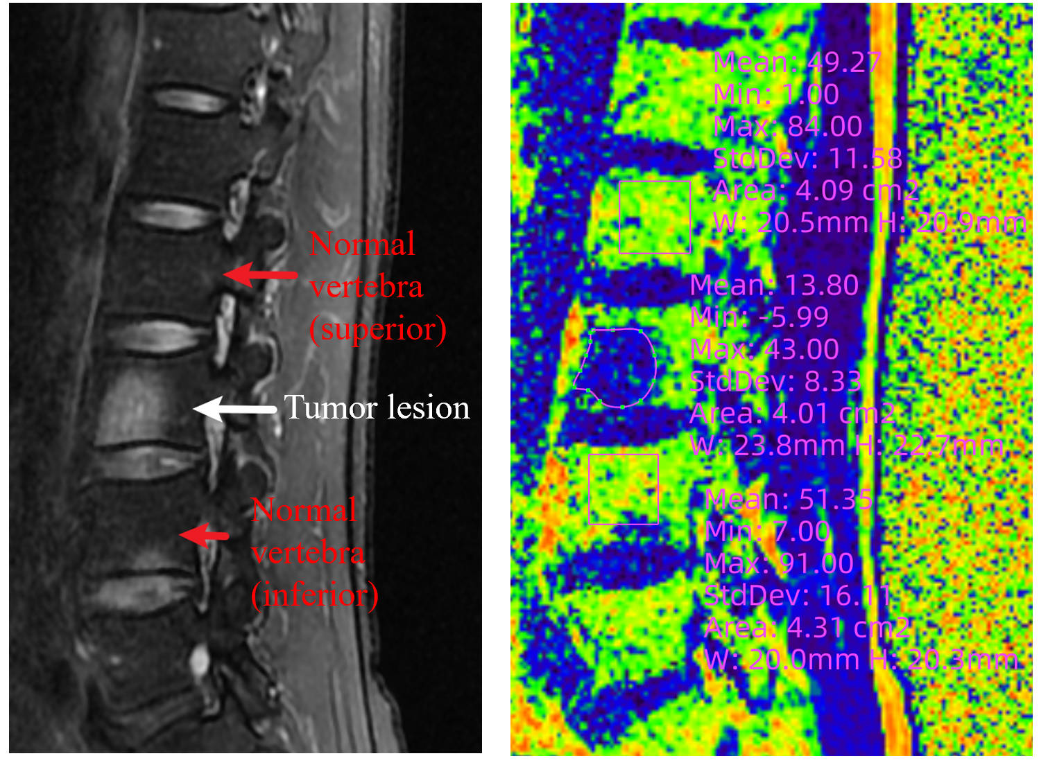
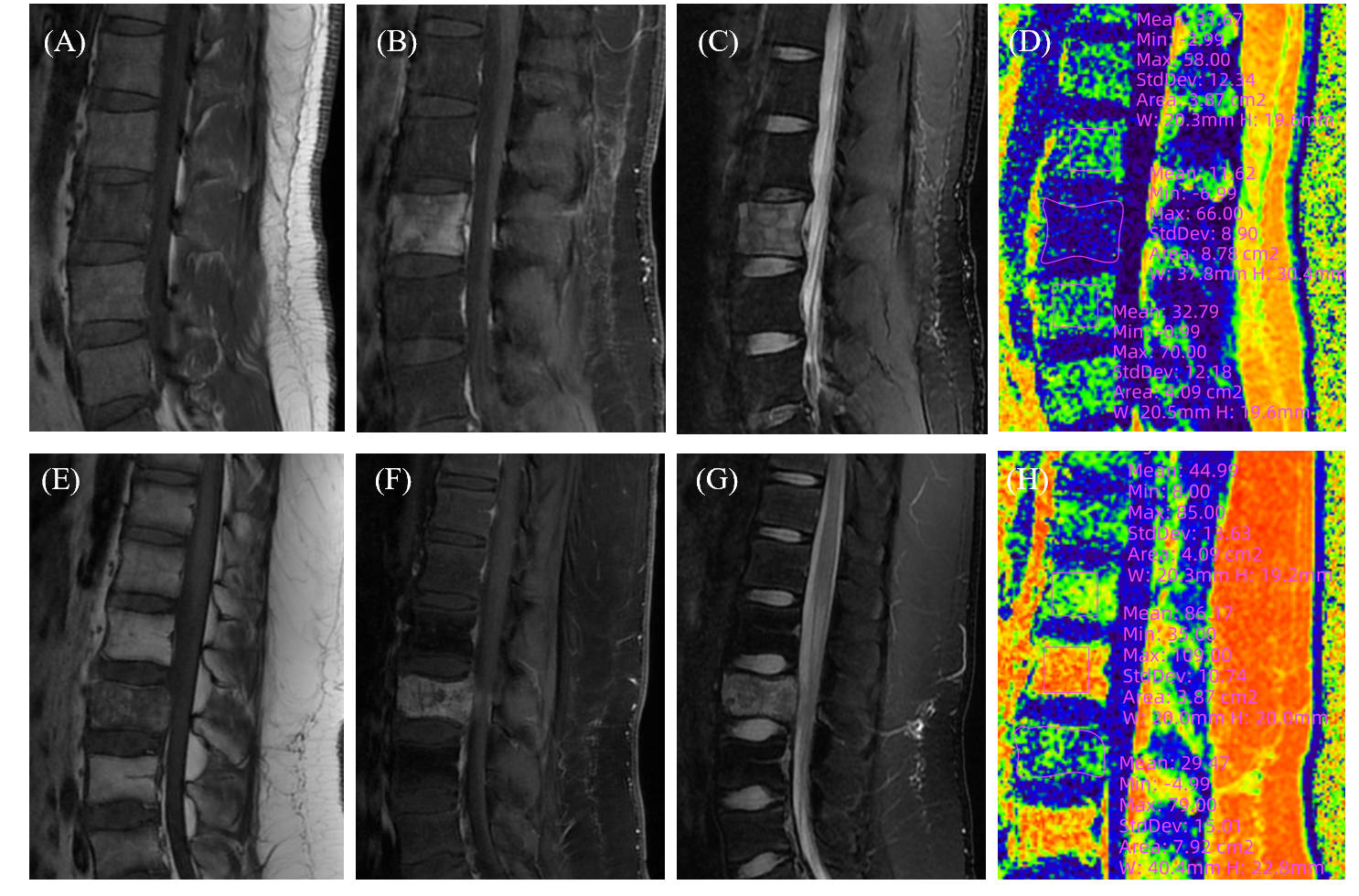
- Quantitative analysis of bone marrow fat fraction (FF) was performed using three regions of interests (ROIs), including tumor lesions (ROI along the edge), upper and lower adjacent vertebra (square ROIs). The FF ratio of tumor lesions and normal vertebrae (FF ratio = FFtumor / FFvertebrae ) was calculated and compared at 0.05 significance level and performed receiver operating characteristic curve analysis.
RESULTS
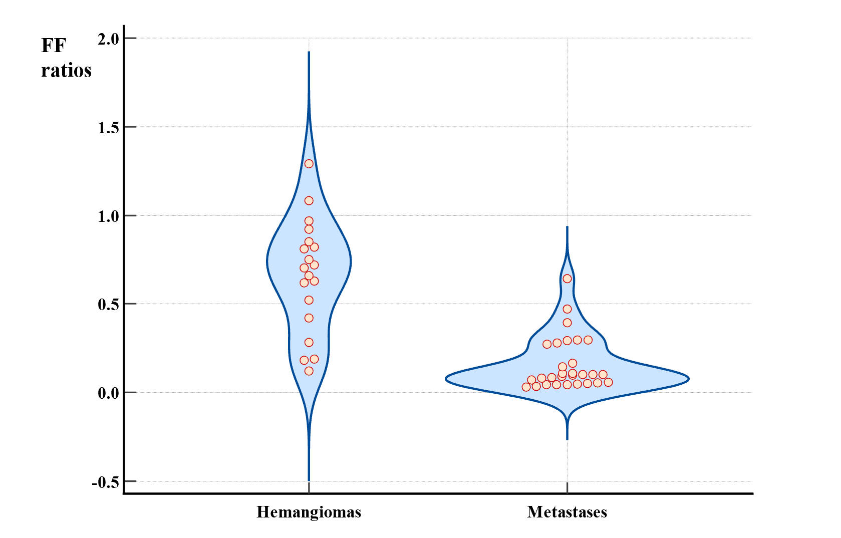
There were 29 patients with metastases and 19 patients with hemangiomas. The mean (± standard deviation) of FF ratio was significantly higher (0.65 ± 0.32) in hemangiomas compared to metastatic lesions (0.16 ± 0.15) with P < 0.001. FF ratio cutoff of ≤ 40.75% had a sensitivity of 78.9% and specificity of 93.1% with the area under the curve is 0.72 in detecting malignant lesions.CONCLUSION
Spinal hemangiomas show a higher fat infiltration than normal vertebral bodies, whereas malignant metastases show a significantly reduced fat fraction, which facilitates efficient and convenient early differential diagnosis. In conclusion, IDEAL-IQ provides reliable quantification of spinal tumor bone marrow infiltration pattern in clinical settings, which may be a valuable tool for providing information for early differential diagnosis in a non-invasive way.DISCUSSION
- Our study shown that proton density fat fraction (PDFF) measurements using IDEAL-IQ enable accurate differential diagnosis of spinal hemangiomas and metastases.
- The following limitations and challenges need to be discussed. Firstly, the sample size was small, and more values are needed to establish a robust normal range. Secondly, patients with metastatic disease in our cohort had diverse primary tumor sites, thus our results may be under-powered and subject to inter-histology variations. Thirdly, we did not compare our findings with diffusion-weighted MR imaging [5,6], which takes a substantial amount of imaging time (4-6 minutes), but now there are more rapid acquisition techniques. At the same time, motion and susceptibility artefacts become much more severe in the spine.
Acknowledgements
We would like to thank the technicians of Peking University Third Hospital for their technical support and efforts.References
[1] Canete AN, Bloem HL, Kroon HM (2016) Primary bone tumors of the spine. Radiologia 58 Suppl 1:68-80
[2] Gaudino S, Martucci M, Colantonio R et al (2015) A systematic approach to vertebral hemangioma. Skeletal Radiol 44:25-36
[3] Bahouth SM, Yeboa DN, Ghia AJ et al (2022) Multidisciplinary management of spinal metastases: what the radiologist needs to know. Br J Radiol 95:20220266
[4] Subramaniam MH, Moirangthem V, Venkatesan M (2023) Management of Aggressive Vertebral Haemangioma and Assessment of Differentiating Pointers Between Aggressive Vertebral Haemangioma and Metastases - A Systematic Review. Global Spine J 13:1120-1133
[5] Hajalioghli P, Daghighi MH, Ghaffari J et al (2020) Accuracy of diffusion-weighted imaging in discriminating atypical vertebral haemangiomas from malignant masses in patients with vertebral lesions: a cross-sectional study. Pol J Radiol 85:e340-e347
[6] Winfield JM, Poillucci G, Blackledge MD et al (2018) Apparent diffusion coefficient of vertebral haemangiomas allows differentiation from malignant focal deposits in whole-body diffusion-weighted MRI. Eur Radiol 28:1687-1691
Figures