5031
Neuromelanin-sensitive MRI of the substantia nigra distinguishes bipolar from unipolar depression1Department of Radiology, Shanghai Municipal Hospital of Traditional Chinese Medicine, Shanghai University of Traditional Chinese Medicine, Shanghai, China, 2Department of Radiology, Ruijin Hospital, Shanghai Jiao Tong University School of Medicine, Shanghai, China, 3GE Healthcare, Beijing, China, 4Division of Mood Disorders, Shanghai Mental Health Center, Shanghai Jiao Tong University School of Medicine, Shanghai, China, 5Department of Radiology, Shanghai Mental Health Center, Shanghai Jiao Tong University School of Medicine, Shanghai, China
Synopsis
Keywords: Psychiatric Disorders, Psychiatric Disorders
Motivation: Depression in bipolar disorder (BD-II) is frequently misdiagnosed as unipolar depression (UD), leading to inappropriate treatment and downstream complications.
Goal(s): We aimed to evaluate neuromelanin (NM) MRI signal and volume changes in substantia nigra (SN) in HC, BD and UD patients.
Approach: NM MRI was acquired and analyzed.
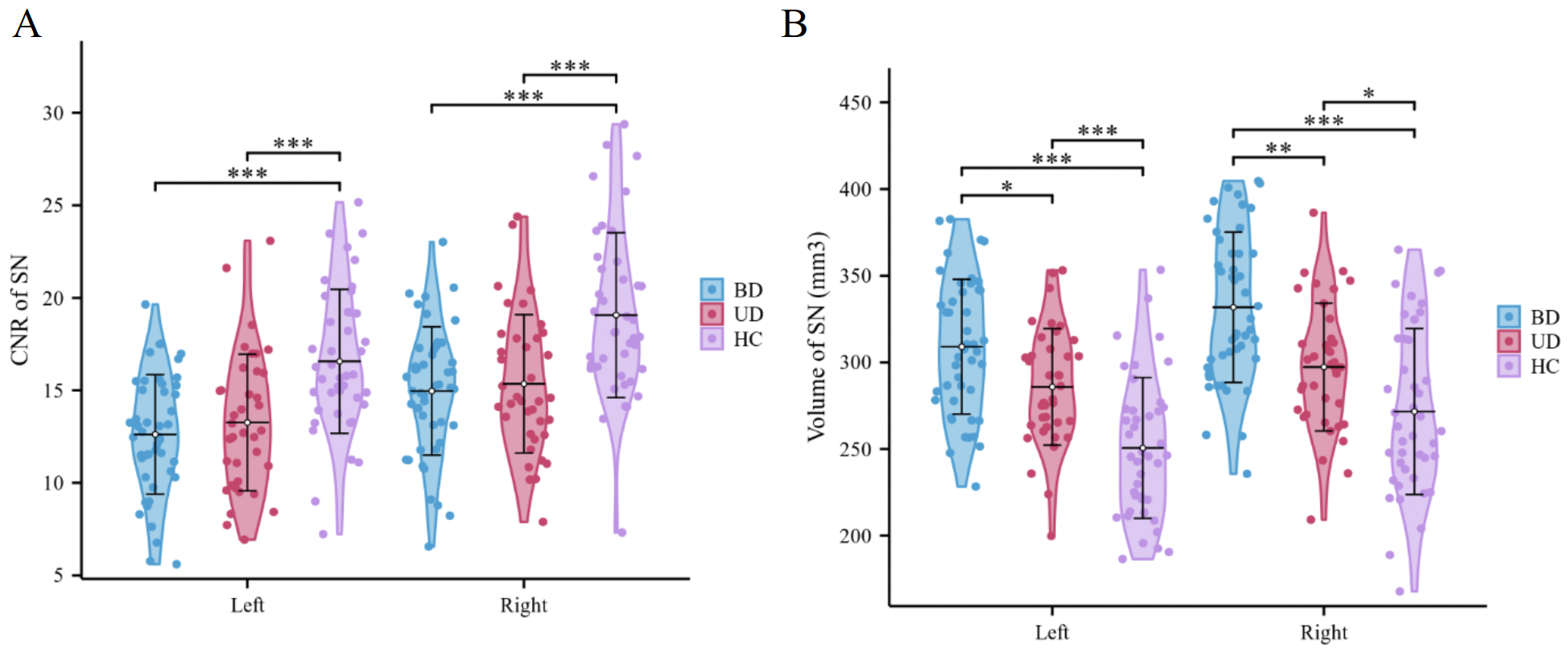
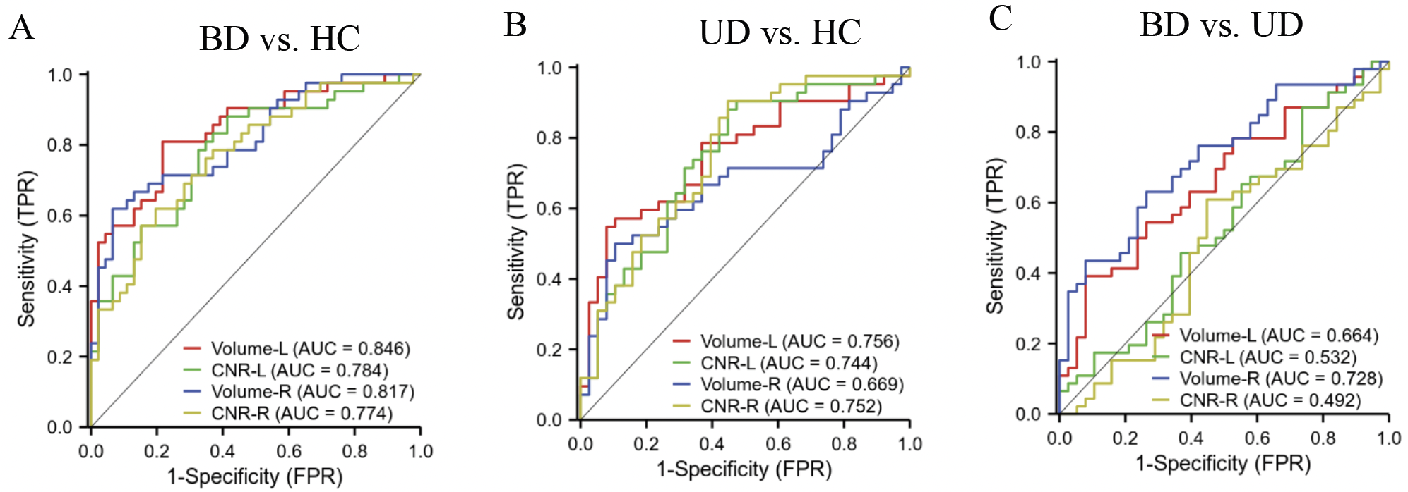
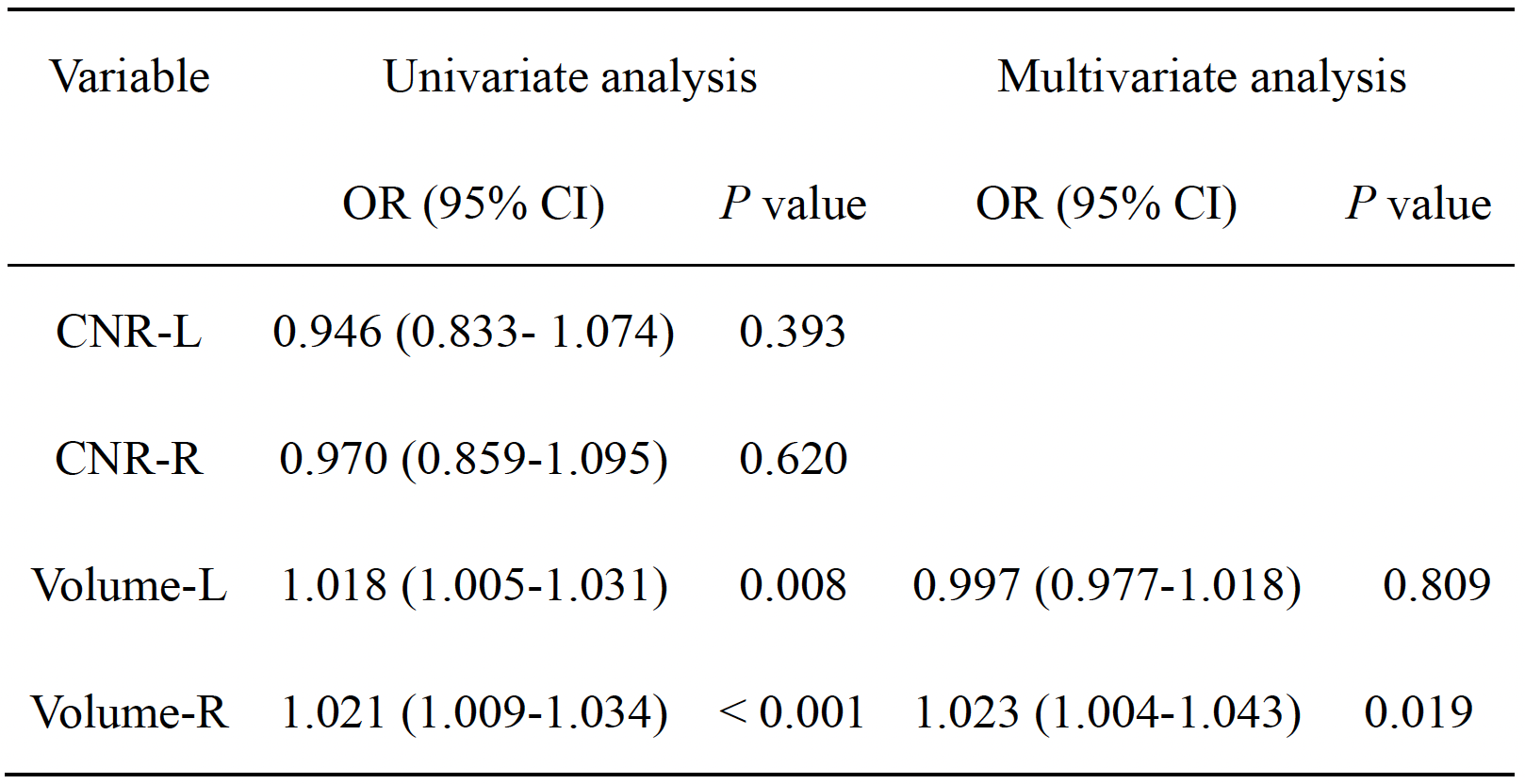
Results: We found that compared with HC, patients with BD-II and UD had decreased CNR and increased volume on bilateral SN. The NM volume in BD-II group was increased compared to UD group. Furthermore, left and right NM volume exhibited the largest AUC for discriminating patient from HC, and BD from UD, respectively.
Impact: Our findings revealed that abnormally decreased CNR and increased volume in SN region might underlie predisposition to BD-II and UD. NM MRI techniques can discriminate BD-II from UD and can be a useful clinical tool in evaluation of mental disorders.
Introduction
Bipolar disorder (BD) is a mood disorder characterized by recurrent episodes of depression and mania or hypomania1. Because of the absence of objective and reliable neurobiological markers, BD, especially BD-II, is commonly misdiagnosed as unipolar depression (UD)2,3. Misdiagnosis leads to inadequate treatment, poor clinical outcomes and long-term medical costs3. Therefore, it is crucial to differentiate BD-II from UD in defining prognosis and treatment decisions. The dopamine hypothesis has been a key theory in the pathophysiology of BD and UD4,5. Two major anatomically defined populations of dopaminergic neurons are located in the substantia nigra (SN) and ventral tegmental area (VTA)6,7. Dopamine neurons provide dense, topographic innervation to the striatum for different functions8. Therefore, it is necessary to map the dopamine system and its discrepancy in BD and UD. MRI, such as neuromelanin-sensitive MRI (NM-MRI)9-11, could be used to delve deeper into depressive behaviors and feature distinctions in pathophysiology between these mood disorders. NM, a dark pigment of dopamine metabolism, accumulates in dopamine neurons of the SN12,13. NM-MRI enables estimation of NM concentration, and has been validated as a marker of dopamine function, with applications to noninvasively interrogate the dopamine system in neuropsychiatric disorders11. Based on the pathophysiology of BD and UD, we hypothesized BD and UD behave differently relative to the neuronal pathology in SN. Therefore, we aimed to determine whether the NM-MRI signal and volume alterations in SN can be used as potential biomarkers to differentiate BD-II from UD.Materials and Methods
The project was approved by the Ethics Committee of Shanghai Mental Health Centre. We recruited 46 patients with the first episode of BD-II, 38 patients with the first episode of UD, and 42 age-matched healthy volunteers. All subjects were evaluated using the structured clinical interview for DSM disorders (SCID) to meet the diagnostic criteria (DSM-IV) for BD-II and UD. Clinical symptoms were assessed according to 24-item HDRS14. Brain MRI examinations were performed on a 3.0 T MRI scanner, and NM-MRI was acquired using a 2D MTC-GRE sequence. ROIs of SN and superior cerebellar peduncles (SCP) were manually delineated on NM-MRI images. CNR of each voxel in SN was calculated by: CNR = (I - mode(ISCP)) / mode(ISCP)10,15. Mean CNR and voxel volume were extracted from bilateral SN. NM parameters among group were compared using one-way ANOVA. ROC curve analysis was conducted to assess discrimination capacity of NM parameters of bilateral SN. Optimal cutoff points were assessed by maximizing both sensitivity and specificity. Diagnostic models were obtained by binary logistic regression. Correlation between continuous variables was performed using Pearson’s correlation coefficient.Results
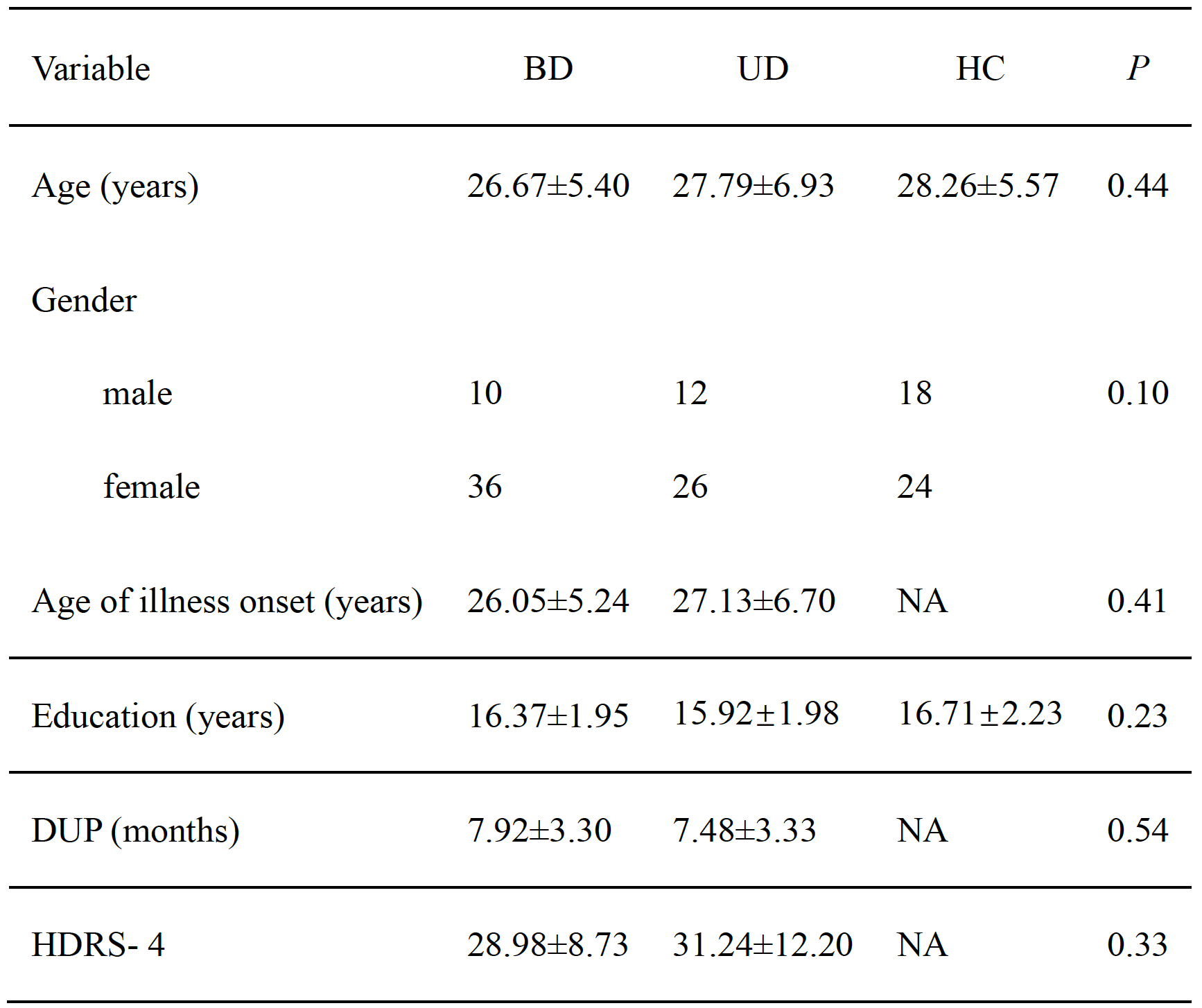
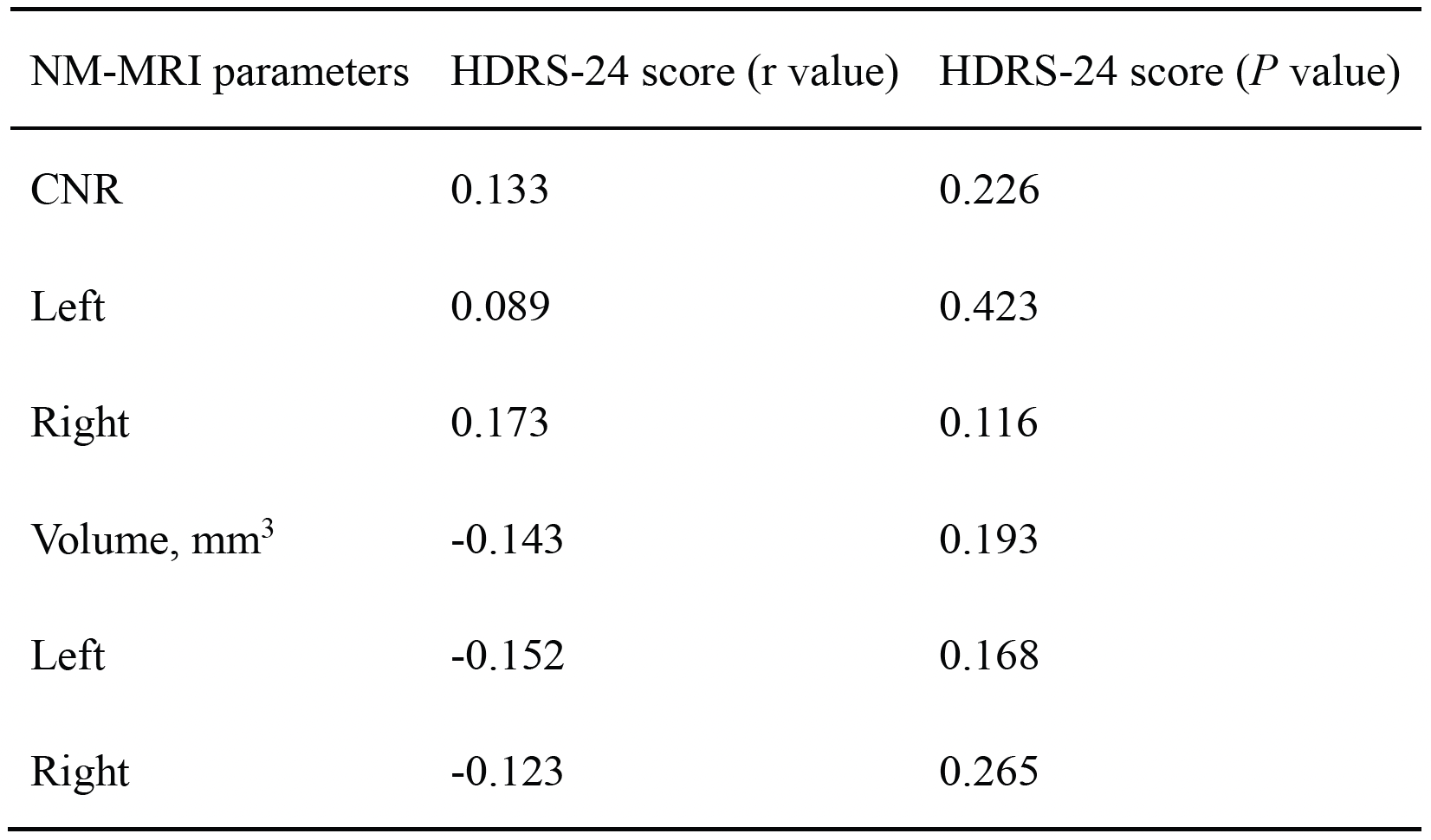
Table 1 showed no significant differences were detected among three groups. In addition, there was no significant difference in the severity of depression between BD and UD groups. Compared with HCs, patients with BD and UD had significantly decreased CNR on both sides (Figure 1A). However, there was no significant difference in the CNR between BD and UD groups. Compared with HCs, patients with BD and UD had significantly increased volume on both sides (Figure 1B). Additionally, the volume in the BD group was significantly increased compared to the UD group. None of NM parameters showed a significant correlation with HDRS-24 score (Table 2). ROC analysis (Figure 2) showed that Volume-L had the largest AUC = 0.846 for discriminating BD from HC, with sensitivity and specificity of 80.9% and 78.3%. Volume-L also had the largest AUC = 0.756 for discriminating UD from HC, with sensitivity and specificity of 54.8% and 92.1%. Volume-R had the largest AUC = 0.728 for discriminating BD from UD, with sensitivity and specificity of 63.0% and 73.7%. Univariate logistic regression demonstrated that the Volume-L and Volume-R were significantly different between BD and UD patients (Table 3). We further calculated predicted probabilities using multivariate logistic regression to explore diagnostic performance of Volume-L (OR = 0.997, P = 0.809), and Volume-R (OR = 1.023, P = 0.019).Discussion
In this study, we found BD and UD patients had reduced CNR and increased volume on bilateral SN compared to HC. Additionally, the volume was higher in patients with BD than in UD and HC. We also demonstrated the potential of NM parameters to provide differential diagnosis of BD and UD patients. To our knowledge, this is the first study to systematically investigate differences between BD-II and UD patients in SN NM parameters. In conclusion, our study revealed NM alterations in SN region might underlie predisposition to BD-II and UD. NM-MRI can serve as biomarkers for discriminating BD-II from UD and might be a useful clinical tool in the evaluation of mental disorders.Acknowledgements
No acknowledgement found.References
1. Hirschfeld RM. Differential diagnosis of bipolar disorder and major depressive disorder. Journal of affective disorders.2014:169 Suppl 1:S12-16.
2. Phillips ML, Kupfer DJ. Bipolar disorder diagnosis: challenges and future directions. Lancet (London, England).2013:381(9878):1663-1671.
3. Siegel-Ramsay JE, Bertocci MA, Wu B, Phillips ML, Strakowski SM, Almeida JRC. Distinguishing between depression in bipolar disorder and unipolar depression using magnetic resonance imaging: a systematic review. Bipolar disorders.2022:24(5):474-498.
4. Grace AA. Dysregulation of the dopamine system in the pathophysiology of schizophrenia and depression. Nature reviews Neuroscience.2016:17(8):524-532.
5. Ashok AH, Marques TR, Jauhar S, Nour MM, Goodwin GM, Young AH, Howes OD. The dopamine hypothesis of bipolar affective disorder: the state of the art and implications for treatment. Molecular psychiatry.2017:22(5):666-679.
6. Gantz SC, Ford CP, Morikawa H, Williams JT. The Evolving Understanding of Dopamine Neurons in the Substantia Nigra and Ventral Tegmental Area. Annual review of physiology.2018:80:219-241.
7. Schulz J, Zimmermann J, Sorg C, Menegaux A, Brandl F. Magnetic resonance imaging of the dopamine system in schizophrenia - A scoping review. Frontiers in psychiatry.2022:13:925476.
8. Cox J, Witten IB. Striatal circuits for reward learning and decision-making. Nature reviews Neuroscience.2019:20(8):482-494.
9. Chen X, Huddleston DE, Langley J, Ahn S, Barnum CJ, Factor SA, Levey AI, Hu X. Simultaneous imaging of locus coeruleus and substantia nigra with a quantitative neuromelanin MRI approach. Magnetic resonance imaging.2014:32(10):1301-1306.
10. Cassidy CM, Zucca FA, Girgis RR, Baker SC, Weinstein JJ, Sharp ME, Bellei C, Valmadre A, Vanegas N, Kegeles LS, Brucato G, Kang UJ, Sulzer D, Zecca L, Abi-Dargham A, Horga G. Neuromelanin-sensitive MRI as a noninvasive proxy measure of dopamine function in the human brain. Proceedings of the National Academy of Sciences of the United States of America.2019:116(11):5108-5117.
11. Wengler K, He X, Abi-Dargham A, Horga G. Reproducibility assessment of neuromelanin-sensitive magnetic resonance imaging protocols for region-of-interest and voxelwise analyses. NeuroImage.2020:208:116457.
12. Zucca FA, Basso E, Cupaioli FA, Ferrari E, Sulzer D, Casella L, Zecca L. Neuromelanin of the human substantia nigra: an update. Neurotoxicity research.2014:25(1):13-23.
13. Horga G, Wengler K, Cassidy CM. Neuromelanin-Sensitive Magnetic Resonance Imaging as a Proxy Marker for Catecholamine Function in Psychiatry. JAMA psychiatry.2021:78(7):788-789.
14. Husain MI, Chaudhry IB, Khoso AB, Kiran T, Khan N, Ahmad F, Hodsoll J, Husain MO, Naqvi HA, Nizami AT, Chaudhry N, Khan HA, Minhas F, Meyer JH, Ansari MA, Mulsant BH, Husain N, Young AH. Effect of Adjunctive Simvastatin on Depressive Symptoms Among Adults With Treatment-Resistant Depression: A Randomized Clinical Trial. JAMA network open.2023:6(2):e230147.
15. Oshima S, Fushimi Y, Okada T, Nakajima S, Yokota Y, Shima A, Grinstead J, Ahn S, Sawamoto N, Takahashi R, Nakamoto Y. Neuromelanin-Sensitive Magnetic Resonance Imaging Using DANTE Pulse. Movement disorders: official journal of the Movement Disorder Society.2021:36(4):874-882.
Figures