5028
Effects of intermittent Theta Burst Stimulation and adjunctive D-Cycloserine on GABA levels in the medial Prefrontal Cortex1University of Calgary, Calgary, AB, Canada
Synopsis
Keywords: Psychiatric Disorders, Brain
Motivation: Non-invasive brain stimulation is an evidenced based treatment for major depression. Change in GABA in the medial prefrontal cortex (mPFC) has been suggested as a mechanism of antidepressant effect of brain stimulation, including intermittent Theta Burst Stimulation (iTBS).
Goal(s): Investigate the effect of iTBS combined with adjunctive D-Cycloserine on magnetic resonance spectroscopy measured GABA levels in the mPFC.
Approach: Participants were randomized to receive adjunctive D-Cycloserine or placebo in addition to two weeks of iTBS treatment, with assessments at baseline and after two weeks of intervention.
Results: There was no effect of iTBS, adjunctive D-Cycloserine or responder status on GABA levels.
Impact: This study provides evidence of dissociation between GABA change and clinical response following the delivery of intermittent Theta Burst Stimulation (iTBS) in major depression. The antidepressant effects of iTBS may not rely on change in GABA within the mPFC.
Introduction
Non-invasive brain stimulation therapies such as intermittent Theta-Burst Stimulation (iTBS) are efficacious for treatment of major depression, however, not all patients benefit from this therapy, leaving a substantial unmet treatment need. One system hypothesized to involved in the pathogenesis and treatment response in major depression is the GABAergic system. GABA levels as measured by magnetic resonance spectroscopy (MRS) have been shown to be lower in the cortex of participants with major depression, particularly within the medial prefrontal cortex (mPFC)1-3. Additionally, lower GABA levels within the mPFC have been shown to associate with anhedonia in major depression2,3 and to increase following successful intervention, including with pharmacological4 and non-invasive brain stimulation5 interventions.Investigating GABA change following non-invasive brain stimulation offers valuable insight into treatment mechanisms and provides direction for improved therapeutics. One avenue of investigation for enhancing the treatment response to non-invasive brain stimulation is the delivery of adjunctive agents to enhance synaptic plasticity and response to stimulation. D-Cycloserine, a NMDA-receptor partial agonist, has been investigated for such enhancement of treatment effects with promising clinical results6.
This study investigated the effects of intermittent theta-burst stimulation (iTBS), with a pharmacological adjunct, on GABA levels in the medial prefrontal cortex.
Methods
Data is drawn from a larger clinical trial6 that investigated iTBS with a pharmacological adjunct, D-Cycloserine, as a treatment for major depression.Clinical response was measured using the Montgomery-Åsberg Depression Rating Scale (MADRS)7, with responders defined as individuals who showed a 50% decrease on MADRS score after two weeks.
MRI data was collected on a 3T GE 750 scanner. A T1-weighted anatomical scan was collected for voxel placement, coregistration and segmentation. GABA-edited MEGA-PRESS data was collected in the mPFC (voxel 30x30x30mm3, TR/TE = 2 s/68 ms, 320 averages, 14 ms editing pulses alternating between 1.9 and 7.46 ppm every two averages, and eight unsuppressed water averages). An exemplar voxel placement and spectrum is shown in Figure 1. Data was processed through the Gannet pipeline8-10 to quantify GABA.
To compare the overall effect of iTBS on GABA levels, the main effect of time was investigated in a repeated measures ANOVA, with adjunct (D-Cycloserine or placebo) as a between groups factor. Similarly, responder status (responder and non-responder) was included as a between groups factor in a separate repeated measures ANOVA.
Results
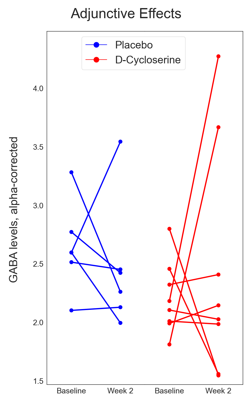
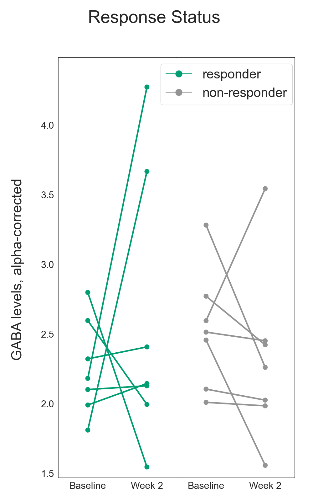
Fourteen participants (mean age=41.93, standard deviation = 14.56) completed MRI scanning at baseline and after two weeks of iTBS with either a pharmacological adjunct, D-Cycloserine (N=8), or placebo (N=6). The MRI sample size is smaller than planned due to Covid-19 related site closures.The MRI sample included 7 responders and 7 non-responders at two weeks of treatment. Of the 6 who received placebo, 2 were classified as responders, while there were 5 individuals identified as responders in the 8 randomized to receive D-Cycloserine.
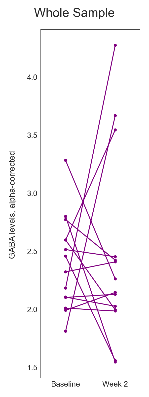
There was no effect of two weeks of iTBS treatment on GABA levels, across the whole sample, (F=0.014, p=0.91, Figure 2) and no differences between the groups who received D-Cycloserine and placebo on GABA levels (F=0.60, p=0.45, Figure 3). When comparing participants who were responders at two weeks to non-responders, there was no significant differences in GABA levels (F=1.10, p=0.32, Figure 4).
Discussion
This study did not evidence change in GABA levels following two weeks of iTBS intervention for major depression. There was no effect at a whole group level, or when comparing those who received adjunctive D-Cycloserine to those who received placebo. Further, there was no significant difference in GABA levels in those who had achieved clinically significant response at two weeks compared to those who were not defined as responders.Finding no effect of iTBS treatment on GABA levels is contrary to previous work investigating both iTBS11 and other repetitive Transcranial Magnetic Stimulation protocols5. It may be that two weeks of intervention may not be long enough for changes in GABA levels. The previous research which has identified changes after treatment are typically on timescales of 4-5 weeks.
In the larger trial, the separation in clinical response between individuals who received adjunctive D-Cycloserine and those who received placebo only became significant at 4 weeks6. Scanning was conducted after two weeks to capture the acute dosage of the adjunct, and not at 4 weeks. It may be that we are not capturing sufficient clinical separation for investigation of GABA levels.
Conclusion
This study provides further evidence for dissociation of clinical response to iTBS from GABA change in the mPFC. It suggests no difference in GABA change for those who receive adjunctive D-Cycloserine to iTBS compared to those who received placebo.Acknowledgements
No acknowledgement found.References
1 Bhagwagar, Z. et al. Low GABA concentrations in occipital cortex and anterior cingulate cortex in medication-free, recovered depressed patients. Int J Neuropsychopharmacol 11, 255-260 (2008). https://doi.org:10.1017/S1461145707007924
2 Gabbay, V. et al. Anterior cingulate cortex gamma-aminobutyric acid deficits in youth with depression. Transl Psychiatry 7, e1216 (2017). https://doi.org:10.1038/tp.2017.187
3 Gabbay, V. et al. Anterior cingulate cortex gamma-aminobutyric acid in depressed adolescents: relationship to anhedonia. Arch Gen Psychiatry 69, 139-149 (2012). https://doi.org:10.1001/archgenpsychiatry.2011.131
4 Brennan, B. P. et al. Acute change in anterior cingulate cortex GABA, but not glutamine/glutamate, mediates antidepressant response to citalopram. Psychiatry Res Neuroimaging 269, 9-16 (2017). https://doi.org:10.1016/j.pscychresns.2017.08.009
5 Dubin, M. J. et al. Elevated prefrontal cortex GABA in patients with major depressive disorder after TMS treatment measured with proton magnetic resonance spectroscopy. J Psychiatry Neurosci 41, E37-45 (2016). https://doi.org:10.1503/jpn.150223
6 Cole, J. et al. Efficacy of Adjunctive D-Cycloserine to Intermittent Theta-Burst Stimulation for Major Depressive Disorder: A Randomized Clinical Trial. JAMA Psychiatry (2022). https://doi.org:10.1001/jamapsychiatry.2022.3255
7 Montgomery, S. A. & Asberg, M. A new depression scale designed to be sensitive to change. Br J Psychiatry 134, 382-389 (1979). https://doi.org:10.1192/bjp.134.4.382
8 Edden, R. A., Puts, N. A., Harris, A. D., Barker, P. B. & Evans, C. J. Gannet: A batch-processing tool for the quantitative analysis of gamma-aminobutyric acid-edited MR spectroscopy spectra. J Magn Reson Imaging 40, 1445-1452 (2014). https://doi.org:10.1002/jmri.24478
9 Harris, A. D., Puts, N. A. & Edden, R. A. Tissue correction for GABA-edited MRS: Considerations of voxel composition, tissue segmentation, and tissue relaxations. J Magn Reson Imaging 42, 1431-1440 (2015). https://doi.org:10.1002/jmri.24903
10 Penny, W. D., Friston, K. J., Ashburner, J. T., Kiebel, S. J. & Nichols, T. E. Statistical parametric mapping: the analysis of functional brain images. (Elsevier, 2011).
11 Diederichs, C. et al. Intermittent Theta-Burst Stimulation Transcranial Magnetic Stimulation Increases GABA in the Medial Prefrontal Cortex: A Preliminary Sham-Controlled Magnetic Resonance Spectroscopy Study in Acute Bipolar Depression. Front Psychiatry 12, 665402 (2021). https://doi.org:10.3389/fpsyt.2021.665402
Figures
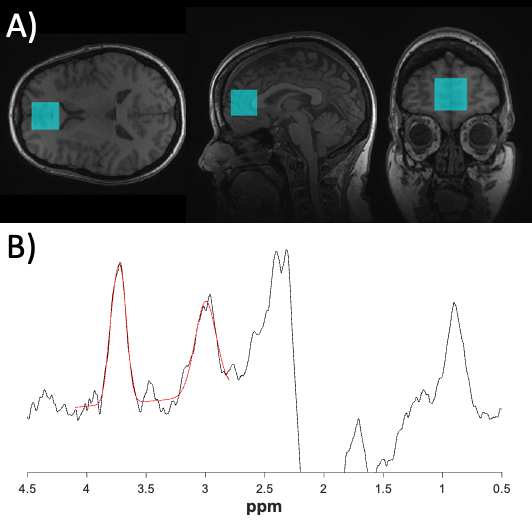
A) an illustrative voxel placement for GABA quantification in the medial prefrontal cortex
B) an exemplar spectrum for the quantification of GABA