5027
Association of peripheral inflammation with disrupted brain functional network topology in bipolar disorder1First Affiliated Hospital of Jinan University, Guangzhou, China, 2MR Research, GE Healthcare, Beijing, China, Guangzhou, China
Synopsis
Keywords: Psychiatric Disorders, Neuroinflammation
Motivation: Increasing evidences show that inflammation might be involved in bipolar disorder (BD), but the association between abnormal brain function and inflammation in BD patients is still unclear.
Goal(s): In this study, we tried to explore the disrupted brain functional network topology, peripheral cytokines and their correlations to demonstrate the role of inflammation in brain functional network topology in BD.
Approach: Graph theory analysis.
Results: The current study demonstrated disrupted topological organization in the whole brain and regional connectivity was associated with inflammatory cytokines of the IL-4, IL-8 and IL-10 levels in BD.
Impact: Our study provided preliminary evidence of the association between disrupted brain functional network topology and neuroinflammation in BD.
Background
Increasing evidences show that inflammation might be involved in bipolar disorder (BD), but the association between abnormal brain function and inflammation in BD patients is still unclear. In this study, we tried to explore the disrupted brain functional network topology, peripheral cytokines and their correlations to demonstrate the role of inflammation in brain functional network topology in BD.Methods
In this study, 53 BD patients and 47 healthy controls (HCs) underwent resting-state magnetic resonance imaging scans. Graph theory analysis was performed to investigate the topological properties of whole-brain functional connectome at both global and nodal levels. Serum levels of interleukin-4 (IL-4), interleukin-6 (IL-6) interleukin-8 (IL-8), interleukin-10 (IL-10), and tumor necrosis factor-α (TNF-α) were measured in all participants. Correlations between topological properties, clinical variables and cytokines levels in BD were calculated. Furthermore, mediation analysis was employed to determine whether inflammatory cytokines affect depressive symptoms by affecting brain network properties.Results
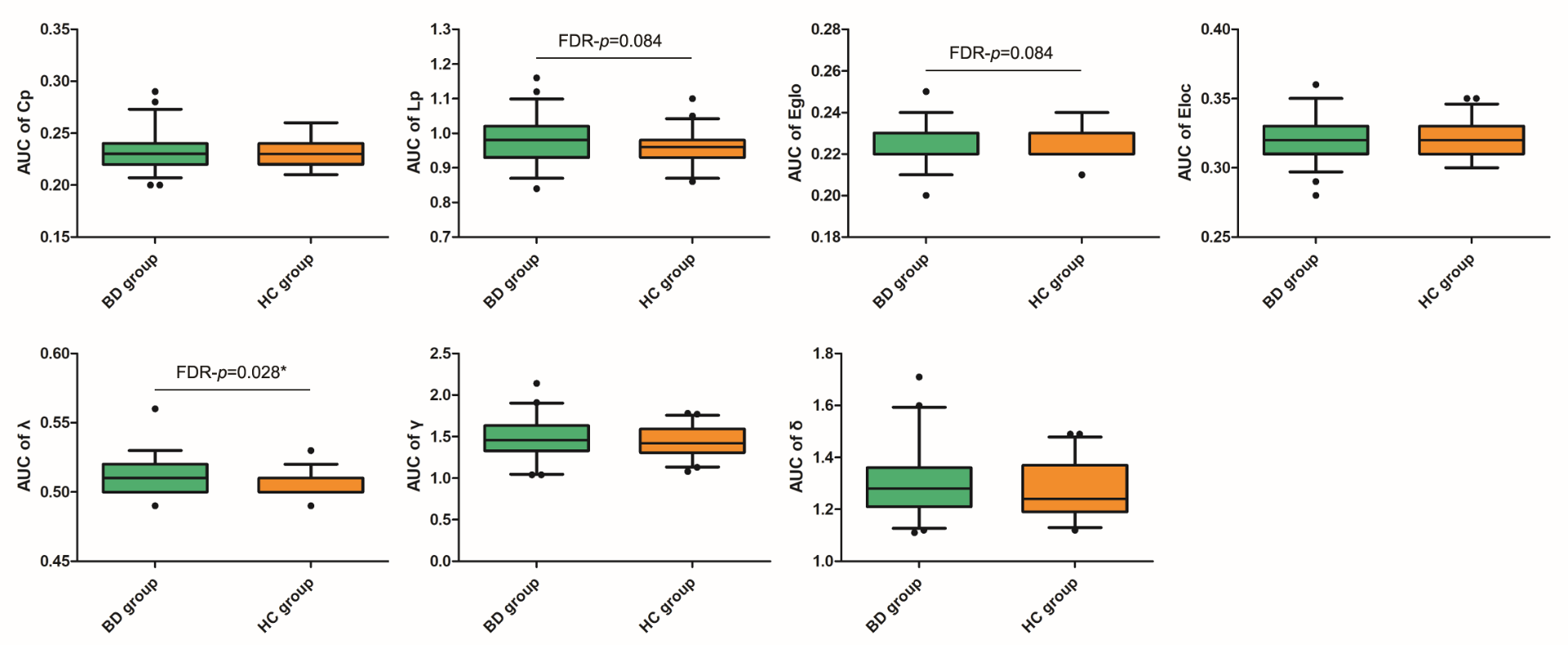
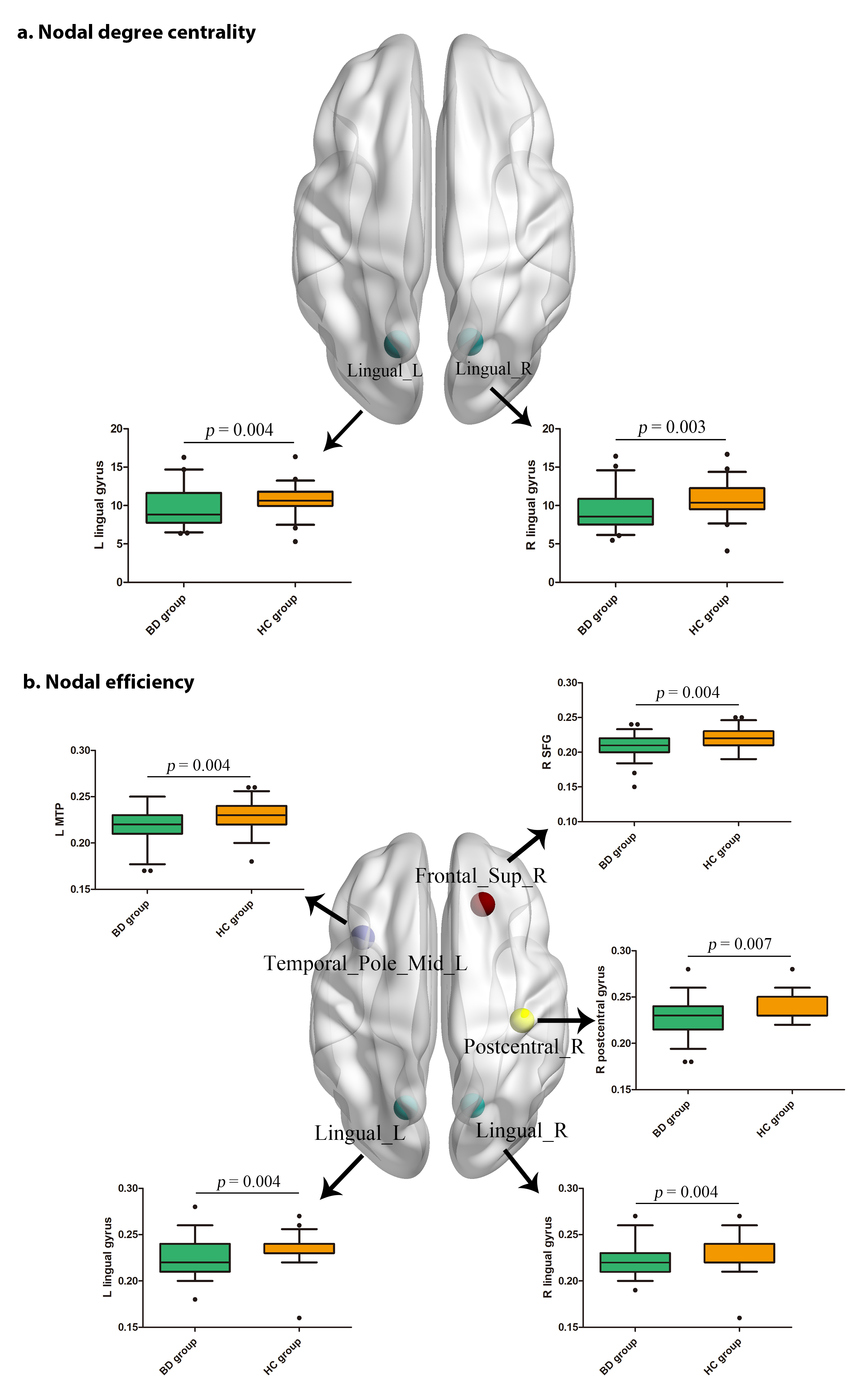
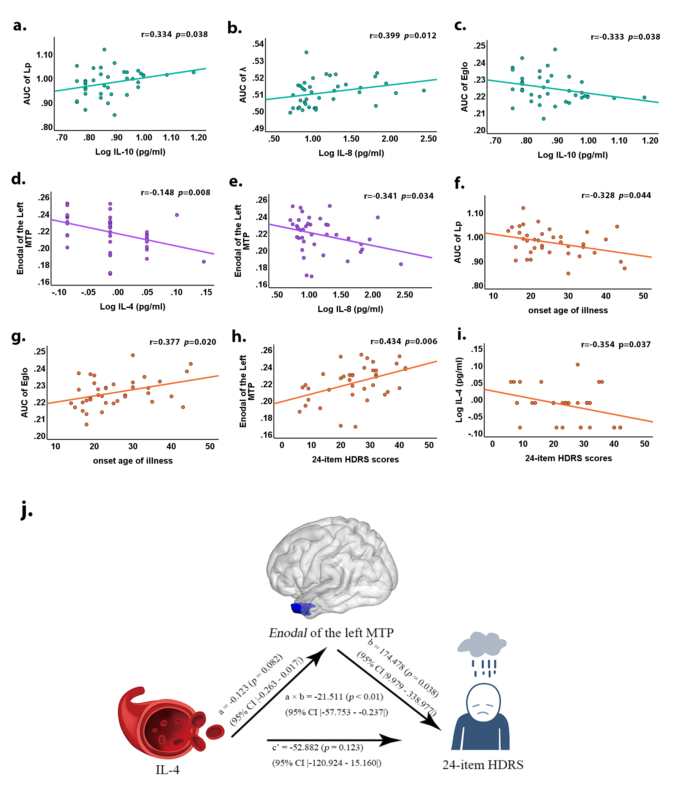
Patients with BD showed significantly greater normalized characteristic path length ( λ ), and marginally greater characteristic path length (Lp) and lower global efficiency (Eglo). Furthermore, the BD group showed lower nodal degree centrality (Degi) in the bilateral lingual gyrus, and lower nodal efficiency (Enodal) in the bilateral lingual gyrus, right postcentral gyrus, right superior frontal gyrus (SFG), and the left middle temporal pole (MTP). Besides, IL-4, IL-6, IL-8, IL-10 levels were higher in BD than HCs. Correlation analyses revealed that elevated levels of IL-10 were correlated with the greater Lp and lower Eglo, and elevated levels of IL-8 was correlated with the greater . Furthermore, the Enodal in the left MTP was negatively correlated with IL-4 and IL-8 levels in BD. Moreover, the Enodal of the left MTP mediated the effect of IL-4 levels on the 24-item HDRS scores.Conclusions
Our study provided preliminary evidence of the association between disrupted brain functional network topology and neuroinflammation in BD. The current study demonstrated disrupted topological organization in the whole brain and regional connectivity was associated with inflammatory cytokines of the IL-4, IL-8 and IL-10 levels in BD. Moreover, our results indicated that higher IL-4 levels and impaired regional connectivity in the temporal pole may be associated with the severer depressive symptoms in BD.Acknowledgements
The study was supported by grants from the National Natural Science Foundation of China (81671670, 81501456, and 81971597). The funding organizations play no further role in study design, data collection, analysis and interpretation, paper writing and decision to publish.References
No reference found.Figures