5026
Motor inhibition in functional paralysis – a task-based fMRI study1Advanced Imaging Research (AIR) Group, Swiss Paraplegic Research, Nottwil, Switzerland, 2Department of Radiology, Swiss Paraplegic Centre, Nottwil, Switzerland, 3Department of Paraplegia, Rehabilitation and Quality Management, Swiss Paraplegic Centre, Nottwil, Switzerland, 4Faculty Health Science and Medicine, University Lucerne, Lucerne, Switzerland, 5Health Services Research, Swiss Paraplegic Research, Nottwil, Switzerland, 6Outpatient Care Unit, Swiss Paraplegic Centre, Nottwil, Switzerland, 7Clinic for Neurology, Cantonal Hospital St.Gallen, St. Gallen, Switzerland, 8Institute for Diagnostic and Interventional Neuroradiology, University Hospital and Inselspital, Bern, Switzerland
Synopsis
Keywords: Psychiatric Disorders, Neuroscience, Functional Neurological Disorder
Motivation: In functional neurological disorders (FND), symptoms (including paralysis) occur despite intact anatomical pathways. The pathophysiology of FND is poorly understood, making diagnosis, treatment and prognosis uncertain.
Goal(s): Assuming that higher cognitive functions may play a role in the manifestation of symptoms, this study investigates motor inhibition in FND.
Approach: Using task-based fMRI, this observational study investigates the blood oxygenation level dependent (BOLD) response of the brain during a motor inhibition task.
Results: Preliminary results indicate reduced behavioral performance and lower BOLD activity in the left caudate nucleus, a key node for motor inhibition, suggesting a disrupted neural pathway in FND.
Impact: Our task-based fMRI results suggest impaired motor inhibition in functional neurological disorders and open new avenues for further investigation on its role in symptom production and potential treatment options.
Introduction
In functional neurological disorders (FND), neurological symptoms occur despite intact anatomical pathways. The symptoms often show similar phenotypes to other neurological conditions, such as stroke or spinal cord injury (SCI). The pathophysiological mechanisms of FND are poorly understood, with uncertain diagnosis, treatment and prognosis [1]. The focus of this study is on the subtype functional paralysis, where symptoms of paralysis is present in certain parts of the limbs. Recent neuroimaging studies have revealed functional alterations in several brain areas in FND. For instance, altered activity in the right temporoparietal junction, sensorimotor areas, and limbic system has been shown in FND [2], as well as abnormal connectivity of fronto-parietal networks [3]. Moreover, impaired cognitive and affective processes have been shown in FND [4], and inhibitory control has been hypothesised to be involved in symptoms’ production [5], [6]. Suppressed activation of frontal areas during a motor inhibition task has been shown in a single patient with functional paralysis [7]. In this observational study, we use event-related functional magnetic resonance imaging (fMRI) with a go/no-go task to reveal the neural correlates of motor inhibition in functional paralysis. The novelty of our approach is that we compare functional paralysis with SCI, i.e., two disorders with a similar phenotype, but different underlying causality, while healthy controls (HC) act as baseline for the comparisons.Methods
So far, 10 FND patients, 8 SCI patients and 11 HC were measured (expected target 25 participants per group). While recording functional images in a 3T Philips Achieva scanner, participants completed a go/no-go task, responding as quickly and accurately as possible to a go stimulus, while inhibiting the response to a no-go stimulus. Functional images were preprocessed using SPM12 [8] as follows: All functional images were realigned, coregistered to the structural images, normalized with a voxel size of 2mm, and smoothed with a 6mm full-width-at-half-maximum kernel. Hit (correct response to go stimulus) and correct rejection (correct inhibition to no-go stimulus) conditions were defined over the entire BOLD timeseries for every participant, and estimated using the hemodynamic response function.Results
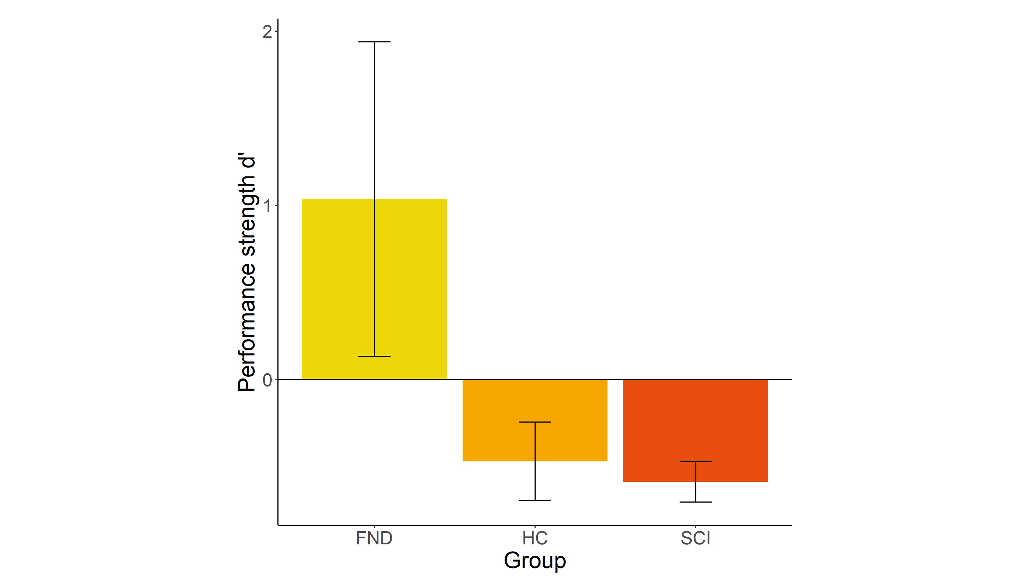
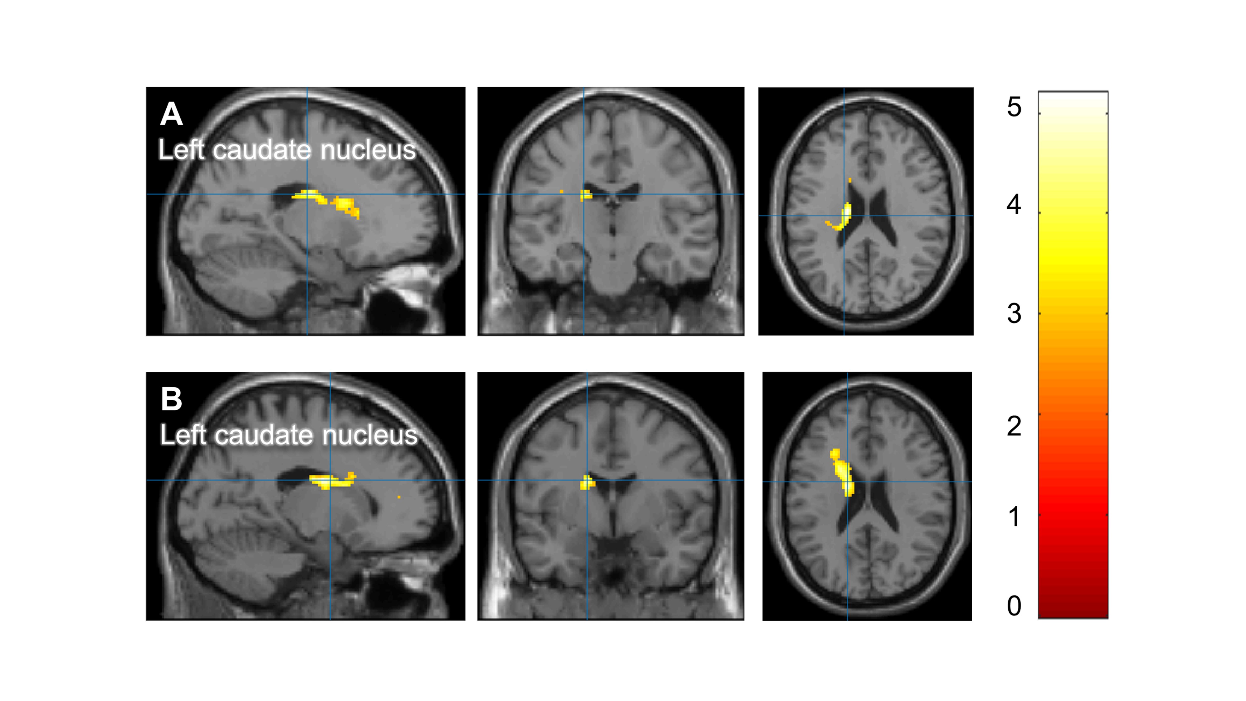
Applying signal detection theory to calculate the strength of behavioral performance (discriminability index d') reveals a trend towards different performance between the groups (F(2,27) = 2.69, p =.085), with FND patients showing lower performance compared to HC and SCI patients (see Figure 1). In the fMRI contrast hit vs. correct rejection, lower BOLD signal was found in the left caudate nucleus in FND patients compared to HC as well as compared to SCI patients (puncorrected <0.001, cluster size>20) (see Figure 2). The SCI patients showed no differences in the left caudate compared to HC.Conclusion
The nucleus caudate is part of the striatum, and is involved in the indirect pathway of motor inhibition [9]. These preliminary findings suggest that hypoactivity of the caudate may be responsible for poor motor inhibition in FND, and possibly correlate with the reduced motor output.Acknowledgements
We would like to thank the Swiss Paraplegic Group for providing us with the infrastructure for the study, the MR technicians of the Radiology Department of the Swiss Paraplegic Centre for performing the MR examinations and especially the study participants.References
Aron, A. R., Durston, S., Eagle, D. M., Logan, G. D., Stinear, C. M., & Stuphorn, V. (2007). Converging Evidence for a Fronto-Basal-Ganglia Network for Inhibitory Control of Action and Cognition: Figure 1. The Journal of Neuroscience, 27(44), 11860–11864. https://doi.org/10.1523/JNEUROSCI.3644-07.2007
Baek, K., Doñamayor, N., Morris, L. S., Strelchuk, D., Mitchell, S., Mikheenko, Y., Yeoh, S. Y., Phillips, W., Zandi, M., Jenaway, A., Walsh, C., & Voon, V. (2017). Impaired awareness of motor intention in functional neurological disorder: Implications for voluntary and functional movement. Psychological Medicine, 47(9), 1624–1636. https://doi.org/10.1017/S0033291717000071
Cojan, Y., Waber, L., Carruzzo, A., & Vuilleumier, P. (2009). Motor inhibition in hysterical conversion paralysis. NeuroImage, 47(3), 1026–1037. https://doi.org/10.1016/j.neuroimage.2009.05.023
Friston, K., Ashburner, J., Kiebel, S., Nichols, T., & Penny, W. (2007). Statistical Parametric Mapping: The Analysis of Functional Brain Images (1st ed.). Academic Press.
Gelauff, J. M., Carson, A., Ludwig, L., Tijssen, M. A. J., & Stone, J. (2019). The prognosis of functional limb weakness: A 14-year case-control study. Brain, 142(7), 2137–2148. https://doi.org/10.1093/brain/awz138
Hammond-Tooke, G. D., Grajeda, F. T., Macrorie, H., & Franz, E. A. (2018). Response inhibition in patients with functional neurological symptom disorder. Journal of Clinical Neuroscience, 56, 38–43. https://doi.org/10.1016/j.jocn.2018.08.005
Maurer, C. W., LaFaver, K., Ameli, R., Epstein, S. A., Hallett, M., & Horovitz, S. G. (2016). Impaired self-agency in functional movement disorders: A resting-state fMRI study. Neurology, 10.1212/WNL.0000000000002940. https://doi.org/10.1212/WNL.0000000000002940
Monsa, R., Peer, M., & Arzy, S. (2018). Self‐reference, emotion inhibition and somatosensory disturbance: Preliminary investigation of network perturbations in conversion disorder. European Journal of Neurology, 25, 888–894. https://doi.org/10.1111/ene.13613
van Wouwe, N. C., Mohanty, D., Lingaiah, A., Wylie, S. A., & LaFaver, K. (2020). Impaired Action Control in Patients With Functional Movement Disorders. The Journal of Neuropsychiatry and Clinical Neurosciences, 32(1), 73–78. https://doi.org/10.1176/appi.neuropsych.19030076
Figures