5017
Multimodal MR identifies the prefrontal cortex as central hub of stress-induced brain alterations in rats1RG Translational Imaging, Central Institute of Mental Health, Medical Faculty Mannheim, University of Heidelberg, Mannheim, Germany
Synopsis
Keywords: Psychiatric Disorders, Preclinical
Motivation: Chronic unpredictable mild stress (CUMS) is utilized to model depression-like symptoms in rodents. Although the prevalence of depression is twice as high in women, 97% of CUMS studies focused on male animals.
Goal(s): We aimed to comprehensively phenotype the effect of CUMS in female rats using MRI.
Approach: Thus, we assessed a CUMS-induced depression-like phenotype with various behavioral tests and multimodal MRI specifically in females.
Results: We found prominent prefrontal and striatal volume loss, together with prefrontal glutamate reduction, and decreased top-down prefrontal control of the nucleus accumbens, corroborating the high sensibility of the PFC to chronic stress in the female population.
Impact: We identified the prefrontal cortex as the central hub of chronic stress-induced changes in brain function in a female population of animals using multimodal MRI, thus creating a model for further investigation of distinct effects in possible treatments.
Introduction
Chronic unpredictable mild stress (CUMS) is used to model depression-like symptoms in rodents, representing currently the most reliable model for the disease. However, 97% of CUMS studies focus on male animals1, while depression has a twice as high prevalence in females. Therefore, our comprehensive study explored the impact of chronic stress on both behavior and brain function with multimodal neuroimaging techniques particularly in female rats.Methods
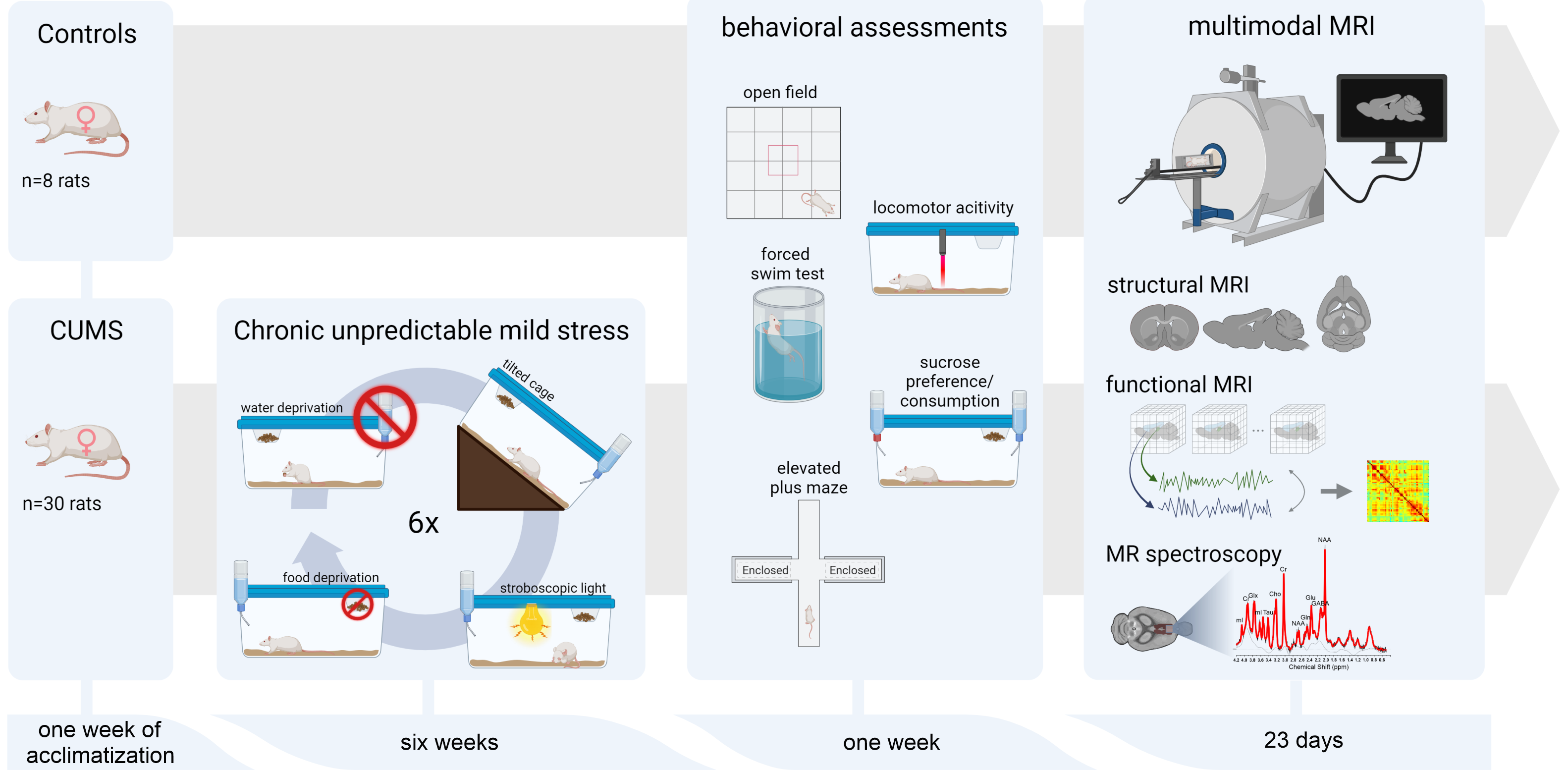
The study involved n=30 Wistar rats, which were exposed to a six-week CUMS procedure to induce a depression-like phenotype through repeated microstressors, and a stress-free control group of n=8 rats. After the CUMS procedure, both groups underwent five behavioral tests to assess the severity of depression-like symptoms. MRI experiments were conducted on a 9.4T MR-scanner with a cryogenically cooled coil. Anesthesia combined 0.5% isoflurane and medetomidine (0.06 mg/kg/h). Respiratory and cardiac signals were monitored to correct functional data for physiological noise. The MRI acquisition protocol included a high-resolution 3D-scan (T2-weighted RARE, (0.15)²×0.288mm³ resolution), MR-spectroscopy (PRESS, 12µl-voxel in the prelimbic-cingulate cortex, TR/TE=4000/10ms, frequency-shift -2ppm) and resting-state fMRI (rsfMRI) (T2*-weighted EPI-FID, TR/TE 1500/17.5ms, (0.365mm)²×0.7mm, 350 acquisitions, 32 slices). An additional field map was acquired to correct for geometrical distortions. Analyses were performed using SPM12, in-house MATLAB scripts, FSL and LCModel.VBM: Preprocessing steps included brain extraction, bias correction, coregistration, segmentation and nonlinear normalization using DARTEL. Normalized and modulated gray matter images were analyzed in a second-level GLM using total intracranial volume and age as covariates.
MRS: Individual frequency and phase correction was done over coils and averages as well as eddy current correction using an unsuppressed water-signal. Spectra were quantified by LCModel using a calibrated phantom basis dataset. Metabolite concentrations were referenced to the unsuppressed water signal, assuming mean water concentration of 46.106mol/L, and corrected for relaxation effects (T1/T2met=1500/300ms, T2water=45ms).
rsfMRI: Preprocessing included correction for field inhomogeneities and movement, regression of physiological noise2, slice-timing correction and spatial normalization to a template using DARTEL-based flow fields from the 3D images, regression of movement parameters and CSF signal, DVARS scrubbing3 and band-pass filtering (0.01–0.1Hz). Regional time courses were extracted using a bilateral anatomical atlas. Preprocessed rsfMRI data was analyzed with NBS4, and seed-based approaches.
Results
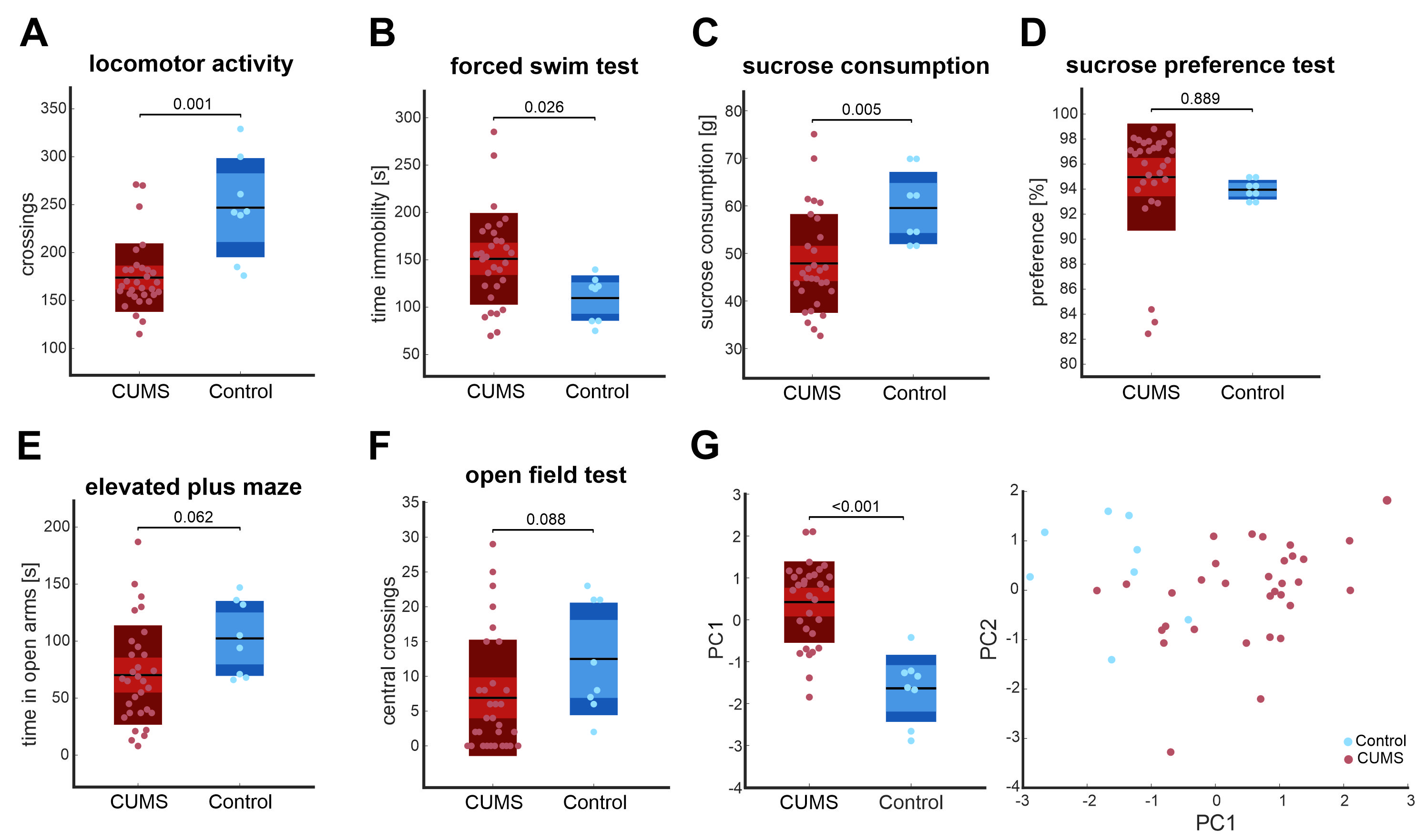
CUMS rats displayed a depression-like phenotypeCUMS-exposed rats demonstrated a reduction of locomotor activity in their home cages (Fig. 2A) and an extended duration of immobility in the forced swim test (Fig. 2B), an indicator of hopelessness. Although the total sucrose consumption was diminished in CUMS-exposed rats (Fig. 2C), no differences in sucrose preference could be detected (Fig. 2D). CUMS-exposed rats exhibited a tendency to spend less time in the open arms of the elevated plus maze (Fig. 2E) and in the central area of the open field (Fig. 2F), implying elevated anxiety.
Chronic stress leads to gray matter volume loss in prefrontal, striatal and somatosensory regions
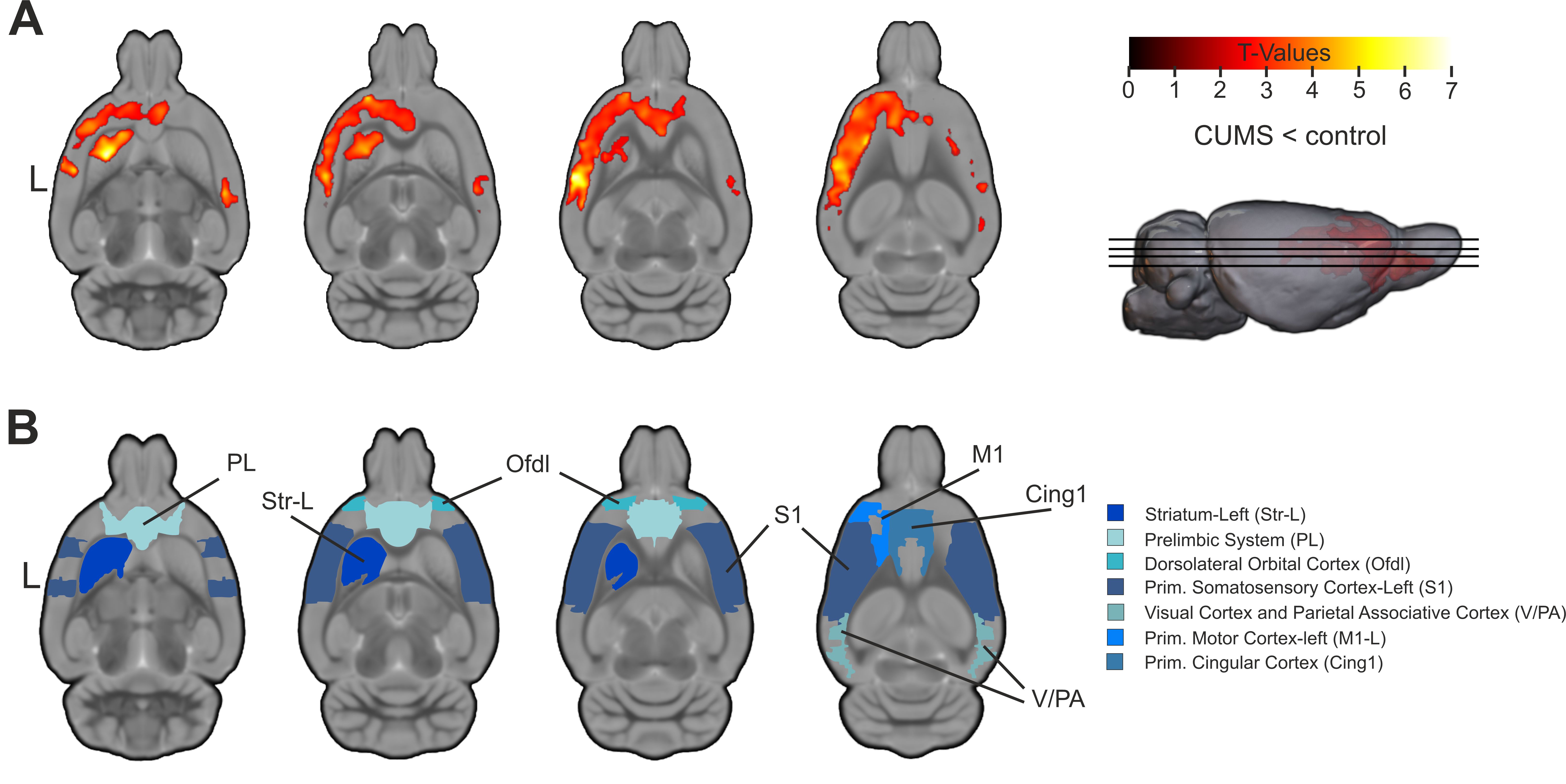
CUMS-exposed rats displayed a wide-spread reduction in gray-matter within prefronto-cortical (PFC) and striatal regions (Fig. 3A). CUMS-induced volume reduction predominantly affected the PFC, encompassing prelimbic (PL) and primary cingulate cortex (Cing1), the left striatum (Str-L), the left and right somatosensory cortex (S1), as well as the primary motor areas (M1) (Fig 3A-B).
MR spectroscopy shows decreased glutamate levels in the PFC after chronic stress exposure
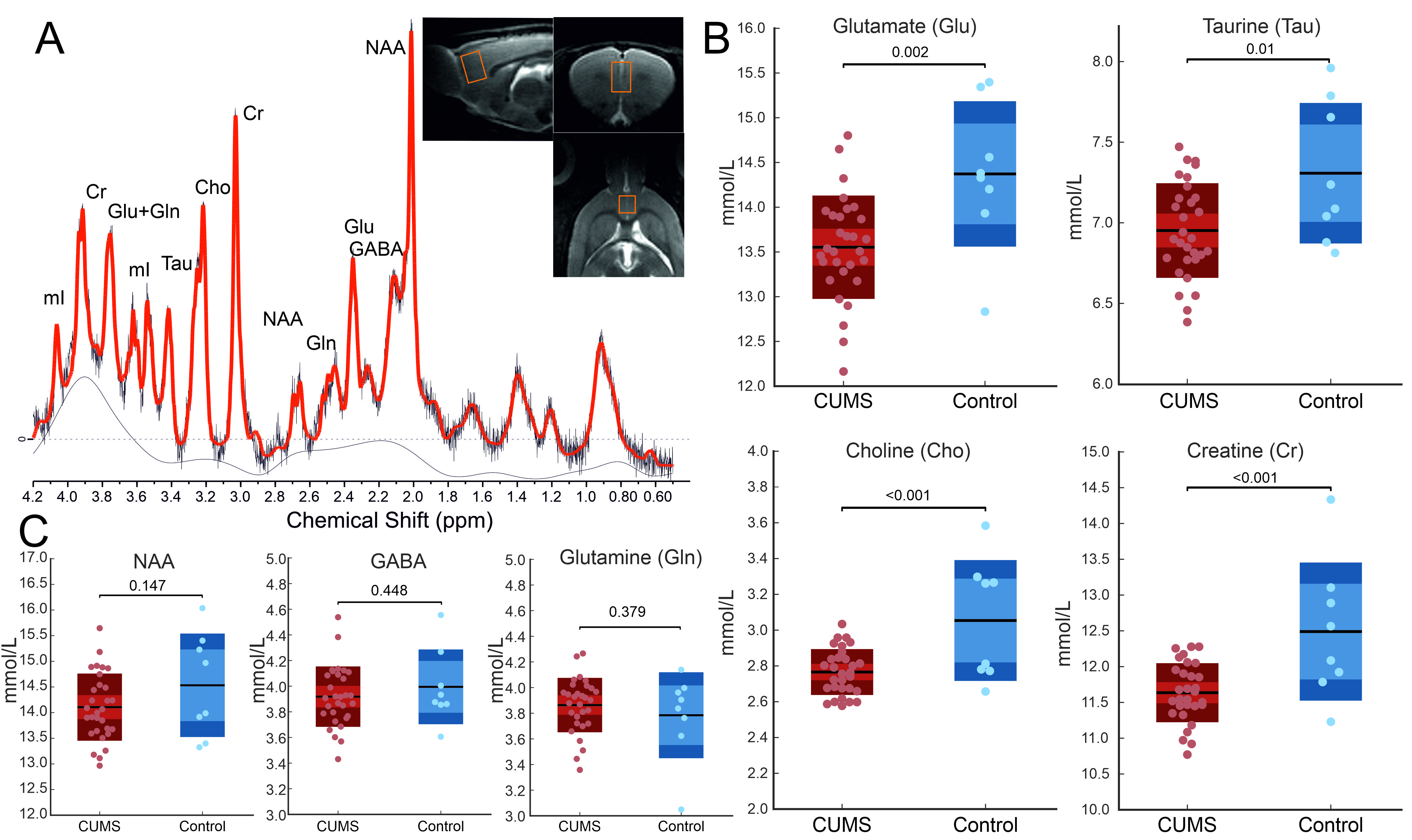
CUMS-exposed rats showed significantly lower levels of the neurotransmitter Glu in the PFC (Fig. 4B). In addition, levels of Tau, Cho, and Cr (Fig. 4B) were also decreased after CUMS exposure. We could not find differences for GABA, Gln, and NAA (Fig. 4C).
Rats exposed to chronic stress show bidirectional alterations of functional connectivity (FC) in th PFC and limbic system
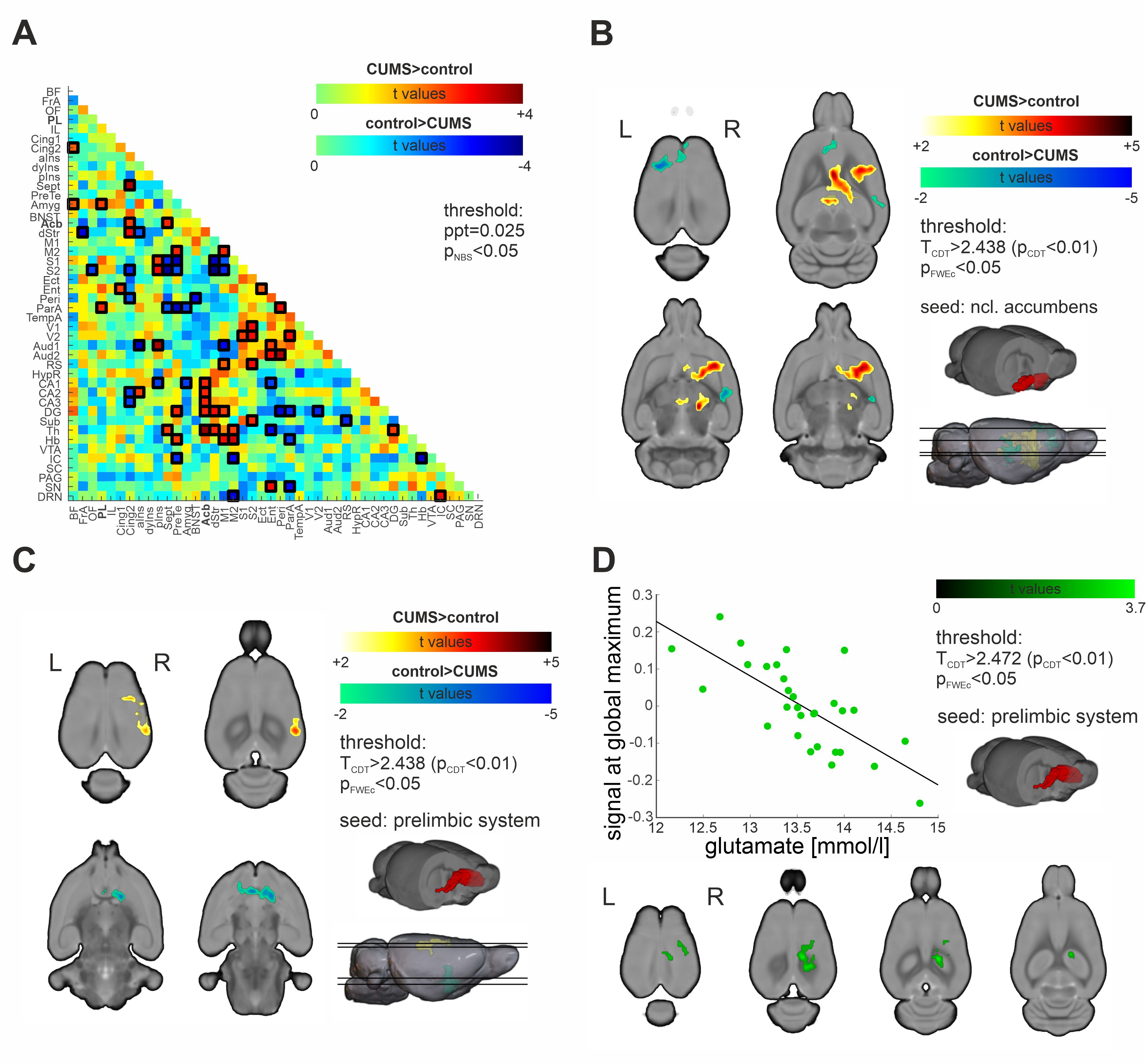
On a whole-brain level, CUMS rats demonstrated increased FC within the striato-hippocampal and the somatosensory-parietal network. Our seed-based FC assessment of the nucleus accumbens (Acb) confirmed higher FC to dorsal striatal and hippocampal regions in CUMS animals (Fig. 5B). In contrast, PL connections to the Acb were decreased (Fig. 5C). The FC between PL and the hippocampus was modulated by the level of glutamate in the PFC (Fig. 5D).
Summary/Discussion
In summary, our results suggest the PFC as a central hub region for stress-induced brain changes in female rats. With that, our data contributes to the current evidence for the PFC describing it as specifically sensible to chronic stress, and hypothesizing a chronic stress-induced enhancement of glutamatergic transmission in the PFC as a crucial factor to changes within the prefrontal GABAergic system. Reported evidence of increased glutamatergic transmission onto prefrontal parvalbumin neurons, particularly in female rodents, is clearly in line with the results of our study5.Acknowledgements
We thank Felix Hörner for his skillfully conducted MR-measurements.References
- Antoniuk S, Bijata M, Ponimaskin E, Wlodarczyk J. Chronic unpredictable mild stress for modeling depression in rodents: Meta-analysis of model reliability. Neurosci Biobehav Rev. 99:101-116 (2019).
- van Buuren M, Gladwin TE, Zandbelt BB, van den Heuvel M, Ramsey NF, Kahn RS, Vink M, Cardiorespiratory effects on default-mode network activity as measured with fMRI. Hum Brain Mapp 30:3031-3042 (2009).
- Power JD , Mitra A, Laumann TO, Snyder AZ, Schlaggar BL, Petersen SE. Methods to detect, characterize, and remove motion artifact in resting state fMRI. Neuroimage 84:320–341 (2014).
- Zalesky A, Fornito A, Bullmore ET, Network-based statistic: Identifying differences in brain networks. Neuroimage 53:1197–1207 (2010).
- Shepard R, Coutellier L. Changes in the Prefrontal Glutamatergic and Parvalbumin Systems of Mice Exposed to Unpredictable Chronic Stress. Mol Neurobiol 55:2591–2602 (2018).
Figures