5014
Multiplexed sensitivity-encoding (MUSE) DWI with deep learning-based reconstruction in breast MR imaging: A comparison with conventional DWI1West China Hospital of Sichuan University, Chengdu, China, 2GE HealthCare MR Research, Beijing, China
Synopsis
Keywords: Breast, Diffusion/other diffusion imaging techniques
Motivation: Conventional DWI has limitations due to low spatial resolution and geometry distortion. Multiplexed sensitivity-encoding (MUSE) DWI can obtain images with higher resolution and less distortion but require longer acquisition time.
Goal(s): Our aim was to apply deep-learning based reconstruction (DLR) in MUSE DWI for breast imaging, and to investigate if DLR can shorten the scan time while maintaining image quality of MUSE.
Approach: We compared quantitative parameters and subjective image quality of MUSE, MUSE-DLR, and conventional DWI.
Results: MUSE-DLR showed improved image quality than MUSE with slightly longer acquisition time compared to conventional DWI.
Impact: MUSE DWI with deep-learning based reconstruction can enhance the accuracy of clinical breast imaging while maintaining an acceptable scanning time, and also has the potential to improve diffusion imaging in other parts of the human body.
Introduction
Diffusion-weighted imaging (DWI) has demonstrated its ability to improve specificity of breast MRI and is widely used in routine clinical applications. DWI offers the advantage of short scan time and no need for exogenous contrast agent1,2. Nevertheless, DWI suffers from distortions, low spatial resolution. Multiplexed sensitivity-encoding (MUSE) DWI can improve resolution, reduce geometric distortions, and exhibit significantly better image quality over conventional DWI3. Nonetheless, MUSE takes longer time to acquire images due to the use of multiple shots for excitation and acquisition. Accelerated techniques, such as parallel imaging and smaller number of excitations (NEX), typically compromise image quality. Deep learning-based reconstruction (DLR) has been employed in MRI to improve image quality and decrease scan time4. This study aimed to combine DLR with MUSE (MUSE-DLR), and compare the performance of MUSE, MUSE-DLR, and conventional DWI in breast MRI.Methods
Patients: This study was approved by the institutional ethics committee. 11 female patients (age: 30-57 years) with suspected breast cancer were enrolled in the study. The MR examinations were performed on a 3.0T MR scanner (SIGNA Premier, GE Healthcare) equipped with a dedicated 8-channel bilateral breast coil.Imaging parameters: The clinical breast MRI protocol was extended to include both conventional single-shot DWI sequence and MUSE DWI with b-values of 0 and 1000s/mm2. The detailed imaging parameters of conventional DWI were: FOV=340×340 mm², TR/TE=4525/53.1 ms, slices=32, matrix=128×168, acquired voxel size=2.7×2.0×4.0 mm³, bandwidth=3906.25 HZ/Px, parallel imaging factor=2, acquisition time=63 s, and NEX=4 for b1000. The imaging parameters of MUSE were: FOV=340×340 mm², TR/TE=3074/53.3 ms, slices=32, matrix=128×168, acquired voxel size=2.7×2.0×4.0 mm³, bandwidth=3906.25 HZ/Px, parallel imaging factor=1, acquisition time=98 s, NEX=2 for b1000, and Num Shots=4. Then a prototype version of DLR (AIR Recon DL) was used to reconstruct k-space data of MUSE to get MUSE-DLR images.
Data processing: To objectively assess the performance of each sequence, signal-to-noise ratio (SNR), and contrast-to-noise ratio (CNR) of lesion, apparent diffusion coefficient (ADC) of lesion values were measured. SNR and CNR were calculated based on the following formulas:
$$SNR=\frac{S_{lesion}}{SD_{background}}$$
$$CNR=\frac{|S_{lession}-S_{tissue}|}{\sqrt{SD_{lesion}^{2}+SD_{tissue}^{2}}}$$
where Slesion and Stissue are the mean signal intensity of lesion and normal tissue. SDbackground, SDlesion and SDtissue stand for the standard deviation of background noise, lesion and normal tissue5.
ROI of lesion was drawn on the slice (b1000) with the largest cross section of the lesion avoiding regions exhibiting necrosis or hemorrhage. ROI of tissue was drawn on the same slice where the glands are homogeneous. ROI of background was drawn in air region on the same slice. ADC parametric maps were calculated using b0 and b1000 images. Subjective image quality was assessed by one radiologist using a 5-point Likert scale (1=poor, 5=excellent) in terms of the overall image quality, lesion conspicuity, artifacts, and geometric distortion6.
Statistical analysis: Statistical analysis was carried out using SPSS, version 22.0. The Likert scales and quantitative parameters were compared between DWI, MUSE and MUSE-DLR using the Kruskal-Wallis test. A p value less than 0.05 was considered significant.
Results
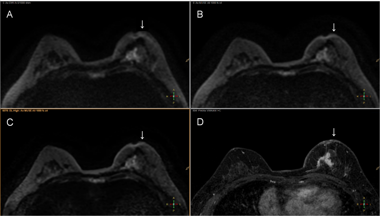
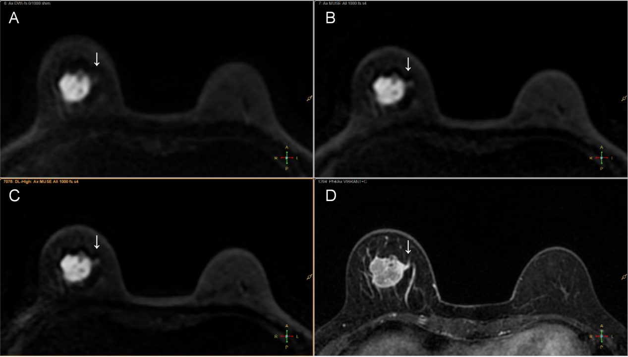
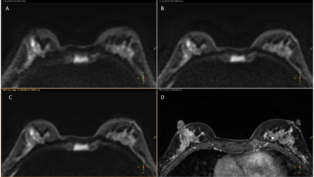
As shown in Table 1, MUSE-DLR showed significant higher SNR than MUSE (P=0.007). Though the CNR did not show a significant difference among three sequences, the values were higher in MUSE and MUSE-DLR. There was no significant difference in the measured ADC values of lesion among three sequences. Image scores in lesion conspicuity and lesion artifacts of MUSE-DLR was higher than those of conventional DWI and MUSE-DLR, but not statistically significant (Table 2). Moreover, MUSE-DLR and MUSE showed less geometric distortion when compared to conventional DWI. MUSE-DLR showed better overall image quality when compared to conventional DWI. Several examples are shown in other Figures.Discussion
This study indicated that MUSE and MUSE-DLR was superior to conventional DWI in terms of distortion. MUSE-DLR showed significant better overall image quality than conventional DWI. But lesion clarity, artifacts and CNR were not significantly different from each other. Furthermore, MUSE-DLR showed greatly improved SNR than MUSE. The results demonstrate that MUSE-DLR in breast MRI provides better image quality within a clinically feasible acquisition time, which can provide more information in diagnosing breast diseases. With the deep-learning based reconstruction technique, it may be possible to use other accelerated techniques in MUSE sequence to achieve shorter scanning time in future. Nevertheless, the limitation of this study is that the sample size was too small.Conclusion
MUSE-DLR allows for less geometry distortion and shows significantly better overall image quality than conventional DWI and higher SNR than MUSE with slightly increased scan time than conventional DWI. This technique can be incorporated into clinical breast MRI protocols used for routine clinical applications.Acknowledgements
No acknowledgement found.References
1. Partridge SC, Nissan N, Rahbar H, Kitsch AE, Sigmund EE. Diffusion-weighted breast MRI: Clinical applications and emerging techniques. J Magn Reson Imaging. 2017 Feb;45(2):337-355. doi: 10.1002/jmri.25479. Epub 2016 Sep 30. PMID: 27690173; PMCID: PMC5222835.
2. Amornsiripanitch N, Bickelhaupt S, Shin HJ, Dang M, Rahbar H, Pinker K, Partridge SC. Diffusion-weighted MRI for Unenhanced Breast Cancer Screening. Radiology. 2019 Dec;293(3):504-520. doi: 10.1148/radiol.2019182789. Epub 2019 Oct 8. PMID: 31592734; PMCID: PMC6884069.
3. Baxter GC, Patterson AJ, Woitek R, Allajbeu I, Graves MJ, Gilbert F. Improving the image quality of DWI in breast cancer: comparison of multi-shot DWI using multiplexed sensitivity encoding to conventional single-shot echo-planar imaging DWI. Br J Radiol. 2021 Mar 1;94(1119):20200427. doi: 10.1259/bjr.20200427. Epub 2020 Sep 9. PMID: 32903028; PMCID: PMC8011253.
4. Wessling D, Gassenmaier S, Olthof SC, Benkert T, Weiland E, Afat S, Preibsch H. Novel deep-learning-based diffusion weighted imaging sequence in 1.5 T breast MRI. Eur J Radiol. 2023 Sep;166:110948. doi: 10.1016/j.ejrad.2023.110948. Epub 2023 Jun 25. PMID: 37481831.
5. Chen M, Feng C, Wang Q, Li J, Wu S, Hu D, Deng B, Li Z. Comparison of reduced field-of-view diffusion-weighted imaging (DWI) and conventional DWI techniques in the assessment of Cervical carcinoma at 3.0T: Image quality and FIGO staging. Eur J Radiol. 2021 Apr;137:109557. doi: 10.1016/j.ejrad.2021.109557. Epub 2021 Jan 21. PMID: 33549900.
6. Fujioka T, Mori M, Oyama J, Kubota K, Yamaga E, Yashima Y, Katsuta L, Nomura K, Nara M, Oda G, Nakagawa T, Tateishi U. Investigating the Image Quality and Utility of Synthetic MRI in the Breast. Magn Reson Med Sci. 2021 Dec 1;20(4):431-438. doi: 10.2463/mrms.mp.2020-0132. Epub 2021 Feb 2. PMID: 33536401; PMCID: PMC8922358.
Figures