5013
Improvement of motion-related misalignments in dynamic contrast-enhanced breast MRI using advanced 3D fast elastic image registration1Department of Radiological Services, Tokyo Women's Medical University, Tokyo, Japan, 2Philips Japan, Tokyo, Japan, 3Department of Diagnostic Imaging and Nuclear Medicine, Tokyo Women's Medical University, Tokyo, Japan, 4Philips Healthcare Iberia, Madrid, Spain, 5Philips India, Bangalore, India, 6Philips Healthcare, Best, Netherlands, 7Philips Research, Humburg, Germany
Synopsis
Keywords: Breast, Breast
Motivation: Motion and breathing artifacts in DCE breast MRI can cause misalignment among each dynamic, resulting in inaccurate tumor assessment.
Goal(s): Our goal was to demonstrate the feasibility of advanced fast elastic image registration (FEIR) for correction of misalignment in breast DCE MRI.
Approach: FEIR was applied and evaluated in 11 patients who underwent breast DCE examinations.
Results: Advanced FEIR clearly improved misalignments among dynamic scans and provided improved accuracy of time intensity curves (TICs).
Impact: FEIR improves motion related misalignments among respective dynamic scans in DCE breast MRI, it could improve the TICs more accurately.
Introduction
Breast MRI has an important role for accurate diagnosis of breast tumors because of its ability to evaluate blood flow as well as qualitative diagnosis of breast tumors and their spread. Dynamic contrast-enhanced breast MRI (DCE-MRI) reflects the state of blood flow in the tumor and is expected to provide qualitative diagnosis.1,2 Furthermore, time intensity curve (TIC) which plots to contrast enhancement signal intensities over time, is used for quantitative analysis of DCE breast MRI, and it has an important role in determining whether breast tumors aggressiveness based on their time enhancement patterns.3,4However, motion-related artifacts can deteriorate the reliable diagnostic performance of DCE breast MRI, it may also alter TIC results. Non-rigid registration algorithms have been reported as motion correction methods for DCE breast MRI.5-7 On the other hand, called fast elastic image registration (FEIR) has gained attention to correct the motion in cardiac MRI.8,9
In this study, we propose a new 3D registration scheme based on FEIR for motion correction in breast DCE MRI.
Methods
All MR imaging was performed on a 3.0T clinical system (Ingenia, Philips Healthcare) using the dStream Breast coil with the patient in prone position.11 patients with breast cancer (mean age, 62.4years±15.9) underwent breast MRI examination, consists of diffusion-weighted imaging, native T1-weighed and T2-weighed sequences, DCE sequence with 5 dynamics (unenhanced, 20s, 70s, 120s, 350s), followed by a high-resolution contrast-enhanced imaging. DCE sequences were performed using fat-suppressed 3D T1-turbo field-echo (T1TFE) with coronal acquisition. After the acquisition, FEIR was applied as a delayed reconstruction.
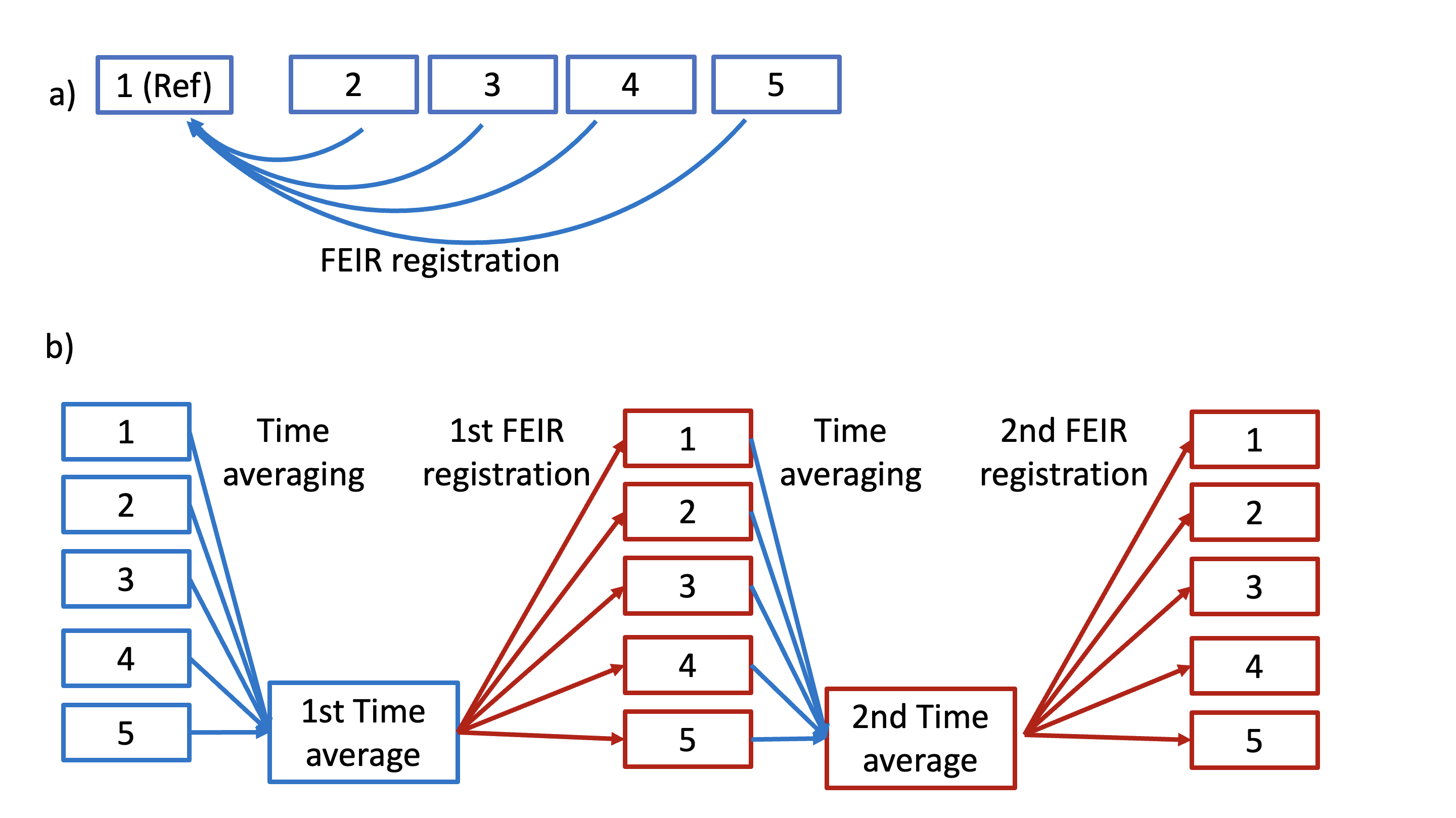
We applied and compared two types of 3D FEIR algorithms, conventional FEIR with arbitrary reference frame, and advanced FEIR, which applies FEIR twice. This advanced approach uses time mean series as a reference frame, to avoid any bias in the reference selection. In the first run all the dynamic series are time average without registration and once all the different dynamic are register to this reference a second reference is build using registered series. This second average frame in used for final second elastic registration (Figure 1). From now on this method will be referred as FEIR Ref.0.
TICs were created and compared for each of the images with and without FEIR. The ROI of the TIC was set along the tumor contour based on BI-RADS-MRI, which states that "a ROI of at least 3 pixels should be set in the area of the lesion that shows the strongest enhancement effect".10
The imaging-parameters of DCE (T1TFE): FOV=250×320mm, voxel-size=1.0×1.0×2.0mm, TR/TE=3.4/1.6ms, flip-angle=11, SPAIR fat suppression, TFE-factor=36, SENSE factor=2, and acquisition time=44sec.
Results and Discussion
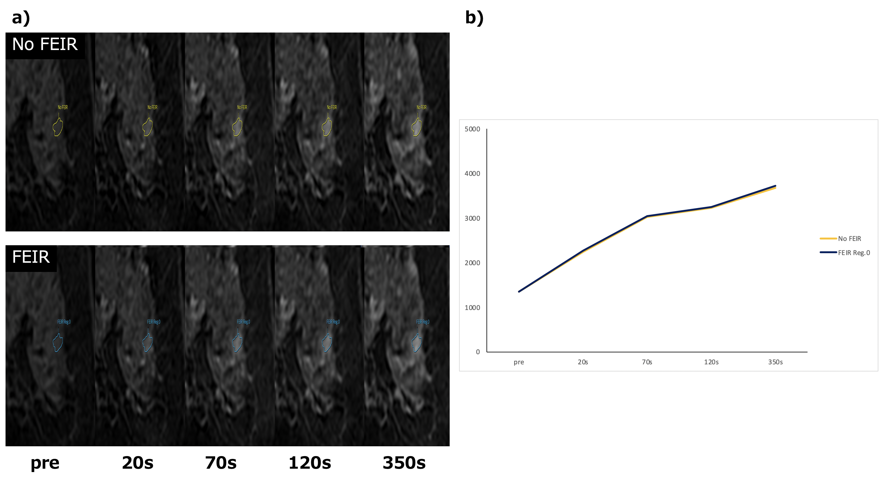
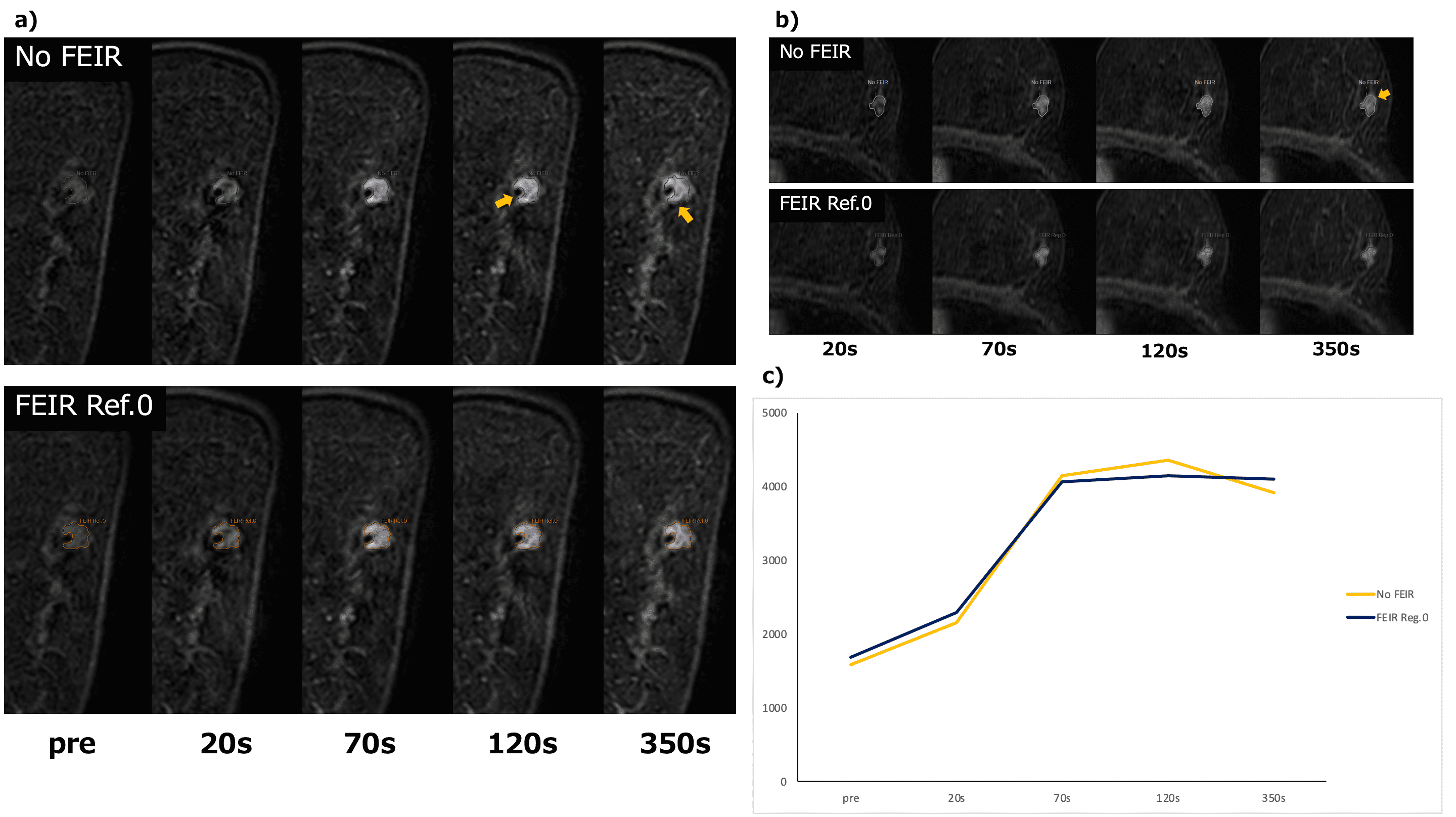
Figure 2a shows representative breast DCE MRI without and with advanced 3D FEIR in a patient without any motion artifacts for each dynamic. Also, TICs created from breast DCE MRI without and with advanced 3D FEIR are shown in Figure 2b. If there is no motion during each dynamic imaging as such case, FEIR does not provide any negative effects.Figure 3a,b shows the comparison of breast DCE images with and without FEIR in a patient with motion during the dynamic scans. Figure 3c shows TICs of breast DCE MRI without and with advanced 3D FEIR. FEIR corrected tumor misalignments well and thus improved the accuracy of TIC.
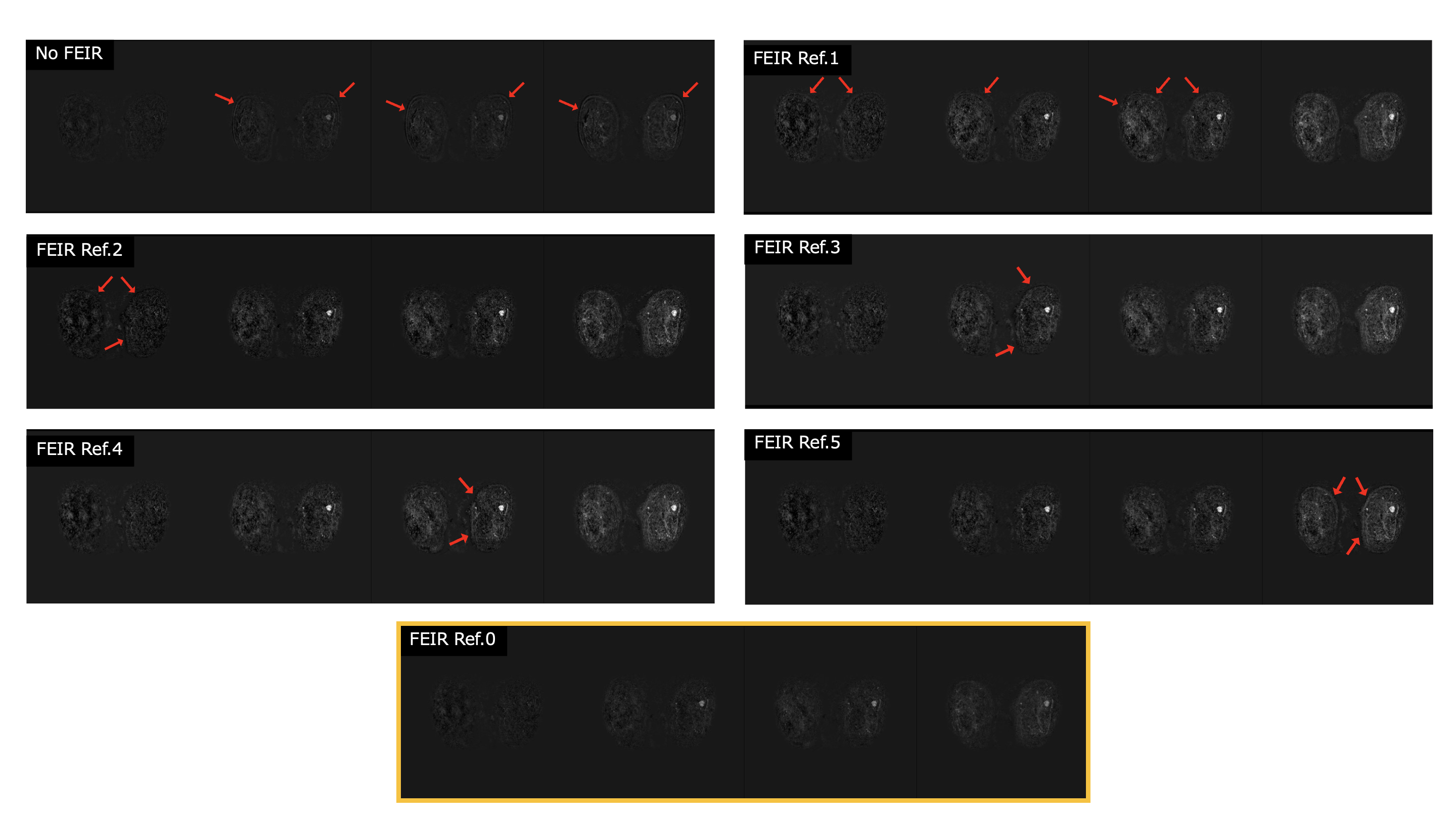
Figure 4 shows subtraction images of DCE breast MRI obtained by different FEIR types (conventional FEIR with each refence frame and advanced double FEIR) in a patient with motion during the dynamic scans. In conventional FEIR images, misalignments were not corrected with some of refence frames, it suggests that the choice of right refence frame is important. On the other hand, using advanced double FEIR showed robust results because it does not require to select the refence frame by the operator, it could prevent FEIR instability due to incorrect setting, resulting in improvement of workflow.
In this study, 4 out of 11 patients were moving during the dynamic scans, their images were misaligned due to movement and/or heavy breathing. In all these cases, advanced FEIR worked well to correct motion-induced misalignments.
Conclusion
We have demonstrated that the advanced double 3D FEIR algorithm clearly improves motion-induced misalignments in breast DCE MRI. It may be promising for robust and accurate assessment of breast tumor enhancement and might be helpful for improving diagnostic performance.Acknowledgements
No acknowledgements found.References
1.Maria Adele Marino, et al. Multiparametric MRI of the Breast: A Review. Journal of Magnetic Resonance Imaging. 2018;47:301–315
2. Doris Leithner, et al. Abbreviated MRI of the Breast: Does It Provide Value? Journal of Magnetic Resonance Imaging. 2019;49:e85–e100.
3.American College of Radiology. Breast Imaging Reporting and Data System. 5th edition, 2013.
4.Kuhl CK, et al. Dynamic breast MR imaging: are signal intensity time course data useful for differential diagnosis of enhancing lesions. Radiology 1999; 211(1): 101-110.
5.Paul M. Hayton, et al, A non-rigid registration algorithm for dynamic breast MR images. Artificial Intelligence. 1999;114:125–156.
6.Yuanjie Zheng, et al. De-enhancing the Dynamic Contrast-Enhanced Breast MRI for Robust Registration. Med Image Comput Comput Assist Interv. 2007;0(Pt 1):933–941.
7.Chiara Mattusch, et al. Development and validation of a four-dimensional registration technique for DCE breast MRI. Insights into Imaging. 2023;14:17
8.Shuo Zhang, et al. Cardiac magnetic resonance T1 and extracellular volume mapping with motion correction and co-registration based on fast elastic image registration. Magnetic Resonance Materials in Physics, Biology and Medicine. 2018;31:115–129
9.Mana kato, et al. Rapid multiple breath-hold myocardial cine T2mapping with dynamic multiple trigger delay acquisition. Proc. ISMRM:2023
10.Akiko shimanai. MRI:Kinetic curve assessment. INNERVISION 2015;30:56-8
Figures