5012
Evaluation of AI Based Reconstruction to Improve image Quality of T2w Images of the Breast1Imaging Physics, MD Anderson Cancer Center, Houston, TX, United States, 2Breast Imaging, MD Anderson Cancer Center, Houston, TX, United States, 3Biostatistics, MD Anderson Cancer Center, Houston, TX, United States
Synopsis
Keywords: Breast, Breast
Motivation: Breast MRI has exceptional sensitivity but is limited by image quality and several artifacts. Advancements in AI-based reconstruction hold promise for improved image quality and efficiency.
Goal(s): This study assesses the impact of applying a vendor AI-based reconstruction on breast MRI at 3T.
Approach: This study retroactively reconstructed 45 series using a commercially available AI-based reconstruction. Three board-certified radiologists scored traditional and reconstructed sequences for quality and improvement.
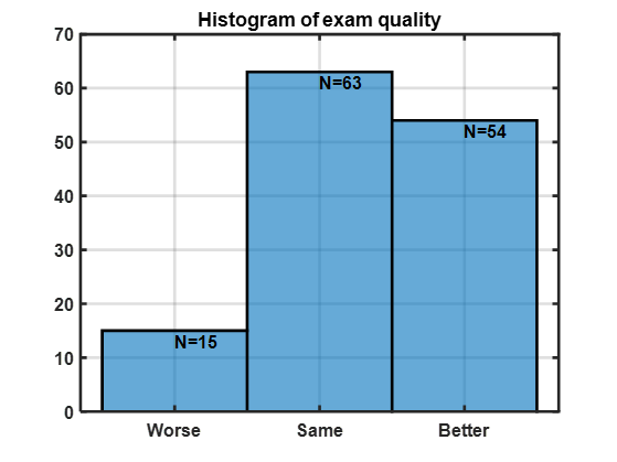
Results: AI reconstruction showed more conspicuous margins as well as an enhanced noise texture. 54 cases showed improvement, 63 showed no change, and 15 exhibited degraded quality. Higher-quality exams were associated with the greatest improvement.
Impact: A retrospective analysis of a recent FDA approved AI based reconstruction method to improve MRI image quality for breast studies.
Introduction
Breast MRI (bMRI) has become an essential modality and standard of care in the practice of breast imaging for a wide spectrum of indications1. Despite the superiority of MRI to other conventional breast imaging modalities (such as mammogram, ultrasound), the technical complexity and variable image quality based on different scanners, magnetic strengths, motion and other artifacts, remain a challenge for breast radiologists. There is ongoing need for improved image quality with thinner slices, reduced artifacts, improved tumor margin conspicuity, and matched slice thickness between T2-weighted images and contrast-enhanced T1-weighted images in addition to shorter scan time. These competing demands often push the technical limitations of MRI2,3.A recent development in the field of MRI reconstruction is the use of convolutional neural network-based reconstruction techniques commonly referred to as deep learning based image reconstruction (AI reconstruction)4,5. These reconstruction methods can reconstruct images from raw k-space data with higher noise suppression and/or improved edge sharpness. The promise of AI based reconstruction is to deliver higher SNR, at better spatial resolution. In this pilot study, a commercially available AI based reconstruction was retrospectively applied to bMRI exams performed at one center on a single scanner to determine if AI can improve image quality.
Methods
Breast MRI exams from a single 3T scanner (Magnetom Vida, version XA30 Siemens Healthcare, Erlangen, Germany) at a cancer center were collected with retrospective AI based reconstruction. Standard of care bMRI protocol included a pre-contrast T1-weighted, contrast-enhanced 3D T1-weighted series with pre and three post-contrasts, 2D T2-weighted two-point Dixon, sagittal delayed 3D-T1-weighted series6, and DWI. AI reconstruction was applied to the T2 Dixon series with ~0.8 x 0.8 x 3.0 mm resolution, 3-fold acceleration, and total acquisition time of ~3 minutes. AI based reconstruction (Deep Resolve Gain and Sharp) was retrospectively applied to the k-space data using the default settings of relative denoising of 0.85 and edge enhancement 2.T2-weighted conventional and AI reconstructed images were reviewed in batch mode and scored by three board-certified breast radiologists with 8 – 22 years of experience. Each series was subjectively scored for image quality using a four-point system: Poor, Average, Good, and Excellent. AI reconstructed images were additionally scored for improvement on a three-point system: -1 for worse, 0 for the same, and 1 for improved. Notably no effort was made to blind the readers to which images were reconstructed using the AI based technique because the noise texture was so different between the two images sets that blinding was felt to be futile.
Results
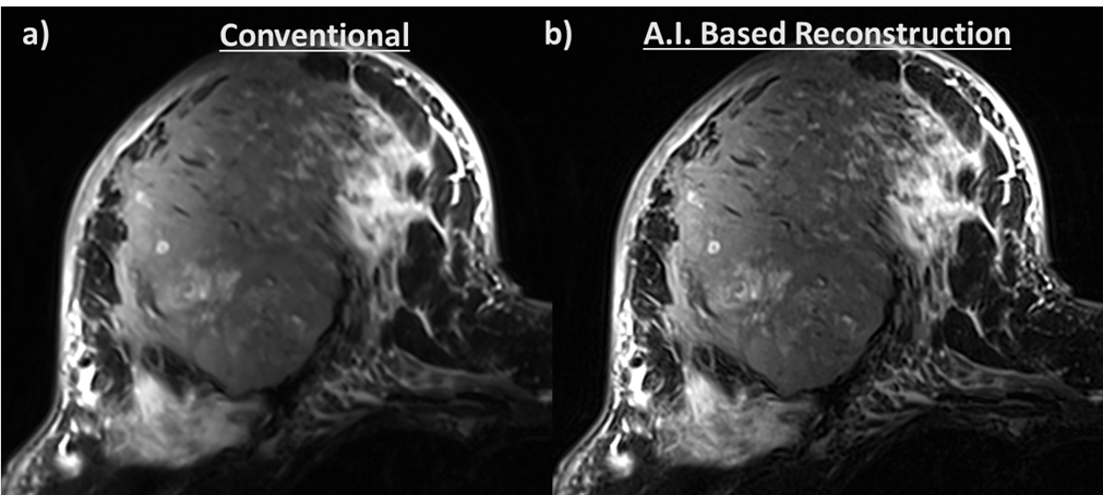
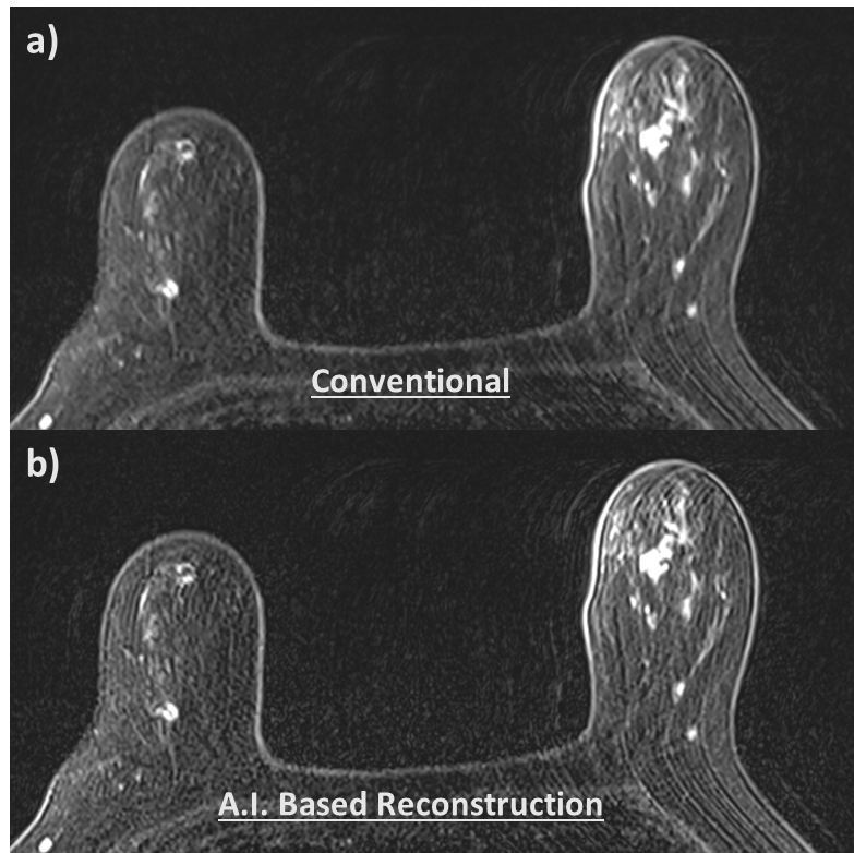
Forty-four of 45 patients completed the bMRI exams, with 88 sets of images available for analysis (one set without AI and one set with AI). Representative images with and without AI reconstruction are shown in Figure 1.Three readers independently provided 132 scores. 63/132 (48%) scores rated no improvement with AI , 54/132 (41%) scores rated improvement with AI based reconstruction (demonstrated in Figure 2).
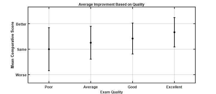
Improvement in margin conspicuity and delineation of the internal heterogeneity in the large tumor or inflammatory breast tumor were observed in Figure 1. In 15/132 (11%) cases, degradation in image quality was observed with AI reconstruction (Figure 2). When stratified by the overall quality of the full bMRI exam, a trend in improvement is observed (Figure 3) with higher quality original bMRI having a greater likelihood of improvement with AI reconstruction. When the original bMRI exam is rated as poor or average, applying AI only minimally improves image quality or did not improve the image quality.
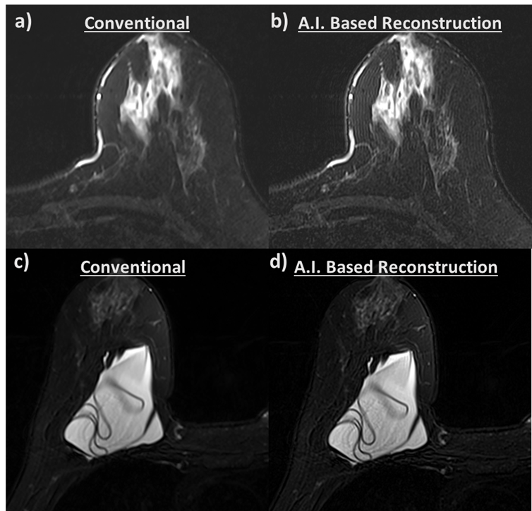
Readers also observed that AI reconstructed images tend to have more “grainy” appearance or pronounced noise texture but allows improved tumor conspicuity and margin delineation (Figures 1, 4, and 5). However, when the exam is of poor overall quality, the AI package and setting employed in this investigation exaggerated the edge enhancement nature of the reconstruction and Gibbs ringing (Figure 4), as well as motion ghosting (Figure 5).
Conclusion
This pilot study indicates that current AI reconstruction can improve the image quality of breast MRI images in studies that are of diagnostic quality at baseline, and potentially may allow scan time reduction. Exams that were considered of non-diagnostic quality could not be improved with the AI package used. Future work will explore the impact of different AI reconstruction setting on these results to determine if there is an optimal tradeoff between noise reduction and edge enhancement, especially given the demonstrated impact on artifacts. Additional prospective work will be aimed at demonstrating if AI reconstructions can allow for reduced acquisition time while still providing diagnostic quality.Acknowledgements
No acknowledgement found.References
1. Ritse M.M., Nariya C., and Linda M., Breast MRI: State of the Art. Radiology 2019 292:3, 520-536
2. Newell MS, Giess CS, Argus AD, et al. ACR practice parameter for the performance of contrast enhanced magnetic resonance imaging (MRI) of the breast. Reston, Va: American College of Radiology, 2018.
3. Warner E, Messersmith H, Causer P, Eisen A, Shumak R, Plewes D. Systematic review: using magnetic resonance imaging to screen women at high risk for breast cancer. Ann Intern Med 2008;148(9):671–679
4. Lin DJ, Johnson PM, Knoll F, Lui YW. Artificial Intelligence for MR Image Reconstruction: An Overview for Clinicians. J Magn Reson Imaging. 2021 Apr;53(4):1015-1028
5. Ren, J., Li, Y., Liu, FS. et al. Comparison of a deep learning-accelerated T2-weighted turbo spin echo sequence and its conventional counterpart for female pelvic MRI: reduced acquisition times and improved image quality. Insights Imaging 13, 193 (2022).
6. Westra C, Dialani V, Mehta TS, Eisenberg RL. Using T2-weighted sequences to more accurately characterize breast masses seen on MRI. AJR Am J Roentgenol 2014;202(3):W183–W190.
Figures