5009
3D MR spirometry in healthy volunteers at 1.5 T using a UTE sequence with a flexible k-space sampling pattern1Université Paris-Saclay, CEA, CNRS, Inserm, BioMaps, Orsay, France, 2Siemens Healthcare SAS, Courbevoie, France, 3Université Paris-Saclay, CEA, CNRS, BAOBAB, NeuroSpin, Gif-sur-Yvette, France, 4Department of Radiology, Foch Hospital, Suresnes, France, 5Université Paris-Saclay, CEA, CNRS, Inria, MIND, NeuroSpin, Gif-sur-Yvette, France
Synopsis
Keywords: Lung, Lung, Acquisition Methods, Biomarkers, New Trajectories & Spatial Encoding Methods
Motivation: To study lung function using 3D MR spirometry.
Goal(s): To optimize 3D dynamic lung MRI with flexible k-space sampling for UTE acquisition.
Approach: A non-Cartesian UTE sequence is developed to execute arbitrary trajectories stored in a readily available external gradient file library. 3D MR spirometry was then performed on freely-breathing healthy volunteers at 1.5 T.
Results: High-quality 4D lung images are obtained, enabling the extraction of relevant respiratory biomarkers. Image quality can then be optimized by directly playing back the shapes and durations of the readout gradients from the files.
Impact: A non-Cartesian, center-out MR sequence that allows out-of-the-box UTE capabilities with flexible k-space trajectories is developed. The result is high-quality 4D lung imaging in free-breathing and supine conditions. The lung functional biomarkers are expected to be sensitive to pathology.
Introduction
The ability to image the lungs using MRI could provide access to local biomarkers describing both their mechanical behavior and ventilatory function, which is of great clinical interest for patients with chronic lung diseases1. However, physiological motion, low proton density and fast signal decay in the lung require the development of specific strategies. Ultra-short TE (UTE) sequences allow acquiring the signal as early as possible, while the implementation of retrospective gating provides the tools to achieve 4D imaging of the lungs. The fast signal decay also limits the k-space sampling to a short readout. An optimized sampling scheme is therefore crucial to maximize image quality.In this work, we developed free-breathing 4D pulmonary imaging to perform 3D MR spirometry using a non-Cartesian center-out MR sequence that supports arbitrary k-space trajectories2. This custom sequence facilitates the study of k-space sampling strategies and their impact on the quantitative assessment of lung ventilation dynamics3.
Methods
Sequence development:The custom MR sequence reads in real time external files containing the temporal data of readout gradients on the 3 axis and thus allows full flexibility in k-space sampling strategy. Short non-selective RF pulses and minimal hardware switching time between RF excitation and reception enabled a minimum TE of 30µs. The center-out radial trajectory is tailored to take the respiratory motion into account by using a specific order of the spokes, called AZTEK4, which ensures highly uniform distributions of the spokes on the k-space sphere for each respiratory-rephased dataset.
MR acquisitions:
Data was acquired on three healthy and freely-breathing volunteers lying supine in a 1.5T MRI system (Magnetom Sola, Siemens Healthineers, Erlangen, Germany). Imaging parameters were: FOV: 280×280×210mm3, resolution: 1.5×1.5×1.5mm3, TE/TR: 0.03/3ms, FA: 4°, NbSpokes: 269325, Tobs: 960µs, with trapezoidal gradient shapes for a total acquisition time of 13min. To evaluate reliability, an additional acquisition was performed on volunteer#1 on a different day. To assess the potential of variable density gradients (Figure1), an acquisition with different encoding trajectories was performed on volunteer#3.
Motion-resolved signal gating and image reconstruction:
A compressed-sensing iterative image reconstruction was performed offline using BART5 with a wavelet decomposition and L1 norm as sparse regularization. The trajectory was computed from the gradient file with an additional calibrated gradient delay correction. The respiratory signal was extracted from the k-space center signal using principal component analysis. Retrospective soft gating3 was applied to distribute the acquired data into 32 respiratory phases of an integrated respiratory cycle over the acquisition. This pipeline allows the reconstruction of a 4D image of the lungs (Figure2).
Spirometry biomarkers evaluation:
The lung parenchyma was segmented using a non-rigid registration of a reference acquisition and associated segmentation mask.The deformation fields between each respiratory phase and the first, chosen as a reference at the beginning of inspiration, were extracted by elastic registration. The Jacobian of the deformation fields was then computed voxelwise and both functional and mechanical biomarkers were derived.
Results
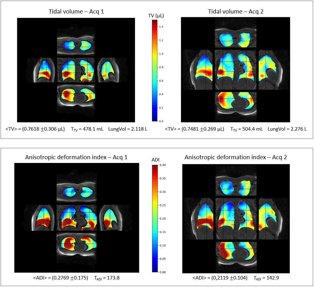
Figure3 presents the temporal dynamics of the reconstructed 3D lung volumes for the three volunteers. The smooth evolution of the respiratory cycle and the absence of motion blurring indicate efficient binning. The UTE approach accounts for the relatively high signal-to-noise ratio with a satisfactory delineation of the vascular tree. Figure4 compares the regular and variable radial sampling schemes. No remarkable image quality improvement was observed when using variable density sampling. Figure5 compares the tidal volume (TV) and anisotropic deformation index (ADI) maps for two different acquisitions from the same healthy volunteer. Both biomarker maps show a relatively high spatial similarity between the two acquisitions. Numerically, total TV, global lung volume and total ADI showed variation rates of 5.5%, 7.5% and 17%, respectively.Discussion
A 3D non-Cartesian center-out UTE MR sequence was developed with the added feature of a flexible k-space trajectory library. The radial spoke order was adapted to the respiratory motion so as to enable a comprehensive uniform k-space spoke sampling for each respiratory phase (AZTEK). The resulting images are of high quality and allow the extraction of relatively reproducible spirometry biomarkers within the expected physiological variability. However, an inherent shortcoming of a 3D radial trajectory is the inhomogeneous sampling density throughout k-space. In addition, due to the very short T2*, the higher frequencies are sampled whereas the signal has already decayed. Variable density gradients6 (as proposed in Figure4) combined with user-defined sampling density (as prescribed in SPARKLING7) might be adequate candidates to mitigate these two issues. Future work will focus on gradient non-linearities correction and trajectory optimization. Finally, given the recently demonstrated clinical opportunities8, this implementation is considered at low-field MRI.Acknowledgements
This project has received funding from the European Union’s Horizon Europe research and innovation program under grant agreement No 101099934. Camille Dubuc and Mathieu Bournac for their support in data acquisition.References
1. Feng L, Delacoste S, et al. Simultaneous Evaluation of Lung Anatomy and Ventilation Using 4D Respiratory-Motion-Resolved Ultrashort Echo Time Sparse MRI. J. Magn. Reson. Imaging 49:411-422 (2019).
2. Lazarus C, Weiss P, El Gueddari L, Mauconduit F, Massire A, Ripart M, Vignaud A, Ciuciu P. 3D variable-density SPARKLING trajectories for high-resolution T2*-weighted magnetic resonance imaging. NMR Biomed. 2020 Sep;33(9):e4349.
3. Boucneau T, Fernandez B, Larson P, Darrasse L, Maitre X. 3D magnetic resonance spirometry. Scientific Reports 10, 9649 (2020).
4. Boucneau T, Fernandez B, Besson FL, Menini A, Wiesinger F, Durand E, Caramella C, Darrasse L, Maître X. AZTEK: Adaptive zero TE k-space trajectories. Magn Reson Med. 2021 Feb;85(2):926-935.
5. Berkeley Advanced Reconstruction Toolbox (BART) https://mrirecon.github.io/bart/ (2023).
6. Johnson KM, Fain SB, Schiebler ML, Nagle S. Optimized 3D ultrashort echo time pulmonary MRI. Magnetic Resonance in Medicine, 70(5), 1241-1250 (2013).
7. Giliyar Radhakrishna C, Daval-Frérot G, Massire A, Vignaud A, Ciuciu P. Improving spreading projection algorithm for rapid k-space sampling trajectories through minimized off-resonance effects and gridding of low frequencies. Magn Reson Med. 2023 Sep;90(3):1069-1085.
8. Campbell-Washburn AE, Ramasawmy R, Restivo MC, et al. Opportunities in Interventional and Diagnostic Imaging by Using High-Performance Low-Field-Strength MRI. Radiology. 2019;293(2):384-393.
Figures
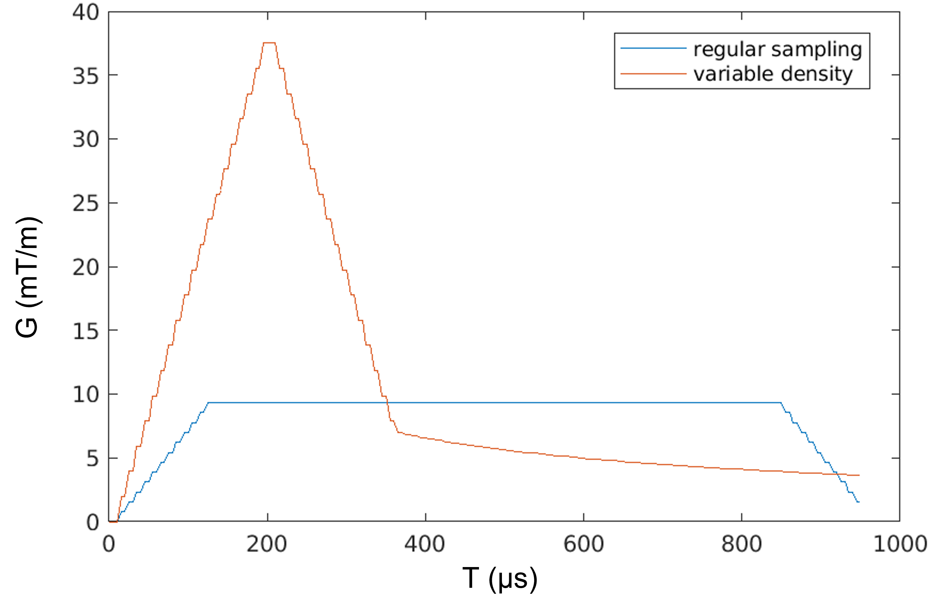
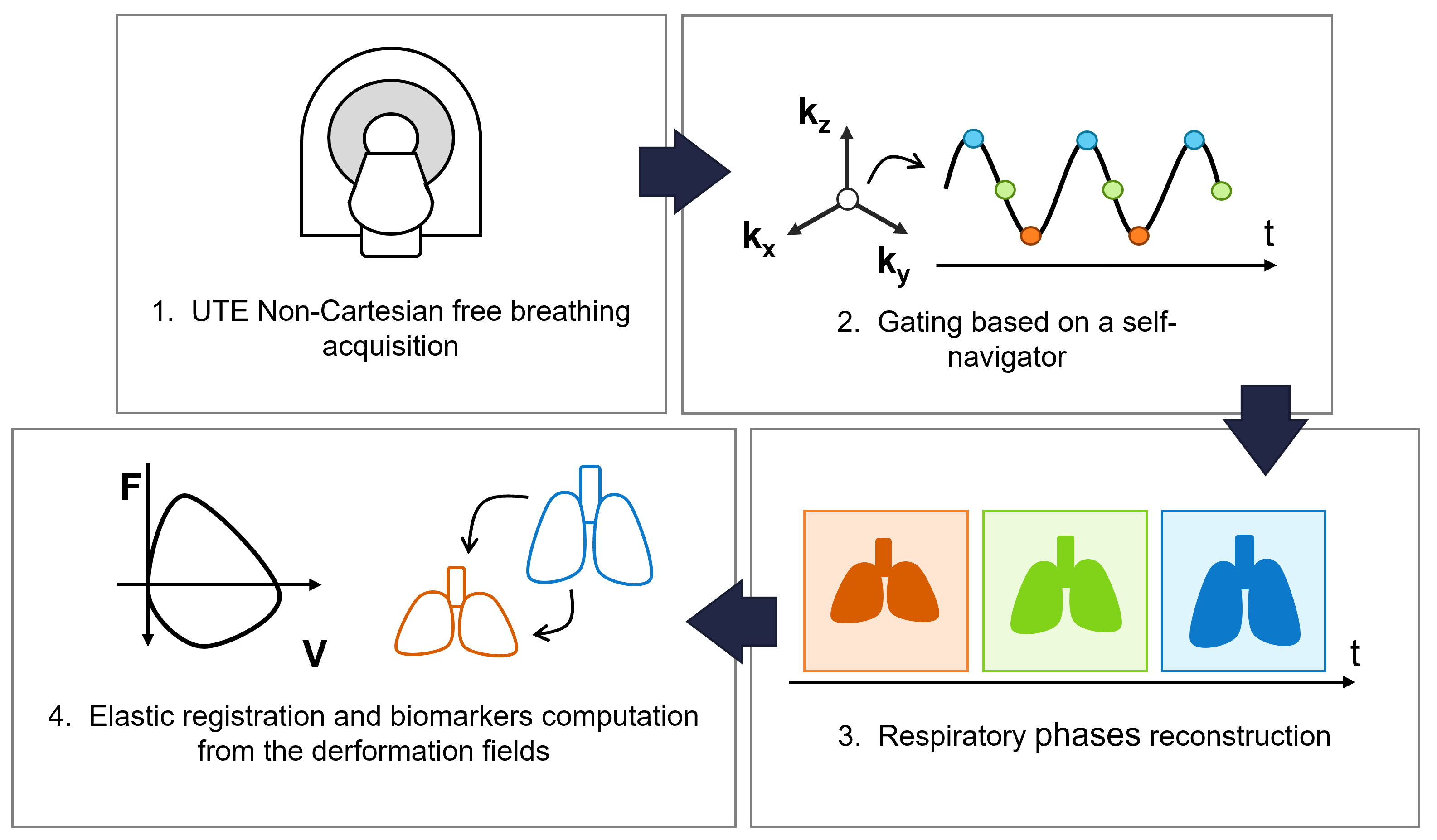
Figure 2: 3D MR lung spirometry workflow. 1/ UTE non-Cartesian 3D MR lung dynamic imaging using a custom k-space trajectory MR sequence at 1.5 T in free-breathing; 2/ Respiratory signal extraction using principal component analysis and sampling spoke gating assignment into 32 respiratory phases; 3/ Offline image reconstruction using BART, 4/ Spirometry biomarker derivation from the extracted deformation fields.
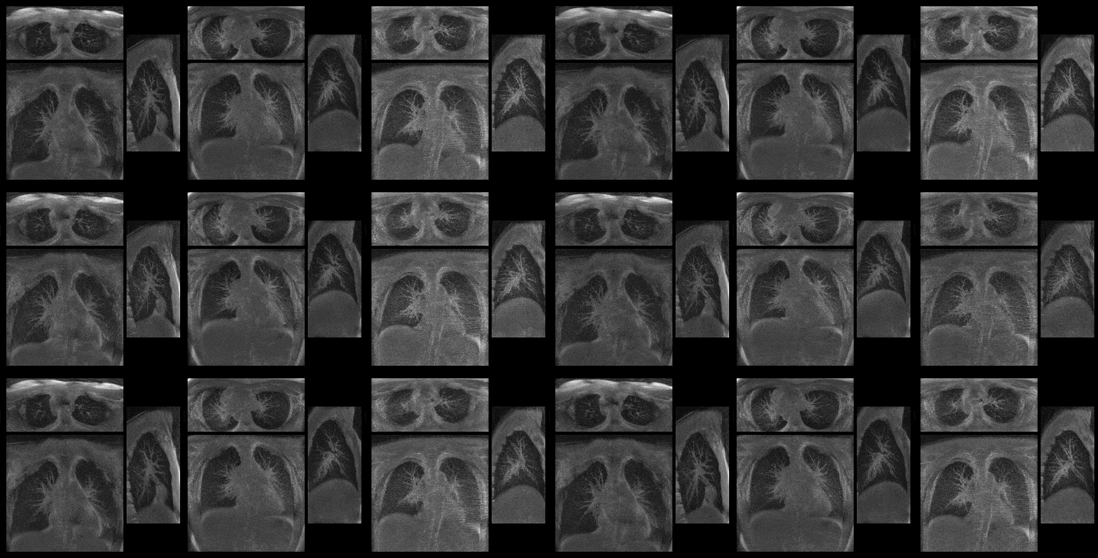
Figure 3: 4D lung MRI in three healthy volunteers for central views in the coronal (bottom), axial (top), and sagittal (right) planes. Data were acquired with a 3D center-out radial trajectory combined with the AZTEK sampling scheme. Signal sampling was uniform along each spoke (gradient trapezoidal shapes). High image quality is achieved, with clear visualization of lung parenchyma and vessels, in addition to smooth temporal resolution.
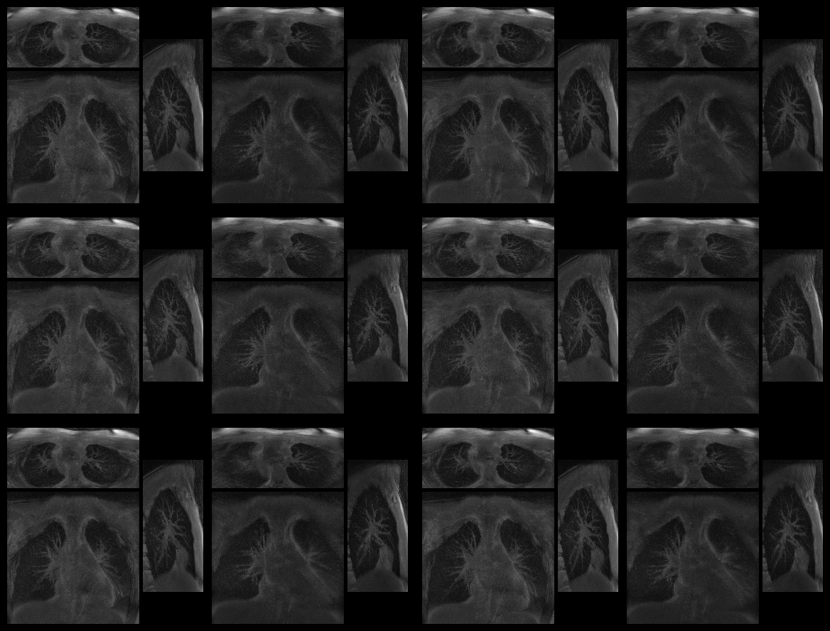
Figure 4: 4D lung MRI on one volunteer with regular radial sampling (left) and variable density radial sampling (right). Central views in the coronal (bottom), axial (top), and sagittal (right) planes are illustrated. Data were acquired with a 3D center-out trajectory combined with the AZTEK sampling scheme. High image quality is achieved, with clear visualization of lung parenchyma and vessels, in addition to smooth temporal resolution.