5008
Use of distortion correction combined with deep learning reconstruction in DWI: how does image quality compare to conventional acquisition?1GE HealthCare, Blacklick, OH, United States, 2GE HealthCare, Washington, DC, United States, 3Fairfax Radiological Consultants, Fairfax, VA, United States, 4GE HealthCare, Boston, MA, United States
Synopsis
Keywords: Prostate, Machine Learning/Artificial Intelligence
Motivation: The use of deep learning reconstruction, combined with Multiplexed Sensitivity Encoding (MUSE), can extend the benefit of distortion robustness in prostate DWI to poor SNR conditions while maintaining a large spatial matrix.
Goal(s): The purpose of this study is to evaluate the quantitative image quality improvement provided by combining MUSE and DLR in DWI of the prostate.
Approach: Quantitative analysis including SNR, CNR and ADC were compared through ROI analysis of MUSE DWI with conventional and DL reconstruction in 50 prostatic cancer patients.
Results: DLR images demonstrated a significantly higher SNR and CNR. ADC values were consistent among methods.
Impact: Deep learning reconstruction in combination with MUSE can be exploited for better prostate DWI image quality in cases of low SNR, or traded for increased resolution or reduced scan time.
Introduction
Diffusion-Weighted Imaging (DWI) is a key component to the latest version of the PI-RADS guidelines for evaluating prostate cancers. An accurate assessment of lesion position and conspicuity on DWI is critical in the treatment pathway, and the adoption of new technology continuously aims to improve image quality, protocol and workflow [1].Due to the prostate’s adjacent anatomy, peristalsis and air in the gastrointestinal tract can induce susceptibility artifacts and geometric distortion on DWI, particularly at high field strengths and in high resolution imaging [2]. Long acquisition times for DWI sequences can also contribute to blurring from patient motion. Multiplexed Sensitivity Encoding (MUSE) reconstruction has shown to be effective in reducing distortion in large FOV scans and in recovering reliable ADC values [3].
The use of deep learning reconstruction (DLR) can potentially improve DWI image quality by boosting the signal-to-noise ratio (SNR) and decreasing acquisition time. Combined with MUSE, it can extend the benefit of distortion robustness to poor SNR conditions while maintaining a large spatial matrix [4]. However, it is important to ensure that the new protocol does not come at a cost of reduced image quality or diagnostic accuracy. The purpose of this study is to evaluate the quantitative image quality improvement provided by combining MUSE and DLR in DWI of the prostate.
Methods
Fifty patients undergoing clinical MRI examination were included in the study. Patients were scanned at 3T (46 patients) and 1.5T (4 patients) (SIGNA Premier, Architect, and Artist, GE HealthCare, Waukesha, WI) with 20-, 21- or 30-channel phased array blanket coils (AIR™ coils). The exam was performed using the standard clinical protocol, including T1-weighted axial whole pelvis, tri-planar T2-weighted (Fast Spin-Echo or PROPELLER), axial DWI with MUSE distortion correction, reduced FOV (FOCUS) DWI, and 3D Dynamic Contrast Enhanced Imaging. The parameters for the DWI were generally as follows: 30 slices, 3.5mm slice thickness, 3 shots, 27cm FOV, 92x92 acquisition matrix, reconstructed to 256x256, TE=69ms, TR=4300ms, b-values=50(NEX=1), 800(NEX=6), acceleration factor = 2x. A synthetically derived set of high B-value images (b=1400s/mm2) was automatically computed and included derived ADC maps. MUSE DWI data were reconstructed with both conventional and deep learning methods. Three slices were selected for each subject from the MUSE dataset and on each slice, six circular regions of interest (ROI) of 2.5mm radius were placed. Two ROIs were drawn on each side of the prostatic transition and peripheral zones, and the remaining two were traced on the obturator internus muscle as the reference for the noise floor. Student t-tests were used to compare signal- and contrast-to-noise ratios as well as quantitative ADC values between the different reconstruction methods.Results
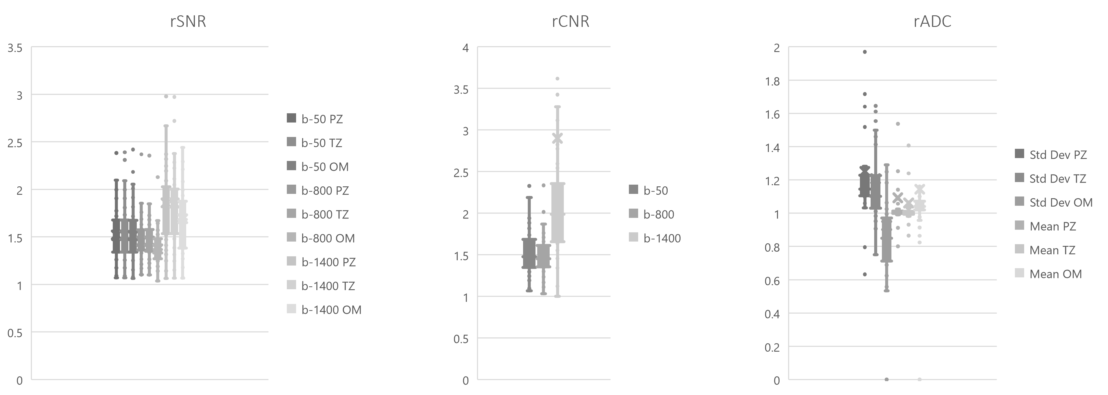
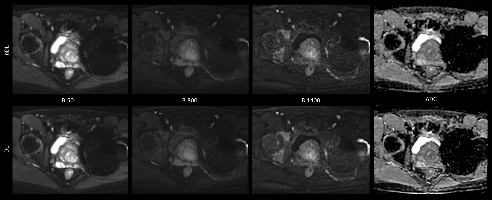
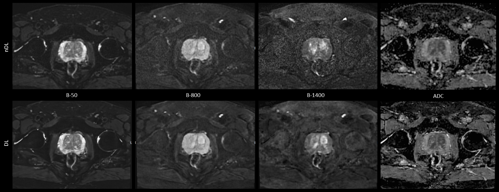
Quantitative analysis revealed that DLR images demonstrated a significantly (p<0.001) higher SNR than conventional MUSE at b-value=50s/mm2 and b-value=800s/mm2 and in the synthetically reconstructed b-value=1400s/mm2. CNR, defined as the difference between SNR in peripheral zone minus SNR in the noise floor, also showed significant (p<0.001) increase with DL Reconstruction. ADC values within each ROI showed no significant differences, but standard deviation over the ROI differed for DL reconstructed diffusion weighted images. Side-by-side comparisons between the two reconstruction methods are shown for two cases acquired at 1.5T and 3.0T in Figure 2 and Figure 3, respectively.Discussion
Deep learning reconstruction led to improved quantitative markers of image quality (SNR, CNR), with strong consistency across systems and anatomical region. Increased SNR and an average of twofold increase in CNR in synthetically derived DWI suggests a reliable fit of DL-reconstructed images. ADC maps appear sharper, albeit with higher standard deviation within the ROI. Nevertheless, the average value remains consistent for both reconstruction methods. Despite a relatively small sample size of clinical examinations acquired thus far, the main take-away of this on-going study is that deep learning reconstruction leads to higher quality DWI images. Additional analysis of prostate lesions is underway to confirm whether high SNR and CNR is achievable while preserving the accuracy of ADC in the lesions.Conclusion
In this study, we demonstrated prostate MUSE DWI image quality improvement with a DL-based image reconstruction method, with no loss in diagnostic information. Deep learning reconstruction can be exploited for better image quality, especially in cases of low SNR, or traded for increased resolution versus reduced scan time.Acknowledgements
No acknowledgement found.References
1. Steiger, P., Thoeny, H.C. Prostate MRI based on PI-RADS version 2: how we review and report. Cancer Imaging 16, 9 (2016). https://doi.org/10.1186/s40644-016-0068-2.
2. Lee K.L., Kessler D.A., Dezonie S., Chishaya W., Shepherd C., Carmo B., Graves M.J., Barrett T. Assessment of deep learning-based reconstruction on T2-weighted and diffusion-weighted prostate MRI image quality. European Journal of Radiology 166 (2023), 111017.
3. Chen NK, Guidon A, Chang HC, Song AW. A robust multi-shot scan strategy for high-resolution diffusion weighted MRI enabled by multiplexed sensitivity-encoding (MUSE). Neuroimage. 2013;72:41-7.
4. Lebel RM. Performance characterization of a novel deep learning-based MR image reconstruction pipeline. arXiv preprint arXiv:2008.06559. 2020.
Figures