5005
Fast and Generalized Motion Correction in Brain MRI using 3D Radial Trajectory and Projection Moment Analysis1School of Biomedical Engineering, Shanghai Jiao Tong University, Shanghai, China
Synopsis
Keywords: Motion Correction, Motion Correction, 3D radial, projection moment, center-of-mass
Motivation: Traditional projection moment analysis in 3D radial MRI failed to get specific rigid-body motion parameters with stationary multichannel coils.
Goal(s): Our goal was to develop a method to extract rigid-body motion parameters directly through projection moment analysis.
Approach: A PCA-based coil compression, together with projection information from different channels were used to estimate rigid-body motion parameters. A recursive least-squares model was used to recursively estimate motion parameters for every single spoke. Simulation and scanning of moving object were performed to demonstrate its capability in brain scan.
Results: The proposed method can correct motion in brain successfully and quickly.
Impact: The proposed method provides an easy, robust, and time-efficient tool for motion correction in brain MRI, which may benefit clinical diagnosis of uncooperative patients like children, in addition to many other applications including extremity MRI.
Introduction
MRI is sensitive to motion, and subject motion during brain MRI has been problematic in clinical practice. Generally, motion causes artifacts manifesting as ghosting, blurring, geometric distortion or decreased signal-to-noise ratio (SNR)1. Study showed that both motion-free time and average displacement of scanning object were highly correlated with brain image quality2. Severe motion may result in non-diagnostic image in clinical scans, which result in repeated scans and additional cost.Several methods have been proposed for motion correction of the brain. One type of methods utilizes the self-navigation property of radial acquisition, and use the analysis of projection moment to detect motion3-5. However, the detection of both translational and rotational parameters using single coil is not robust to noise. Projection moment analysis in 3D radial MRI was able to detect motion occurrence, but failed to get specific rigid-body motion parameters with stationary multichannel coils.
In this study, we proposed a generalized rigid-body motion correction method using 3D radial acquisition and projection moment analysis. The motion pattern can be continuous, and the calculation is relatively fast that it can be easily implemented in clinical scans. The preliminary results demonstrate that the proposed method is a powerful tool for motion correction in the brain.
Methods
Motion Estimation and CorrectionThe center-of-mass (COM) of an image is the ratio of its first and zeroth moments4. In 3D radial acquisition, the projection of COM of an object onto a radial spoke (P) is:
$$P=\frac{\int r\left(F^{-1}s\right)dr}{\int\left(F^{-1}s\right)dr}\tag{1}$$
where s is the k-space data of a spoke, F-1 is 1D inverse Fourier transform operator, and r is the radius vector along spoke direction. Let $$$v=\left(v_x,v_y,v_z\right)^T$$$ denote a unit vector pointing at spoke direction, and the relationship of P and COM can be expressed as5:
$$\left(\begin{array}{c}P_1 \\P_2 \\\vdots \\P_n\end{array}\right)=\left(\begin{array}{ccc}v_{1,x} & v_{1,y} & v_{1,z} \\v_{2,x} & v_{2,y} & v_{2,z} \\\vdots & \vdots & \vdots \\v_{n,x} & v_{n,y} & v_{n,z}\end{array}\right)\left(\begin{array}{c}\operatorname{COM}_x \\\operatorname{COM}_y \\\operatorname{COM}_z\end{array}\right)\tag{2}$$
where n is the number of spokes involved in the calculation. P can be calculated after 1D inverse Fourier transform of k-space data. By introducing variable forgetting-factor recursive least-squares (VFF-RLS) algorithm6, the above least-squares problem can be extended to recursively estimate subject COM with a nominal temporal resolution of one TR.
To estimate rigid-body object displacement directly from k-space data, we first assume that a flexible coil is used that it moves with the head. For translational motion estimation, a PCA-based coil compression is performed that its first virtual coil contains information which is an overall representation of object position, as shown in Figure 1. By calculating the evolution of COM in this virtual coil using VFF-RLS, the translational motion can be estimated and the original k-space data is aligned accordingly for translation correction. After this operation, the COM of each coil was calculated using VFF-RLS separately, as shown in Figure 2(A). Rotational parameters can be calculated using a least-squares estimation between sets of vectors acquired at different time (Figure 2(B))7.
Experiments
One healthy subject was scanned using a 3D radial sequence with multi-dimensional golden-means trajectory8 with two flexible coils (totally 20 channels) on a 3T MRI scanner (uMR790, United Imaging Healthcare, Ltd., Shanghai, China). Written consent was obtained before scan. The scanning parameters were: FOV = 240×240×240 mm3, matrix = 240x240x240, flip angle = 17°, TR = 7.1 ms, TE = 3 ms, bandwidth = 250 Hz/pixel, spoke number = 90,000. The total scan time was 10.65 min. Two scans were made. In one scan the volunteer was asked to remain still, while in the other scan he was told to perform some random movements.
A simulation was made that a sinusoidal periodic motion was added to the motion-free k-space data to create motion-corrupted image. The proposed method was applied, and the motion corrected image was compared with original image. It was then tested on k-space data with real motion.
Results
Simulation showed that for continuous motion, the proposed method successfully eliminated motion artifacts (Figure 3). Motion artifacts in prospective motion-corrupted scan were also eliminated with the cost of slightly-reduced SNR (Figure 4). The estimated motion pattern in prospective motion-corrupted scan is shown in Figure 5, which consists of a large-scale fast motion and a small-scale random position change, which successfully mimics head motion pattern commonly seen in clinical scans9.Discussion and Conclusion
Preliminary results have demonstrated that the proposed method is an easy, robust, and time-efficient tool for motion correction in brain MRI. The prerequisite is that a flexible coil attached to the head needs to be used rather than a stationary coil, which may require a specialized coil design.Acknowledgements
This work was supported by the Shanghai Science and Technology Commission Explorer Program under Grant 22TS1400300.References
1. Godenschweger F, Kägebein U, Stucht D, et al. Motion correction in MRI of the brain. Physics in medicine & biology. 2016;61(5):R32.
2. Afacan O, Erem B, Roby DP, et al. Evaluation of motion and its effect on brain magnetic resonance image quality in children. Pediatric radiology. 2016;46:1728-1735.
3. Welch EB, Rossman PJ, Felmlee JP, et al. Self‐navigated motion correction using moments of spatial projections in radial MRI. Magnetic Resonance in Medicine. 2004;52(2):337-345.
4. Anderson III AG, Velikina J, Block W, et al. Adaptive retrospective correction of motion artifacts in cranial MRI with multicoil three‐dimensional radial acquisitions. Magnetic resonance in medicine. 2013;69(4):1094-1103.
5. Lee H, Zhao X, Song HK, et al. Self-navigated three-dimensional ultrashort echo time technique for motion-corrected skull MRI. IEEE transactions on medical imaging. 2020;39(9):2869-2880.
6. Paleologu C, Benesty J, Ciochina S. A robust variable forgetting factor recursive least-squares algorithm for system identification. IEEE Signal Processing Letters. 2008;15:597-600.
7. Umeyama S. Least-squares estimation of transformation parameters between two point patterns. IEEE Transactions on Pattern Analysis & Machine Intelligence. 1991;13(04):376-380.
8. Chan RW, Ramsay EA, Cunningham CH, et al. Temporal stability of adaptive 3D radial MRI using multidimensional golden means. Magnetic Resonance in Medicine. 2009;61(2):354-363.
9. Ma S, Wang N, Xie Y, et al. Motion‐robust quantitative multiparametric brain MRI with motion‐resolved MR multitasking. Magnetic resonance in medicine. 2022;87(1):102-119.
Figures
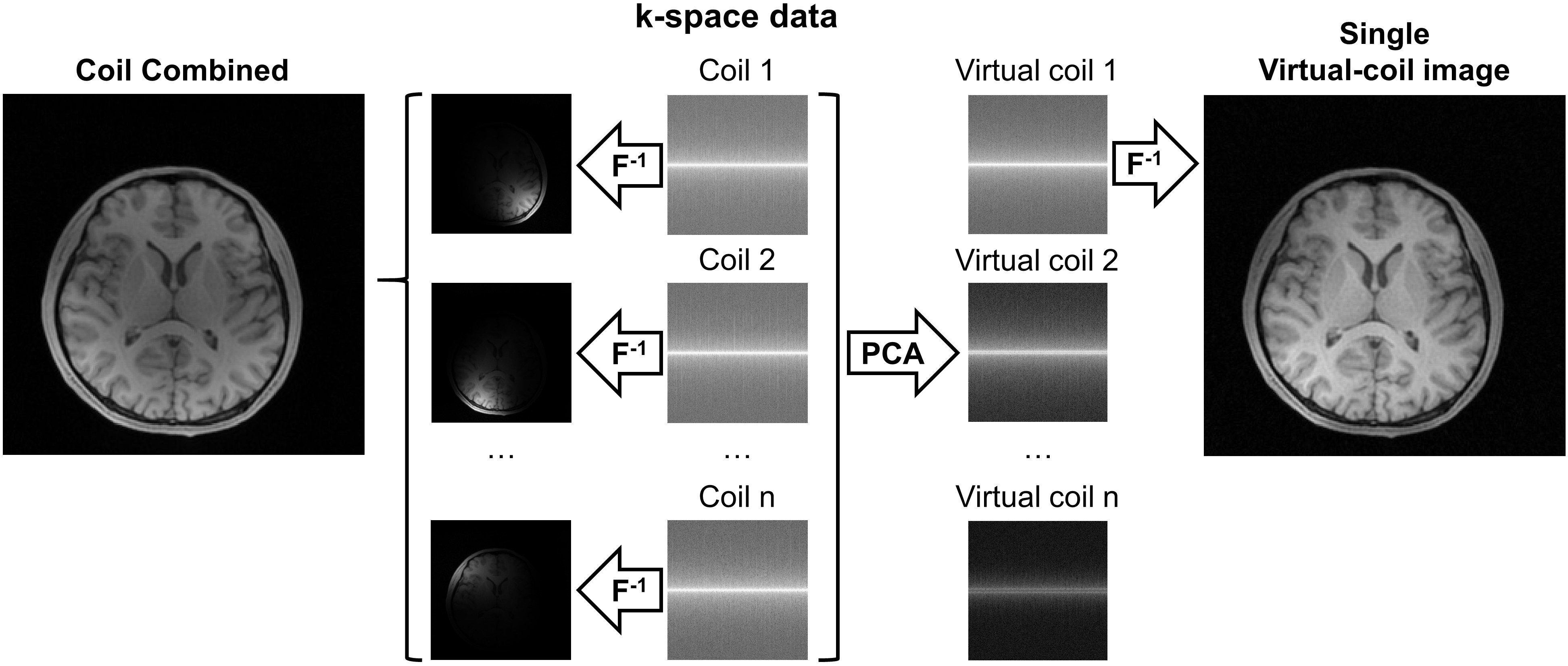
Figure 1. An illustration of the effect of PCA coil compression. Although each coil only contains sensitivity of a limited area, the sensitivity of the first virtual coil after PCA coil compression is global, and the reconstructed image approximates coil-combined image which can be treated as an overall representation of object position. Information of this virtual coil can be used to calculate translational parameters for each coil.
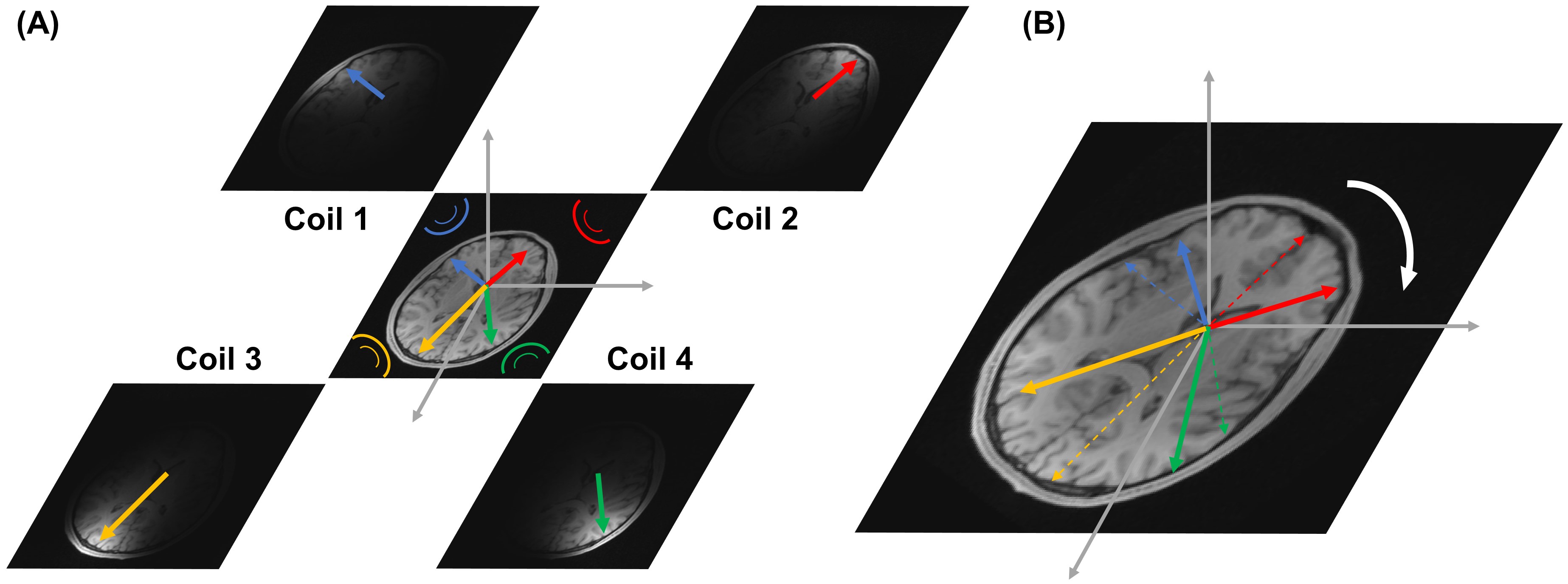
Figure 2. An illustration of rotation estimation scheme. For simplicity, only one slice and four coils are shown. (A) For each coil, a COM can be calculated. Since each coil has a unique sensitivity, these COMs are linearly independent. (B) Assuming coils move with the scanning object. After rotation (indicated by white arrow), these COMs all rotate correspondingly. A least-squares estimation can be used to get rotational parameters between different patterns (solid and dashed colored arrows).
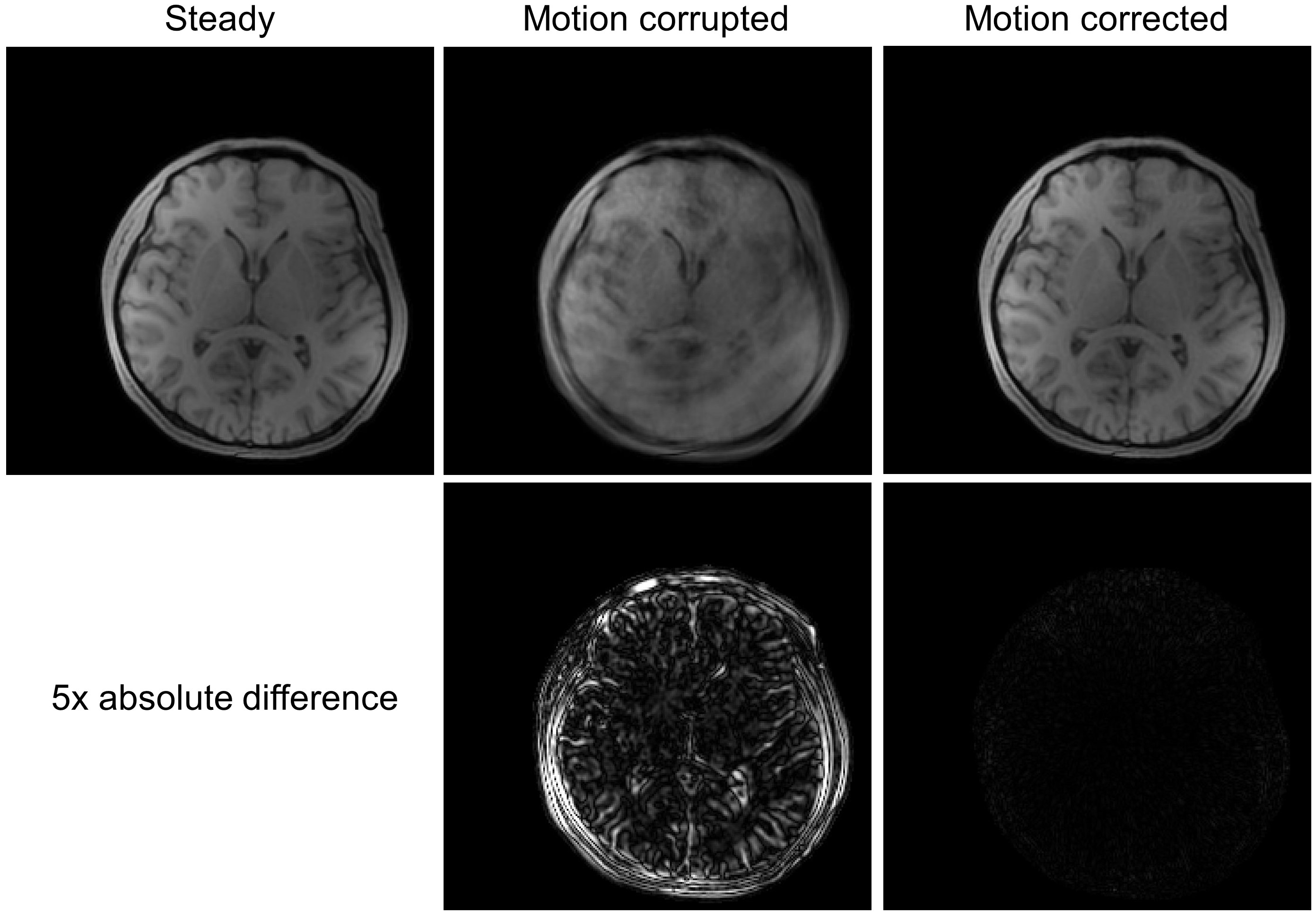
Figure 3. Effects of the proposed correction method on simulated motion-corrupted data. A sinusoidal periodic motion with amplitude = ±5˚ and frequency = 0.1 Hz was added to the motion-free data. Motion artifacts were successfully eliminated.
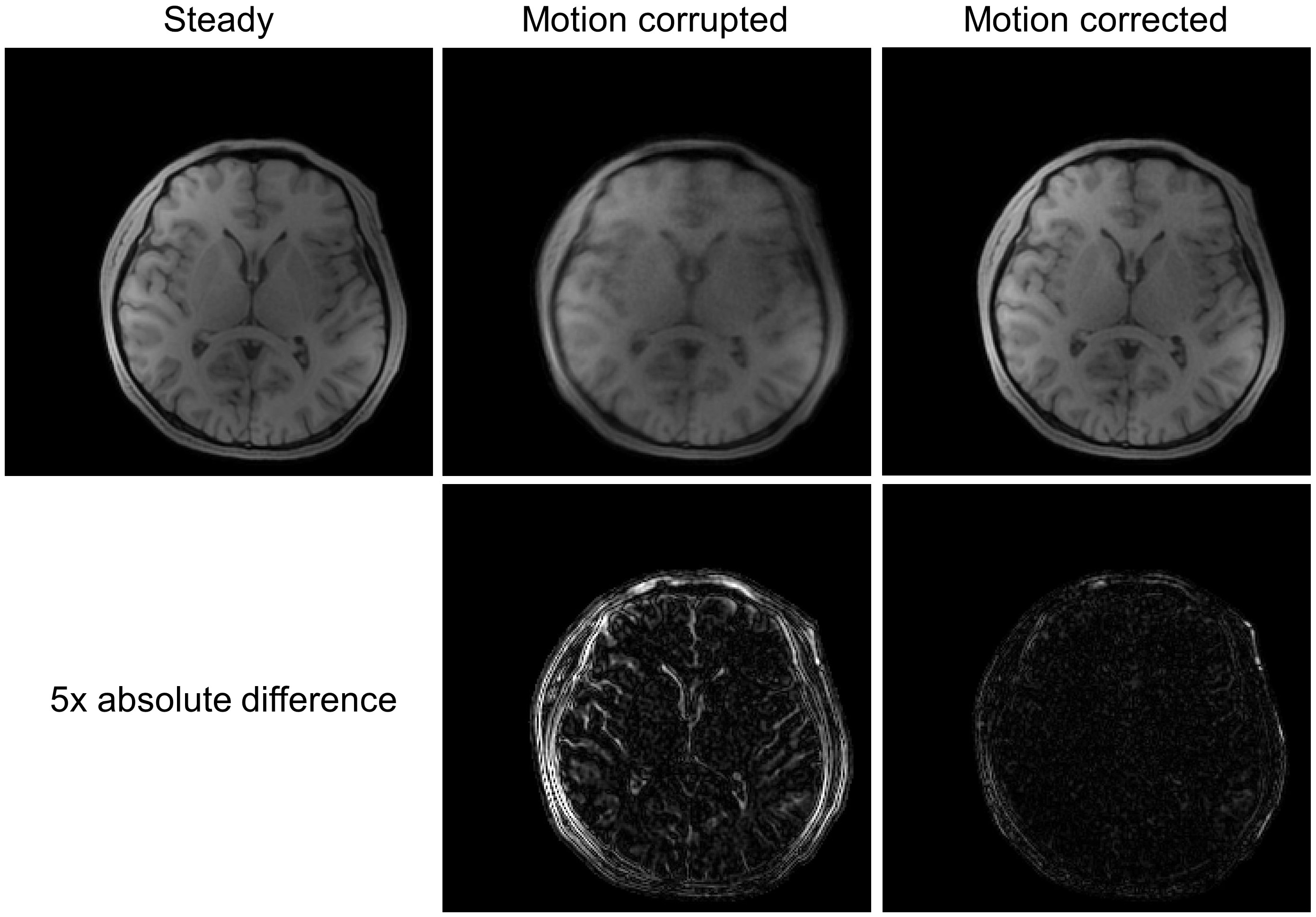
Figure 4. Effects of the proposed correction method on prospective motion-corrupted data. Motion artifacts were successfully eliminated with the cost of slightly-reduced SNR.
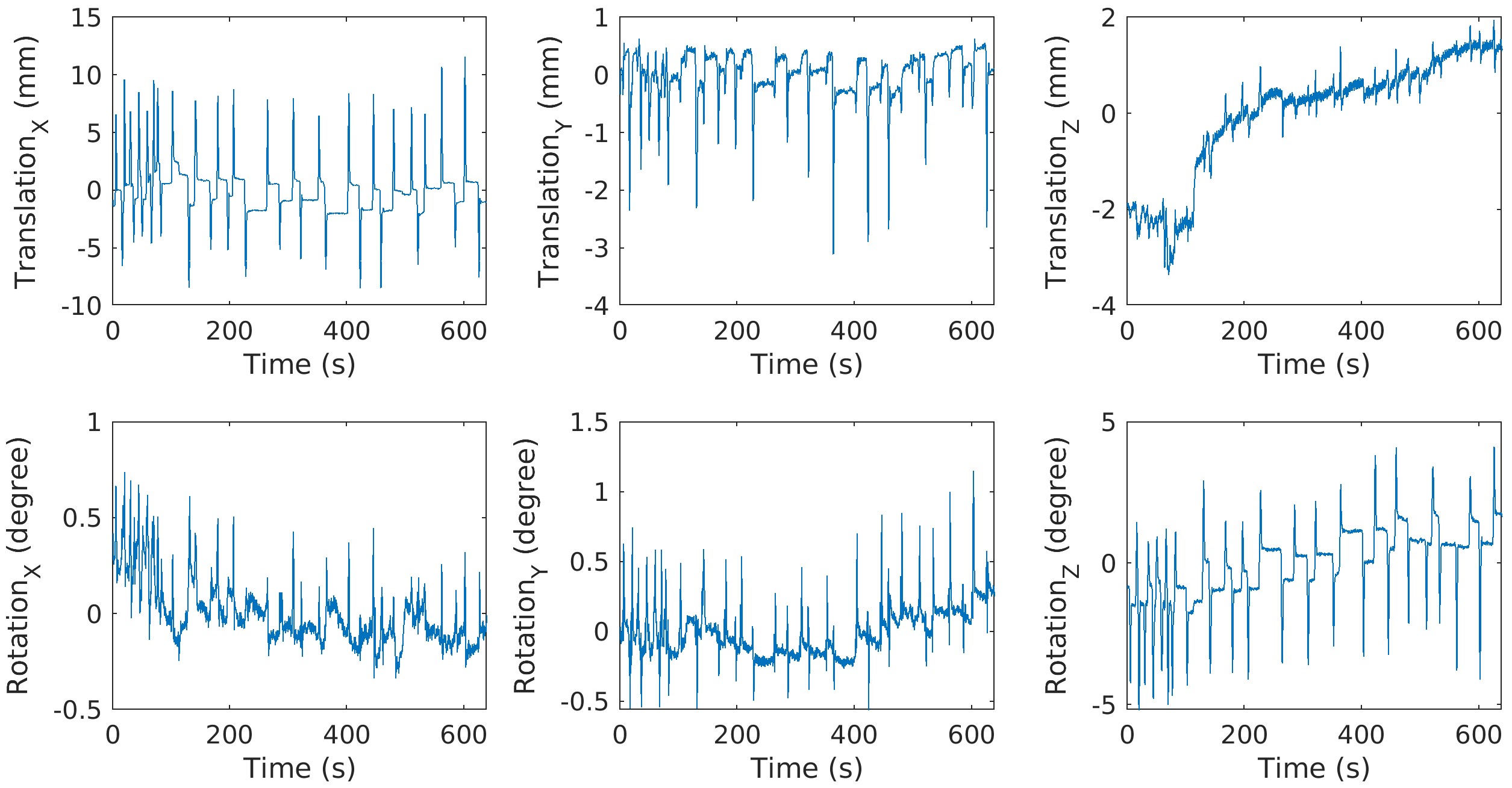
Figure 5. Estimated motion pattern in prospective motion-corrupted scan. It consists of a large-scale fast motion and a small-scale random position change.