5004
Susceptibility anisotropy imaging from single-orientation MRI with a training-free physics-informed autoencoder1Department of Computer Science and Automation, Technische Universität Ilmenau, Ilmenau, Germany, 2Buffalo Neuroimaging Analysis Center, Department of Neurology at the Jacobs School of Medicine and Biomedical Sciences, University at Buffalo, The State University of New York, Buffalo, NY, United States, 3Center for Biomedical Imaging, Clinical and Translational Science Institute, University at Buffalo, The State University of New York, Buffalo, NY, United States
Synopsis
Keywords: Quantitative Imaging, Quantitative Susceptibility mapping, Susceptibility Tensor Imaging
Motivation: To overcome the clinical limitations of susceptibility tensor imaging (STI) due to the requirement for multiple head orientations.
Goal(s): Develop a method to isolate χ13 and χ23 of the magnetic susceptibility tensor, from a single head orientation, enhancing the clinical viability of STI.
Approach: Employing a deep learning-based autoencoder, calibrated via STI and optimized for each dataset to separate the tensor components without the need for training or data rotation.
Results: The method successfully extracted χ13 and χ23 components comparable to the gold standard multi-orientation STI, showing potential for improved brain tissue characterization in conditions like multiple sclerosis.
Impact: We present a simplified STI approach, extracting critical tensor components from a single orientation scan. The new technique allows to assess structural tissue integrity, particularly in white matter. Requiring only a single orientation renders it clinically feasible.
Introduction
The magnetic susceptibility of brain tissue depends on the orientation of the tissue in the magnetic field.1 Imaging tissue anisotropy can yield relevant information about structural integrity.2 In white matter, for example, the anisotropy is strongly related to the myelin sheaths, which play a key role in early brain development and diseases like multiple sclerosis.2 The 3×3 susceptibility tensor can be mapped via susceptibility tensor imaging (STI).3 STI, however, requires measuring the brain under multiple different head orientations, which inflicts discomfort to subjects and at today’s acquisition durations renders the method clinically infeasible.Under a single orientation, the apparent susceptibility depends on three out of the nine tensor components:
f = f33 + f23 + f13 = (χ33 * d33) + (χ23 * d23) + (χ13 * d13) (Eq. 1)
The field perturbations fij induced by each of these three components follow a characteristic spatial fingerprint (Fig. 1).
In this work, we present a deep learning-based technique that separates these three components of the magnetic susceptibility tensor that are apparent under a single acquisition. We focused on χ13 and χ23, as these off-diagonal elements of the susceptibility tensor are potentially more sensitive to structural breakdown of aligned fibers than the bulk isotropic component on the main diagonal, and imaging of χ33 is already widely established through quantitative susceptibility mapping (QSM).4
Methods
Architecture: Fig. 2 illustrates the architecture of our physics-informed autoencoder. As a key building block, we used the U-Net architecture5 that has previously been demonstrated to solve the dipole inversion in QSM. Each susceptibility component is estimated by its own U-Net. The results are convolved with their respective unit response (dipole kernel) and summed up to the total field. The network’s weights are optimized with backpropagation on the application dataset until the output f matches the input f (autoencoder).Ill-posedness, regularization, and calibration: Separating the sources is underdetermined because the kernels (d13, d23, d33) are not orthogonal. This means, that f33 could be explained with χ23 * d23 and so forth. Typically, such false attributions lead to streaking artifacts and noise amplification. We used the Frobenius norm on the susceptibility estimates to steer the source separation to a plausible solution. Since in real tissue, χ33 is vastly larger then the other two components, we added weighting factors. Using a dataset with recordings from 26 orientations,6 these weighting factors where calibrated such that the single-orientation estimates matched the STI solution.
Validation: We validated the method on another STI dataset with (i) the original phase measurements and (ii) a phantom with perfect phase data simulated from the STI solutions (Fig. 3).
Results
Phantom experiment and comparison to STI gold standard: Fig. 4 shows how the single-orientation estimates of χ13 from a simulated f resemble the ground truth andthe single-orientation estimates of χ13 from a real, measured f resemble the STI estimates. Results for χ23 were similar. We found the largest errors from the ground truth or deviations from the STI reference in regions with notoriously large χ33, for example, the iron-rich basal ganglia.Discussion
Unlike today’s near-perfect QSM estimates in phantom experiments7, our χ13 and χ23 estimates visibly deviated from the ground truth. Still, the presented approach offers an alternative to susceptibility tensor imaging by facilitating the isolation of only specific tensor components, χ13 and χ23, from a single acquisition orientation. A key challenge addressed by this approach is the ambiguity inherent in this source separation problem, especially given the small magnitude of χ13 and χ23 relative to χ33. The method provides a novel basis for structural integrity assessment in white matter.Moreover, the training-free individual optimization of the technique for each dataset eliminates the need for data rotation or the computational burden from orientation-specific trainings.
Conclusion
This study provides a novel approach to STI that sidesteps the need for multiple head orientations, optimizing patient comfort and clinical utility. Despite the potential influence of χ33 on the smaller tensor components, our strategy for source separation demonstrates a high degree of precision in estimating χ13 and χ23. Validated against established STI protocols and through phantom experiments, the method shows promise for studying white matter integrity.Acknowledgements
The research was supported by the Free State of Thuringia grant ThiMEDOP (2018 IZN 0004) with funds of the European Union (EFRE), the German Federal Ministry of Education and Research (BMBF) grant AVATAR (16KISA024, funded by the European Union - NextGenerationEU), the German Academic Exchange Service (DAAD PPP 57599925), and an ISMRM Research Exchange Grant awarded to T.J. Research reported in this publication was partially supported by the National Institute of Neurological Disorders and Stroke of the National Institutes of Health under Award Number R01NS114227 (F.S.) and the National Center for Advancing Translational Sciences of the National Institutes of Health under Award Number UL1TR001412 (F.S.). The content is solely the responsibility of the authors and does not necessarily represent the official views of the National Institutes of Health.
References
1. He, X. & Yablonskiy, D. A. Biophysical mechanisms of phase contrast in gradient echo MRI. Proceedings of the National Academy of Sciences 106, 13558–13563 (2009).
2. Yablonskiy, D. A., Luo, J., Sukstanskii, A. L., Iyer, A. & Cross, A. H. Biophysical mechanisms of MRI signal frequency contrast in multiple sclerosis. Proceedings of the National Academy of Sciences 109, 14212–14217 (2012).
3. Liu, C. Susceptibility tensor imaging. Magnetic Resonance in Medicine 63, 1471–1477 (2010).
4. Schweser, F., Deistung, A., Lehr, B. W. & Reichenbach, J. R. Differentiation between diamagnetic and paramagnetic cerebral lesions based on magnetic susceptibility mapping. Medical Physics 37, 5165–5178 (2010).
5. Ronneberger, O., Fischer, P. & Brox, T. U-Net: Convolutional Networks for Biomedical Image Segmentation. Preprint at https://doi.org/10.48550/arXiv.1505.04597 (2015).
6. Shi, Y. et al. Towards in vivo ground truth susceptibility for single-orientation deep learning QSM: A multi-orientation gradient-echo MRI dataset. NeuroImage 261, 119522 (2022).
7. QSM Challenge 2.0 Organization Committee et al. QSM reconstruction challenge 2.0: Design and report of results. Magnetic Resonance in Medicine 86, 1241–1255 (2021).
Figures
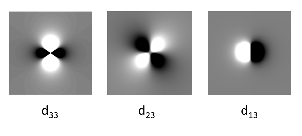
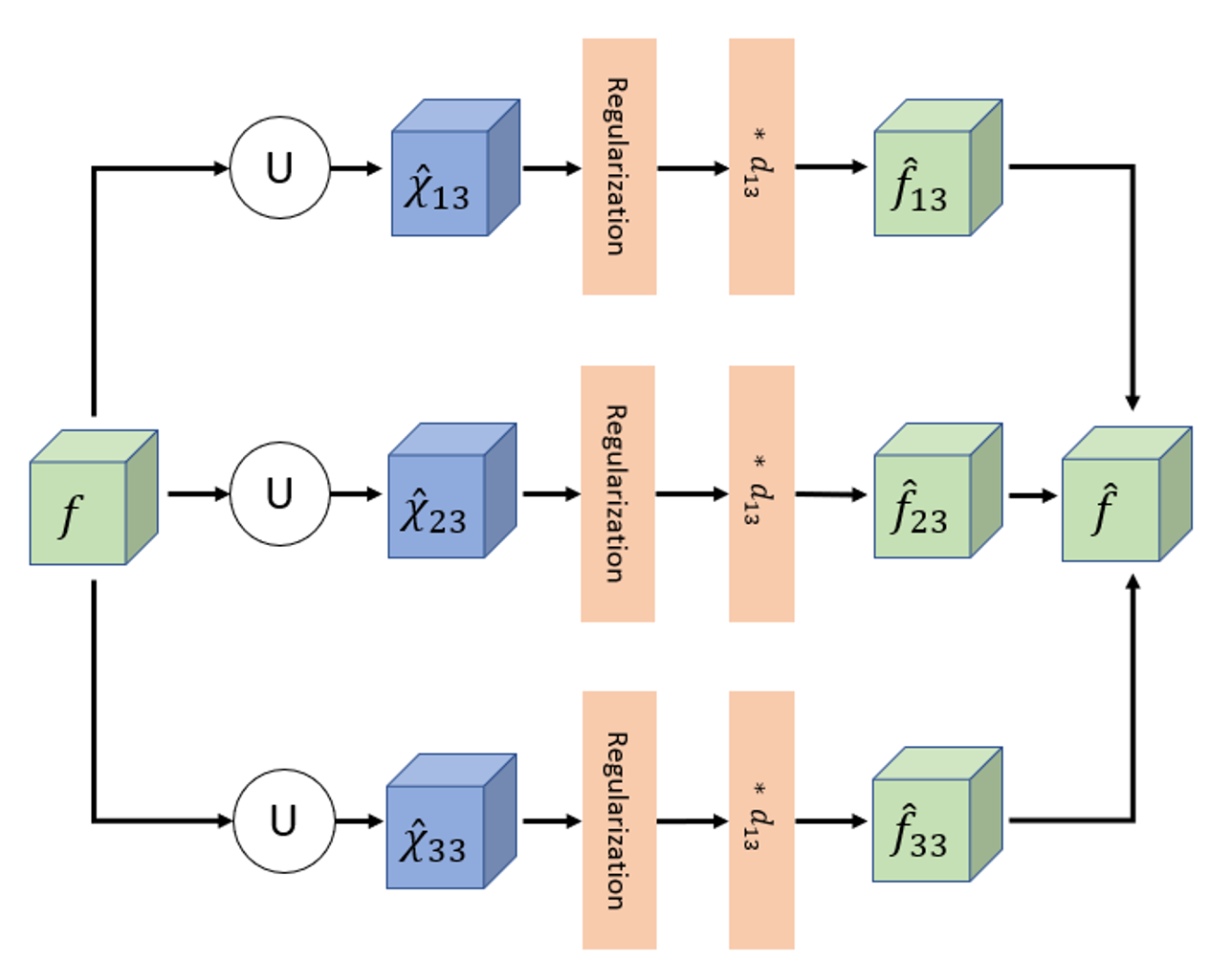
Fig. 2. Physics-informed autoencoder network. The measured frequency shift f (from MRI GRE phase) is split into three branches. U-Net neural networks learn to produce intermediate activations (these are the susceptibility tensor components) that yield an output (f) similar to the input, when the forward model is applied.
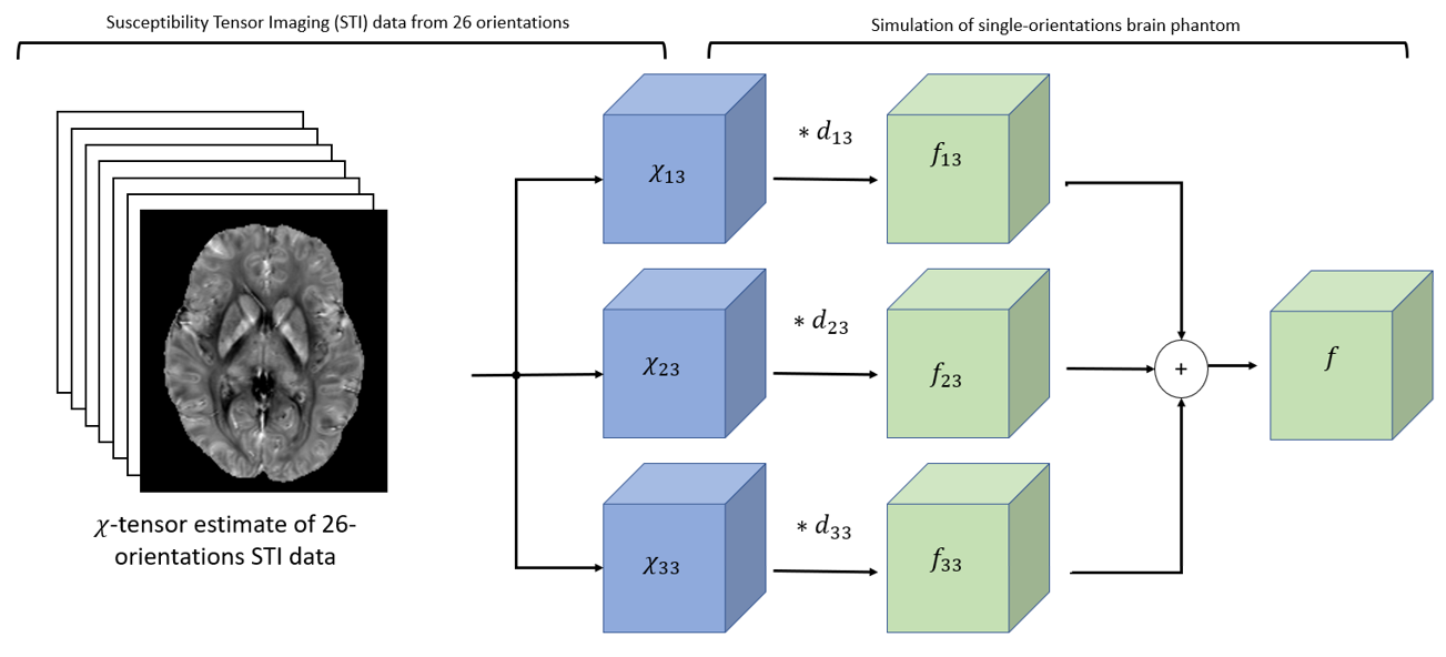
Fig. 3. Susceptibility tensor imaging and phantom simulation. From MRI scans under different head orientations (in our study 11-29 orientations), STI estimates the full susceptibility tensor. We took the elements that are apparent under a single orientation in z direction, 𝝌13, 𝝌23, 𝝌32, and simulated their corresponding frequency shifts. Then, their sum yields the observable frequency shift, f.
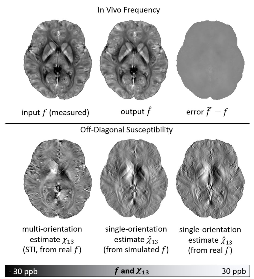
Fig. 4. Estimated off-diagonal susceptibilities. The top shows strong similarity between the in- and output of our autoencoder. The methods finds valid solutions to the ill-posed inverse problems of disentangling the sources and deconvolving the dipole kernels. The bottom shows strong similarity between the three off-diagonal susceptibility estimates. The STI estimate acts as a gold standard for solving the problem from real data (f) and acts as a ground truth for simulated data. The solutions of our method are similar between the real and simulated case and the STI solution.