4998
Quiet Fat-suppressed T1-weighted MRI by Dual-IR PETRA1Faculty of Science and Engineering, University of Nottingham Ningbo China, Ningbo, China, 2Xingaoyi Medical Equipment Co. Ltd, Ningbo, China, 3Nottingham Ningbo China Beacons of Excellence Research and Innovation Institute, Ningbo, China
Synopsis
Keywords: Pulse Sequence Design, Brain
Motivation: MRI produces considerable acoustic noise. PETRA sequence can solve this problem but has limited contrast.
Goal(s): This study improves PETRA sequence by magnetization preparation to achieve widely used T1-weighted imaging with fat suppression and sound reduction.
Approach: Double asymmetric adiabatic RF pulses are designed to invert aqueous tissues for T1-weighted contrast creation and invert lipidic tissues for fat suppression and combined with PETRA gradient trajectories.
Results: The brain and knee experiments are conducted to verify the feasibility of the proposed sequence. The fat is decreased by above 70%. The gray-to-white matter contrast and knee cartilage visualization are enhanced compared with non-prepared PETRA sequence.
Impact: The significantly reduced SPL of 67.4 dBA using limited gradient switching, providing better patient scanning comfort, can promote its acceptance to pediatric imaging. The double RF preparation design improves the limited contrast of PETRA.
Introduction
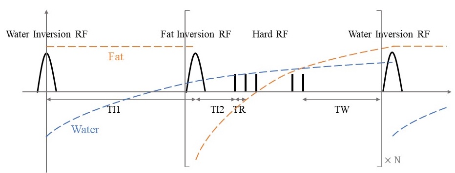
T1-weighted image is of high significance in MRI applications and fat suppression is also vital to avoid obscuring essential tissues or lesions. MRI has a disadvantage of its high acoustic noise, which is commonly 80-110 dBA, causing patient anxiety.1 The primary source of the MRI sound is the gradient coil oscillation resulting from changing Lorentz forces when the switching current goes through. In this study, we proposed a sequence combining double inversion recovery RF pulses to achieve both T1 weighting and fat suppression. An asymmetric adiabatic water-selective inversion pulse with high B1 sensitivity is designed to invert aqueous tissues, and after an inversion time TI1, an asymmetric adiabatic fat-selective pulse is applied to invert lipidic tissues with a gradient spoiler followed as shown in Figure 1.2, 3 Time parameters are optimized to acquire the signals at fat nulling and high tissue contrast point. 3D pointwise encoding time reduction with radial acquisition (PETRA) sampling trajectory is used to reduce acoustic noise by ramping up the gradient before excitation with a small amount of switching, which initially has limited contrasts of proton density to light T1 weighting due to its short TR and TE.4 Its ultrashort TE is also beneficial for the visualization of short-T2 tissues such as bone and cartilage.5-7Methods
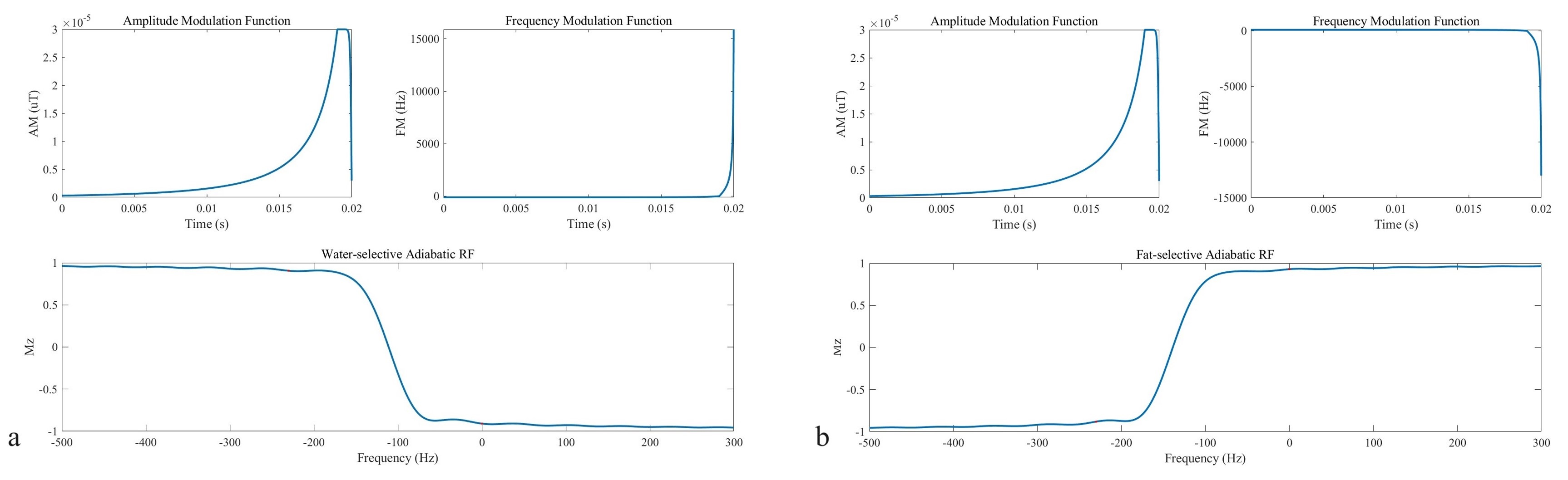
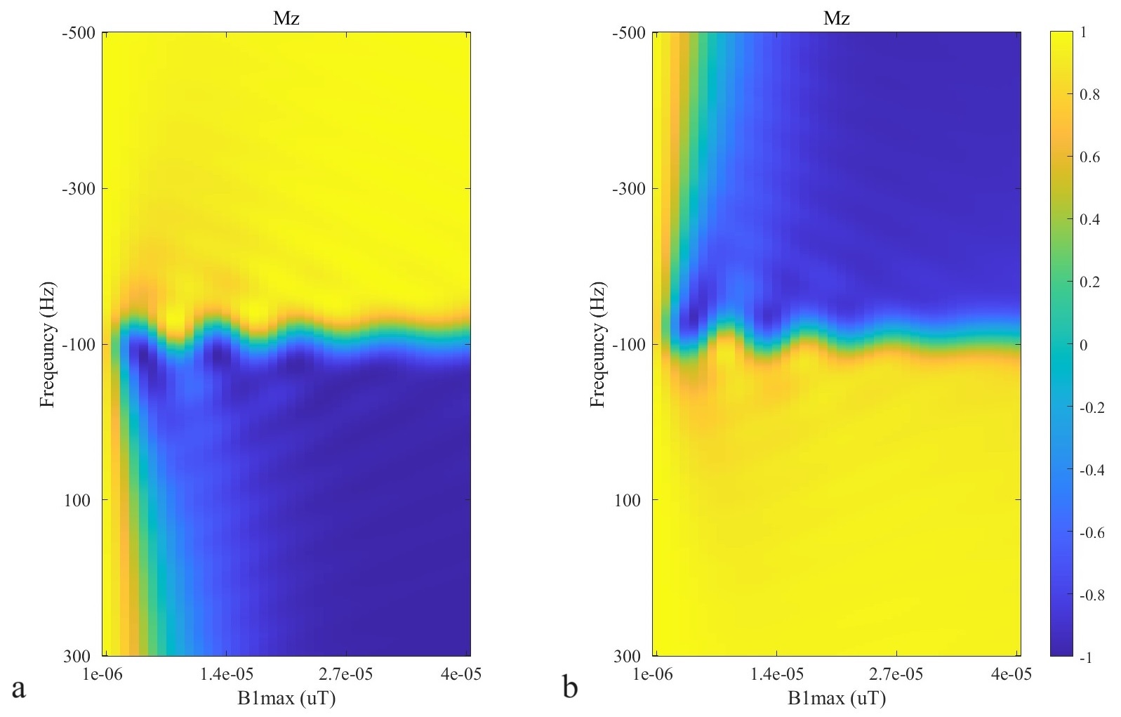
The asymmetric adiabatic RF pulse contains a wide half hyperbolic secant (HS) 1/2 of 95% duration for a sharper transition band and a narrow time-reversed tanh/tan of 5% duration for a shorter time, as shown in Figures 2 and 3. Since fat has a relatively fast T1 recovery, the fat suppression module is repeated after one water inversion section to improve signal acquisition time efficiency. The sequence is applied for brain and knee imaging on an XGY SuperScan-1.5T MR scanner (Xingaoyi Medical Equipment Company, China). Its parameters are: asymmetric adiabatic RF pulse duration=20 ms, number of fat suppression section N=4, hard RF flip angle=12 degrees, TI1=500 ms, TI2=5 ms, TW=40 ms, TR=3.5 ms, TE=100 us, FOV=280 (for brain) and 220 (for knee) mm, matrix=256*256*256 and sampling frequency=10 kHz. The 3D non-uniform Fourier transform (NUFFT) is applied for image reconstruction in Matlab (The MathWorks, USA). The scanning sound pressure level (SPL) is recorded by a 4955 microphone (Bruel Kjaer, Denmark) and a 2250 sound level meter (Bruel Kjaer, Denmark) placed in the isocenter of the scanner bore.Results
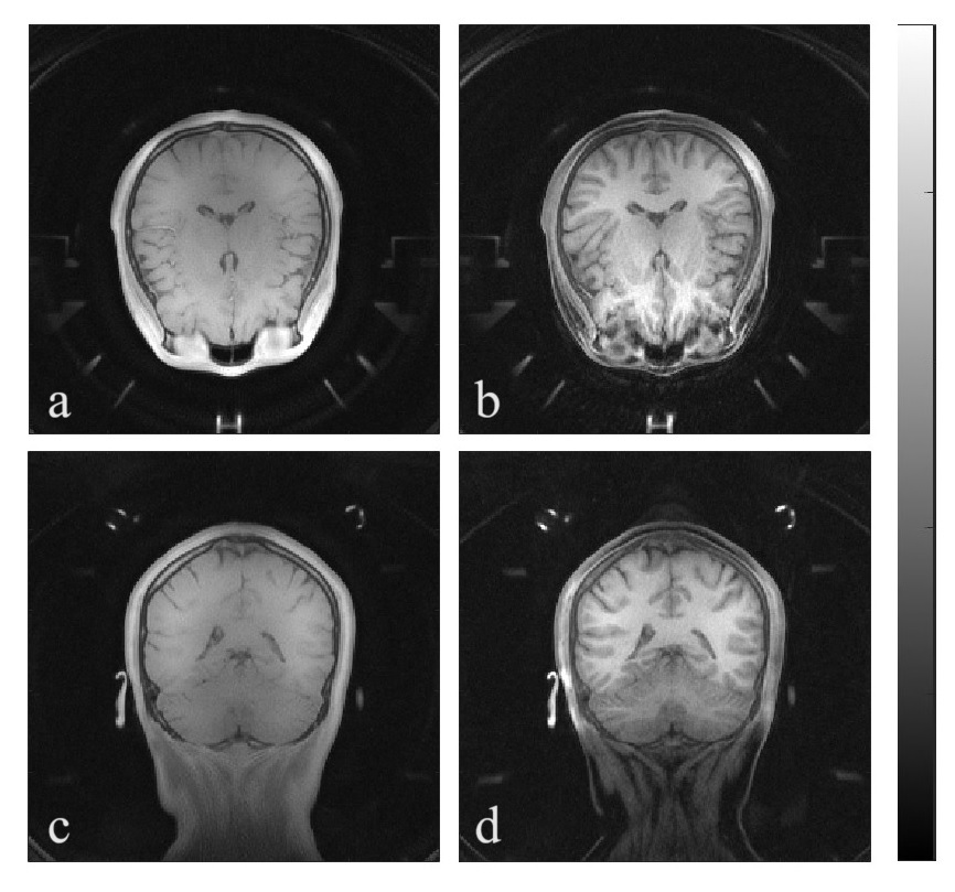
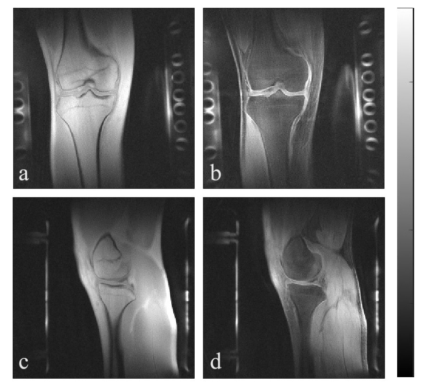
The 3D isotropic images of the brain and knee scanned by the normal PETRA sequence without preparation module and by the proposed T1-weighted fat-suppressed sequence are demonstrated in Figures 4 and 5. In brain imaging, fat is suppressed by 85.48% and the gray-to-white matter contrast to noise ratio calculated by $$$(SIgm-SIwm)/(SDair)$$$ is enhanced significantly from 2.06 to 9.74. The knee fat and bone marrow are decreased by 70.74 and 50.21%. The SPL is reduced to around 67.4 dBA by using the PETRA trajectory, which is only 2.5 dBA above the background noise.Discussion
The lipidic tissue contrast and T1-weighted contrast are observed in both brain and knee in vivo experiments for clearer delineation of structures. The brain image shows a higher difference between white and gray matter, and the knee cartilage can be seen with fat and marrow signals decreased. The TI1 value can be flexibly controlled to null specific tissue.Conclusion
We designed and validated the feasibility of the T1-weighted fat-suppressed 3D PETRA sequence based on asymmetric adiabatic preparation RF pulses on the brain and knee with low acoustic noise of 67.4 dBA, which greatly enhances comfort and safety during scanning. The silent sequence is also potentially beneficial for neonatal applications due to their larger risks of hearing injury under high SPL for diseases like tumors, cartilage injuries, bone marrow disorders, etc.Acknowledgements
No acknowledgement found.References
1. Bjorn H, Martin O, and David G. Sequence-based acoustic noise reduction of clinical MRI scans. Magn. Reson. Med. 2015;73(3):1104-1109.
2. Michael D, Thomas L. Flexible MEGA editing scheme with asymmetric adiabatic pulses applied for T2 measurement of lactate in human brain. Magn. Reson. Med. 2021;85(3):1160-1174.
3. Tsang-Lin H, Peter M, Van Z, et al. Asymmetric adiabatic pulses for NH selection." J. Magn. Reson. 1999;138(1):173-177.
4. Grodzki M, Peter J, and Bjoern H. Ultrashort echo time imaging using pointwise encoding time reduction with radial acquisition (PETRA). Magn. Reson. Med. 2012;67(2):510-518.
5. Markus W, David B, Benjamin D, et al. ZTE imaging in humans. Magn. Reson. Med. 2013;70(2):328-332.
6. Richard K, Azadeh S, Marcelo W, et al. Age‐dependent changes in knee cartilage T1, T2, and T1p simultaneously measured using MRI fingerprinting. J. Magn. Reson. Imaging. 2023;57(6):1805-1812.
7. Marius M, Martin B, Josef K, et al. STIR vs. T1-weighted fat-suppressed gadolinium-enhanced MRI of bone marrow edema of the knee: computer-assisted quantitative comparison and influence of injected contrast media volume and acquisition parameters. J. Magn. Reson. Imaging. 2005;22(6):788-793.
Figures