4995
Towards Practical SEMSI at 0.55 Tesla1Department of Biomedical Engineering, University of Southern California, Los Angeles, CA, United States, 2Department of Electrical and Computer Engineering, University of Southern California, Los Angeles, CA, United States, 3Siemens Healthcare USA, Los Angeles, CA, United States, 4Department of Radiology, Stanford University, Stanford, CA, United States
Synopsis
Keywords: Artifacts, Low-Field MRI
Motivation: Spectrally-encoded multi-spectral imaging (SEMSI) is an approach for distortion-free MRI around metallic implants, but requires multiple-readouts per TR to be clinically feasible.
Goal(s): To determine the benefits of SEMSI over SEMAC at 0.55T, using parameters that are consistent with a future multi-readout SEMSI implementation.
Approach: We used single-readout SEMSI to prospectively mimic the performance of multi-readout SEMSI with high slew rate and readout bandwidth.
Results: We observe that SEMSI simultaneously achieves the expected SNR improvement and artifacts reduction compared to SEMAC.
Impact: SNR improvement is important at 0.55T to examine the tissues near the metallic implants. This prospective study confirms the improvement performance of multi-readout SEMSI which will further improve SNR and scan efficiency.
INTRODUCTION:
Low-field MRI systems, including 0.55 Tesla, are favorable for imaging around metallic implants, because of the substantially reduced susceptibility artifacts and shorter scan times achievable with multispectral imaging methods1. However, SNR decreases at lower field, and while lower readout bandwidth can partially mitigate this, it can exacerbate readout artifacts and introduce more blurring effects in conventional multi-spectral imaging, such as SEMAC or MAVRIC-SL2,3.Spectrally-encoded multi-spectral imaging (SEMSI) is an alternative approach that has shown promise at 3T and 0.55T4,5, but has not yet been demonstrated in clinically feasible scan times. SEMSI attempts to achieve the SNR of a low-bandwidth readout, but with the low artifact of a high-bandwidth readout.
In this study, we make one step towards multi-readout SEMSI. We investigated the performance of single-readout SEMSI with high readout bandwidth (rBW) with timing that matches multi-readout SEMSI (remains to be implemented). We demonstrate, in a spine phantom, the expected improvement in artifact compared to low rBW SEMAC and in SNR compared to high rBW SEMAC.
METHODS:
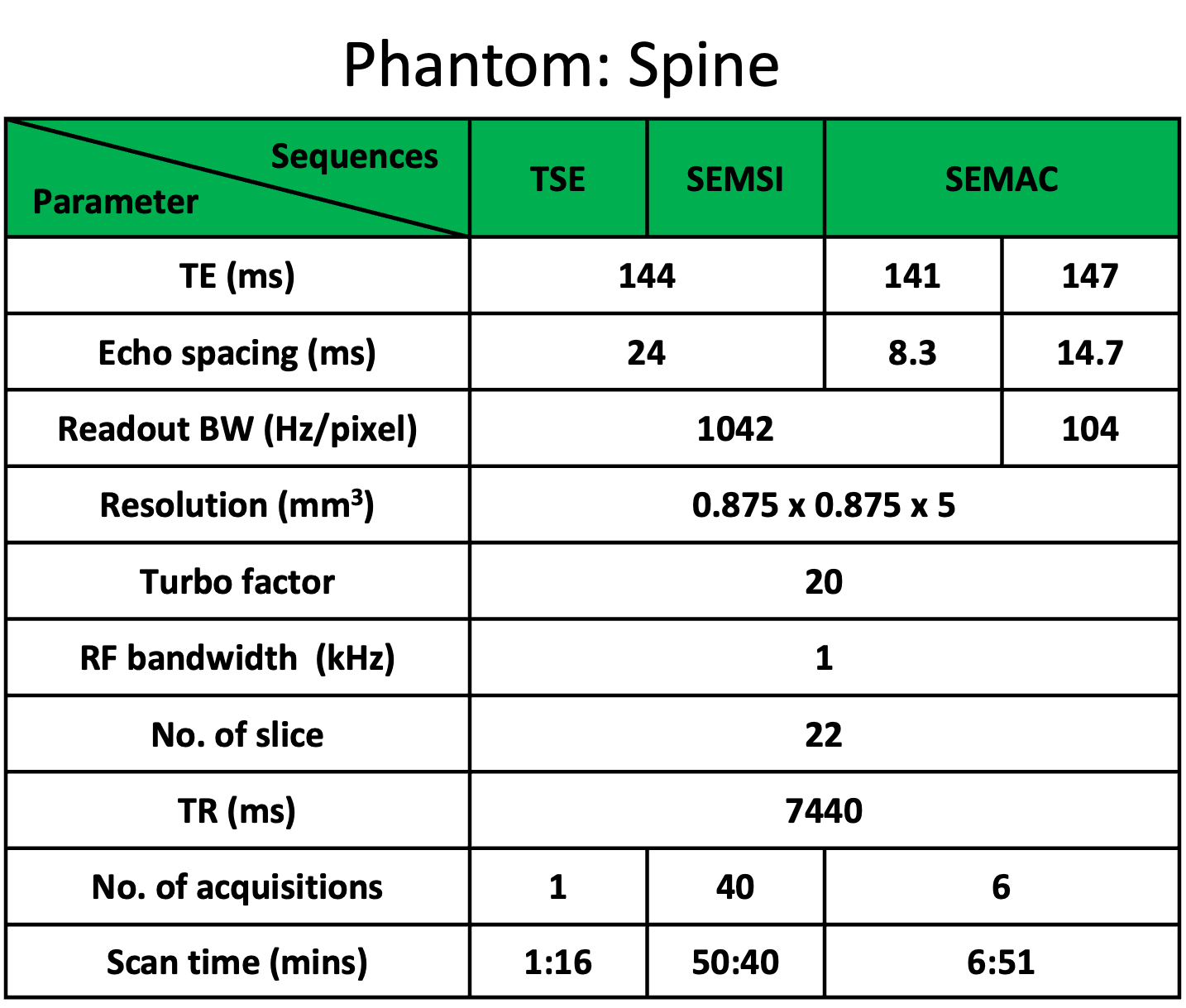
Experiments were performed using a 0.55T whole-body system (prototype MAGNETOM Aera, Siemens Healthineers, Erlangen, Germany) equipped with high-performance shielded gradients (45 mT/m amplitude, 200 T/m/s slew rate). A six-element body coil (anterior) and six elements from an 18-element spine coil (posterior) were used for signal reception. We imaged a spine phantom composed of cobalt-chromium/titanium screws and titanium rods on a meshed pattern, in a water bath.We mimic multi-readout SEMSI, with 4 gradient echo shifts per TR, rBW = 1 kHz/pixel (tRO = 1.5 ms), 10 readouts shifts. The resulting spectral resolution is 167 Hz, and spectral FOVf is 6.7 kHz. This is done using single-readout SEMSI with 40 tshift values [-3000:150:2850] µs. High rBW of 1042 Hz/pixel was used and high slew rate was turned on. A hamming window was applied along the tshift dimension to suppress Gibss ringing and sidelobes. Readout translation was applied to each spectral image by ∆f/rBW. Every spectral image was shifted along the slice direction by applying a linear phase (slope = ∆f/RFBW) along kz. Only the spectral bins that have largest signal intensity contributing were chosen for spectral combination. Spectral summation was performed using root-sum-square (RSS). For SEMAC, six slice encoding was applied with two rBWs (1042 Hz/pixel and 104 Hz/pixel), and combined with RSS. Other scan parameters are listed in Table 1.
RESULTS:
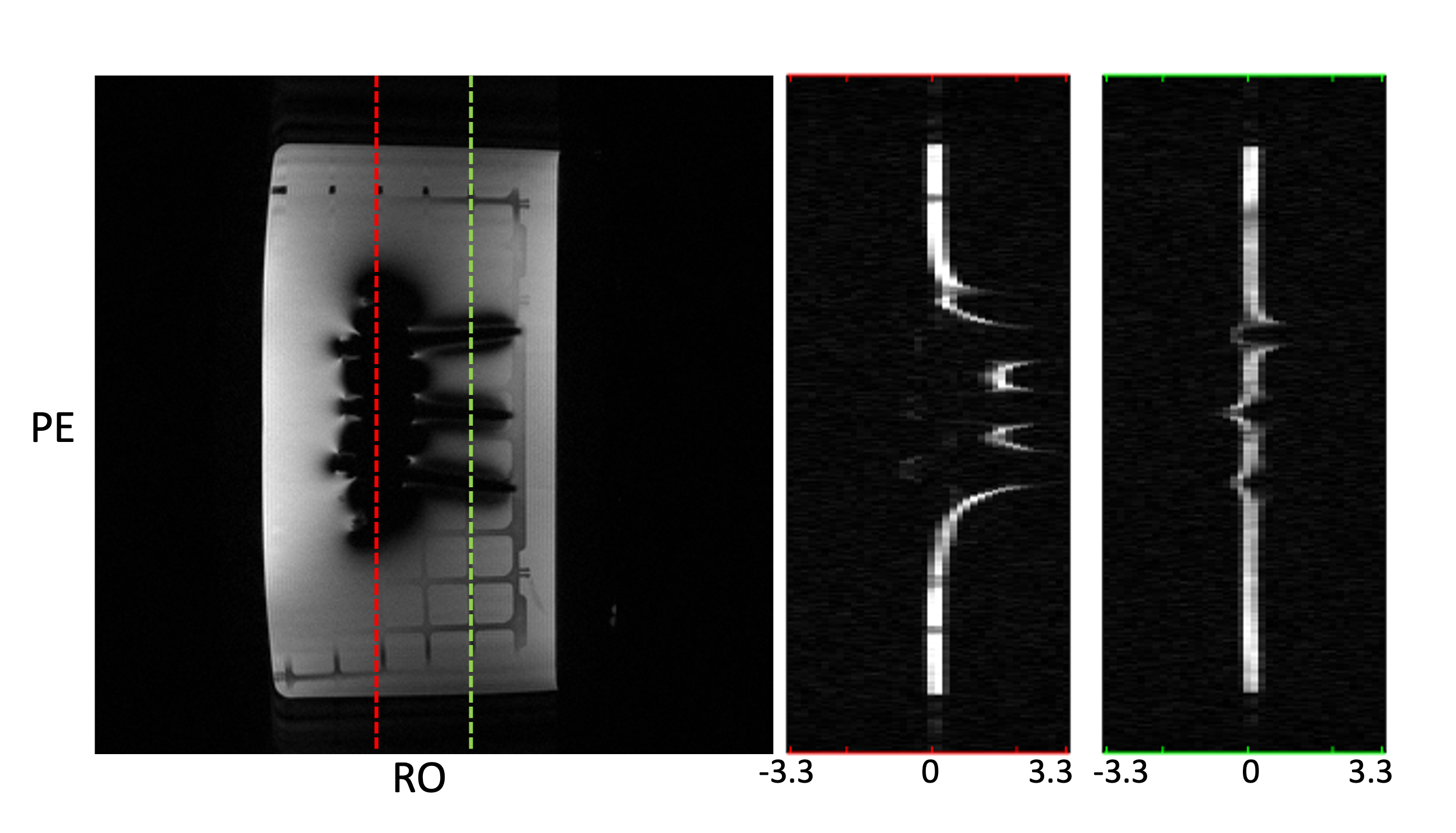
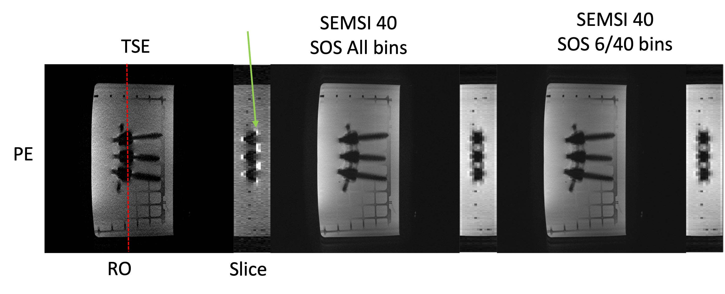
Figure 1 illustrates the narrow-band signal for each pixel. The high signal intensity only occurs in 4 - 5 spectral bins (out of 40) for each pixel. Therefore, we selected the 6 spectral bins with the largest magnitudes for combination.Figure 2 shows in-plane and reformatted SEMSI images. This demonstrates that when using the 6 bins with highest intensities for each pixel, SEMSI exhibits no discernible difference compared to using all 40 bins.
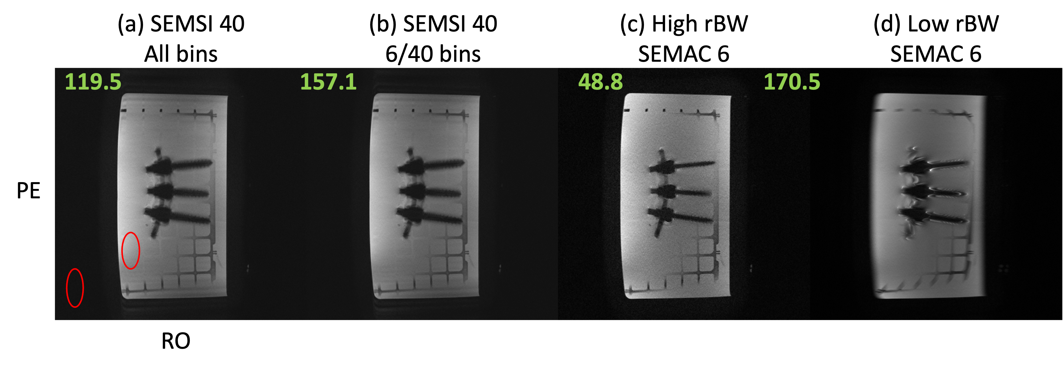
Figure 3 shows the corrected in-plane image and SNR values for SEMSI and SEMAC. High-bandwidth SEMSI demonstrates 2.44- and 3.2-fold higher SNR compared to high rBW SEMAC with 6 bins. This is consistent with the expected improvement in SNR based on the number of acquisitions. Moreover, high-bandwidth SEMSI maintains an SNR comparable to that of low rBW SEMAC, while avoiding blurring and substantially reducing artifacts.
DISCUSSION:
We have demonstrated single-readout SEMSI with parameters that mimic a clinically feasible four-readout SEMSI with reasonable scan time, but has not yet been implemented. Our tests have demonstrated the expected ~3 times higher SNR with comparable artifact compared to high rBW SEMAC, and the expected similar SNR and substantially reduced artifact compared to low rBW SEMAC.The next step, of course, is to implement the multi-readout SEMSI sequence and perform this comparison prospectively in phantoms and then in patients with orthopedic metallic implants. This will require some additional consideration of the oscillating readout gradient polarity and will also require mitigation of concomitant field effects that will accumulate during the echo-planar spectroscopic readout.
CONCLUSION:
Our study has made one important step towards a practical SEMSI implementation for multi-spectral imaging at 0.55 Tesla. We demonstrate the expected SNR improvement compared to high rBW SEMAC, and the expected artifact improvement compared to low rBW SEMAC.Acknowledgements
We acknowledge grant support from the National Institutes of Health (R01-AR078912) and National Science Foundation (#1828736) and research support from Siemens Healthineers.References
1. Khodarahmi I, Brinkmann IM, Lin DJ, et al. New-Generation Low-Field Magnetic Resonance Imaging of Hip Arthroplasty Implants Using Slice Encoding for Metal Artifact Correction. Investigative Radiology. 2022;57(8):10.
2. Lu W, Pauly KB, Gold GE, Pauly JM, Hargreaves BA. SEMAC: Slice Encoding for Metal Artifact Correction in MRI. Magn Reson Med. 2009;62(1):66-76. doi:10.1002/mrm.21967
3. Koch KM, Brau AC, Chen W, Gold GE, Hargreaves BA, Koff M, McKinnon GC, Potter HG, King KF. Imaging near metal with a MAVRIC-SEMAC hybrid. Magn Reson Med. 2011 Jan;65(1):71-82. doi: 10.1002/mrm.22523. PMID: 20981709.
4. Yoon D, Lee PK, Nayak KS, Hargreaves BA, Spectrally-encoded Multi-spectral Imaging (SEMSI) for Off-resonance Correction Near Metallic Implants. ISMRM. May 2021, Vancouver.
5. Li B, Keskin K, Yoon D, Lee NG, Hargreaves BA, Nayak KS, “Spectrally-Encoded Multi-Spectral Imaging (SEMSI) At 0.55T Provides Improved Imaging Adjacent To Metallic Implants”, ISMRM. May 2023, Toronto.
Figures