4989
Assessing the Effect of Universal Pulses on EDGE-MP2RAGE at 7T1NINDS, National Institutes of Health, Bethesda, MD, United States, 2CEA, NeuroSpin, Universite Paris-Saclay, Gif-sur-Yvette, France
Synopsis
Keywords: Parallel Imaging, High-Field MRI
Motivation: To optimize an EDGE-MP2RAGE acquisition at 7T for epilepsy studies.
Goal(s): To determine if it is beneficial to use Universal Pules within EDGE-MP2RAGE at 7T and to find the optimum TI1 to produce the best visulation of the gray-white matter boundary.
Approach: Collect EDGE-MP2RAGE data with conventional RF and Universal Pulses and analyze the gray and white matter signal across the brain.
Results: Overall, it does seem to be beneficial to use Univeral Pulses within EDGE-MP2RAGE, although it does not produce more consistent gray and white matter signal in all brain regions.
Impact: Utilizing Universal Pulses within EDGE-MP2RAGE with the determined optimum TI1=760ms at 7T can improve continuity of the gray-white matter boundary compared to conventional RF pulses, which may allow for detection of focal cortical dysplasia in epilepsy patients.
Introduction
Edge-Enhancing Gradient Echo (3D-EDGE) MRI is a novel contrast that directly images the gray-white matter boundary by acquiring images at an inversion time (TI) with comparable gray and white matter signal intensity but opposite phases. The result is a cancelation of longitudinal magnetization which appears as a thin dark line at the gray-white matter boundary1. 3D-EDGE MRI has been shown to be a valuable tool for detecting focal cortical dysplasia in epilepsy patients and has potential in applications for other neurological diseases1. The 3D-EDGE technique can benefit from the increased net magnetization in 7T imaging as the technique is inherently low in signal-to-noise ratio (SNR). An increase in SNR could be utilized for increased image resolution. Tao et. al. optimized scanning efficiency and acuisition parameters for 7T with the EDGE-MP2RAGE sequence2. However, 7T imaging is challenging due to the inhomogeneous transmit B1 field, which will produce an inconsistent edge between the gray-white matter boundary due to non-uniform flip angle across the brain. Recently introduced, Univeral Pulses (UP) is a calibration-free parallel transmission (pTx) technique that utilizes the GRAPE method3,4,5 to mitigate inhomogeneous B1 transmit fields. In this study, we assess the effects of utilizing UP to improve EDGE-MP2RAGE contrast over the whole brain. We also invesigate the optimum TI for gray and white matter signal cancellation to produce the best visualization of the gray-white matter boundary with UP.Methods
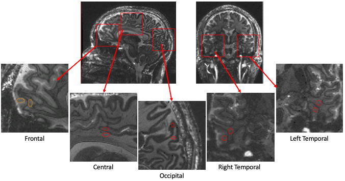
MP2RAGE data were acquired on healthy adults (n=3) under an IRB approved protocol using an investigational 7T MRI system with a 8-Tx/32-Rx head coil in pTx mode with the MP2RAGE sequence from NeuroSpin’s PASTeUR package3,4,5. The following scan parameters were used for EDGE-MP2RAGE data: TE/TR=3.38/4500ms, FA=5°/4°, iPAT = 3, 0.77x0.77x0.77mm, FOV=230, scan time = 9min 10s. For the convential RF pulse acquisition in circularly polarized mode (CP-mode), TI1/TI2 = 800/2700ms, as optimized by Tao et. al. For the UP RF pulse acquisiton, TI2=2700ms and TI1=720, 740, 760, 780, 800, and 820ms. Cortical gray and white matter signal intensities were measured on the TI1 images in ROIs in different brain regions: frontal, central, occipital, left and right temporal lobes. Figure 1 shows the ROIs from one subject. Gray and white matter signal ratios (GM/WM) were calculated for each region and subject. The GM/WM was averaged across subjects for each region. A whole brain GM/WM was calculated for each subject by averaging across regions.Results
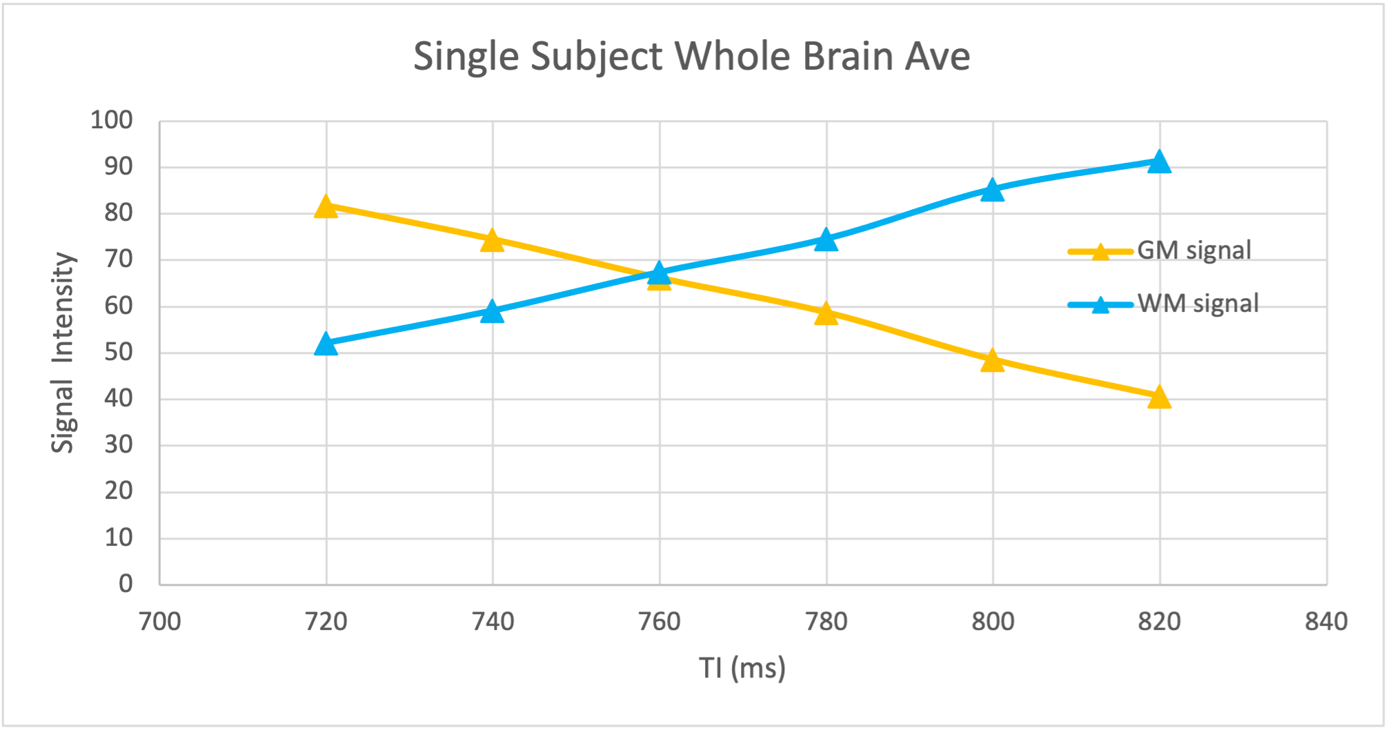
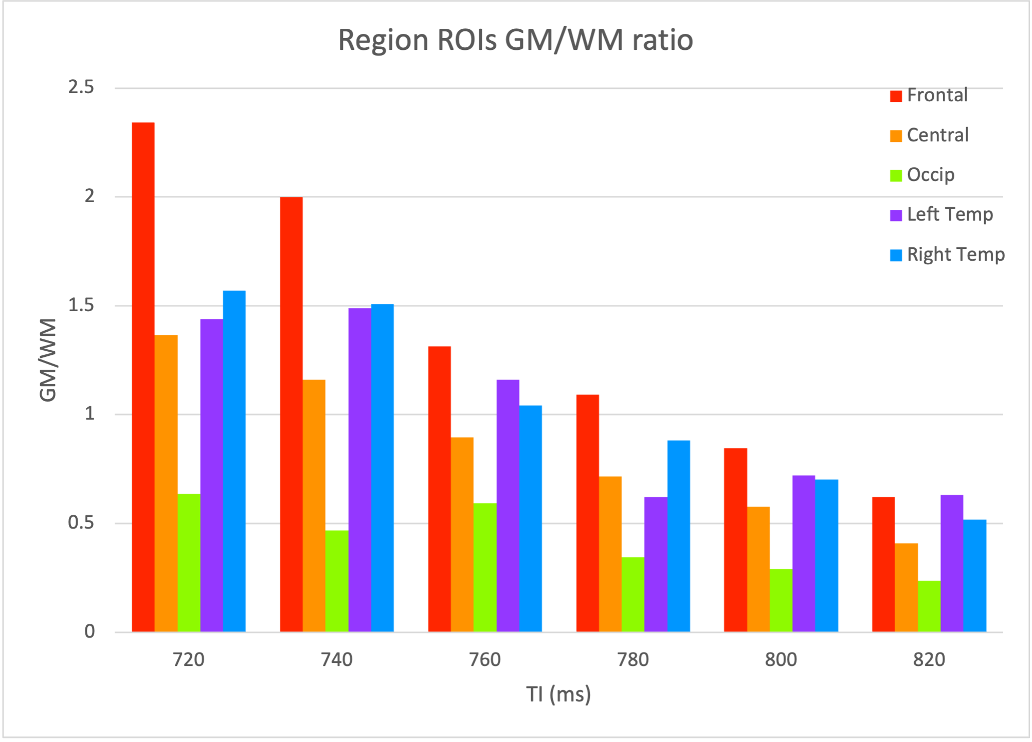
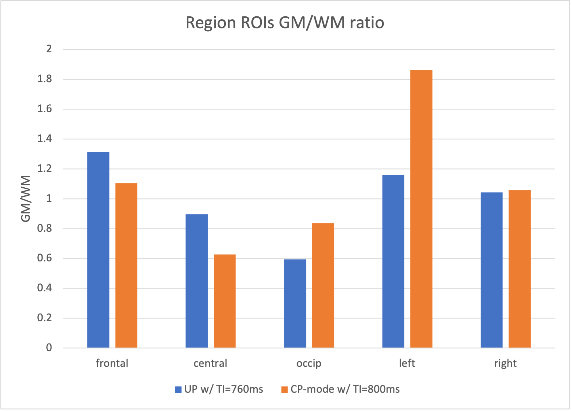
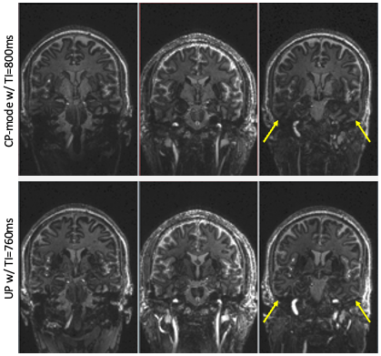
Figure 2 shows a representative plot of the whole brain averaged GM and WM signal intensities over the different TI1s using UP. The optimum TI time is when GM/WM = 1. For all subjects, the optimum TI time for the whole brain using UP was TI=760ms. GM/WM was 0.95, 0.98, and 0.94 for the three subjects with UP. For CP-mode, the optimum TI=800ms. GM/WM was 0.96, 1.15, and 0.86 for the three subjects with CP-mode. To assess the effect of inhomogeneous B1 transmit, GM/WM was analyzed for each region. Figure 3 shows the GM/WM averaged over subjects for each region across TIs. The optimum TI time varies slightly between regions, but is closest to 1 for all regions when TI=760ms. Figure 4 shows GM/WM with the UP acquisition and TI=760ms, for frontal, central, occipital, left and right temporal ROIs were 1.3, 0.9, 0.6, 1.2, and 1.0, respectively. Figure 4 also shows the corresponding GM/WM values with CP-mode were 1.1, 0.6, 0.8, 1.8, and 1.1, respectively. UP produced similar GM/WM in the left and right temporal lobe, being 1.2 and 1.1, unlike the CP-mode data with GM/WM being 1.8 and 1.1, respectively. The difference in consistent contrast in the temporal lobes can be seen in Figure 5.Discussion
This study explores the effect of the UP pTx technique on EDGE-MP2RAGE images. 760ms is the optimum TI for producing the best visualization of the gray-white matter boundry on TI1 images in EDGE-MP2RAGE using UP. The optimum TI was consistent across all three subjects. The results show that UP was effective in reducing the inhomogeneous B1 transmit field in the left and right temporal lobes seen with conventional CP-mode. Conversely, UP produced less optimal GM/WM than conventional CP-mode in the frontal and occipital lobes. However, the deviation from optimal TI for UP in these regions is less than that for conventional CP-mode in the temporal lobes. Overall, UP produced more consistent GM/WM across the brain than conventional CP-mode, improving continuity of the gray-white matter boundary, which will be beneficial for epilepsy patient studies. Further analysis is needed across more regions to fully assess the effect of UP on EDGE-MP2RAGE contrast in the whole brain.Acknowledgements
No acknowledgement found.References
1. Middlebrooks EH, et. al. (2020) Improved detection of focal cortical dysplasia using a novel 3D imaging sequence: Edge-Enhancing Gradient Echo (3D-EDGE) MRI. NeuroImage Clinical 28:102449.
2. Tao S, et. al. (2023) Edge-Enhancing Gradient-Echo MP2RAGE for Clinical Epilepsy imaging at 7T. AJNR Adult Brain 44 (3):268-270.
3. Gras V et. al. (2017). Universal Pulses: A new concept for calibration-free parallel transmission. Magn Reson Med. 77:635–643.
4. Mauconduit F. et al. (2022) Traveling Pulses Visit 7T Terra Sites: Getting Ready For Parallel Transmission In Routine Use, In Proceedings of the 31th Annual Meeting of ISMRM, London, UK, p. 2091.
5. Van Damme L, et al. (2021) Universal non-selective excitation and refocusing pulses with improved robustness to off-resonance for Magnetic Resonance Imaging at 7 Tesla with parallel transmission. Magn Reson Med. 85: 678-693.
Figures