4986
Evaluation of pituitary tumor texture using Synthetic MRI1Department of Radiology, Taihe Hospital, Hubei University of medicine, Shiyan, Hubei, China, 2GE Healthcare,MR Research, Beijing, China, 3Department of Radiology, Taihe Hospital, Hubei University of Medicine, Shiyan, Hubei, China, 4Department of Nuclear Medicine, Taihe Hospital, Hubei University of Medicine, Shiyan, Hubei, China, 5Hubei University of Medicine, Shiyan, Hubei, China
Synopsis
Keywords: fMRI Analysis, Quantitative Imaging
Motivation: Surgery selection mainly relies on texture of pituitary adenomas. There is rarely a non-invasive imaging method to identify texture of pituitary adenomas that greatly determines surgical selection and outcomes.
Goal(s): To explore the predictive performance of synthetic MRI in types of pituitary tumors.
Approach: T1, T2 and PD values of every tumor were measured and divided into the solid and soft tumor groups according to the histology results of surgical samples.
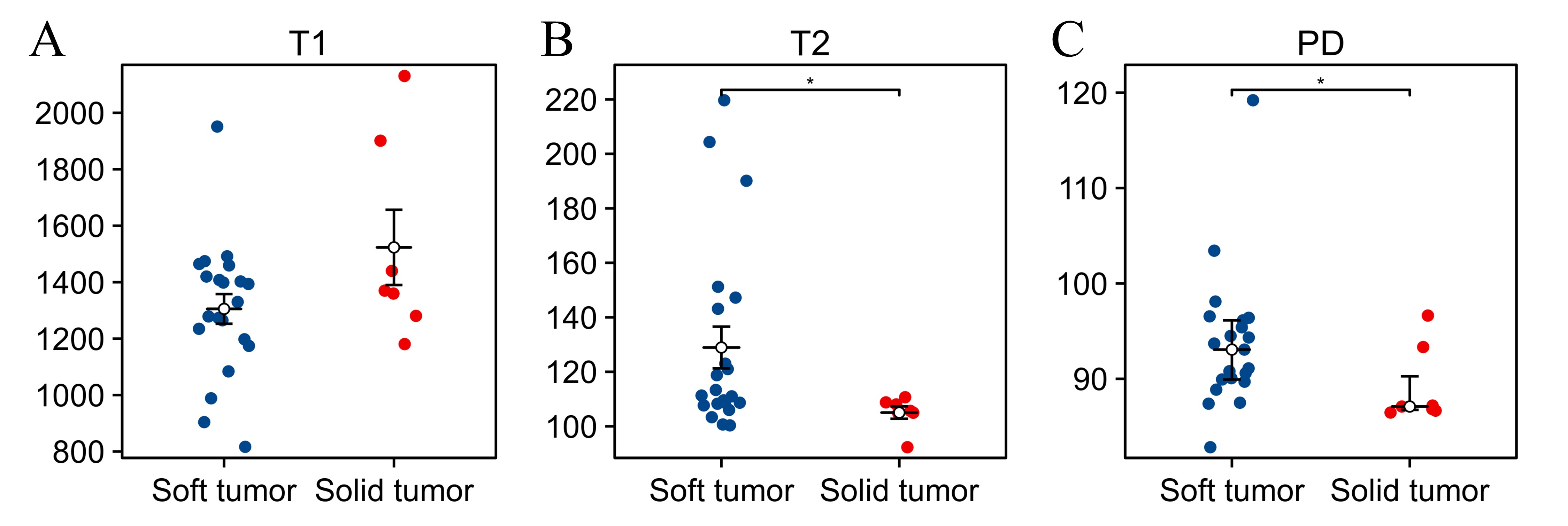
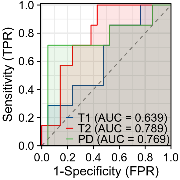
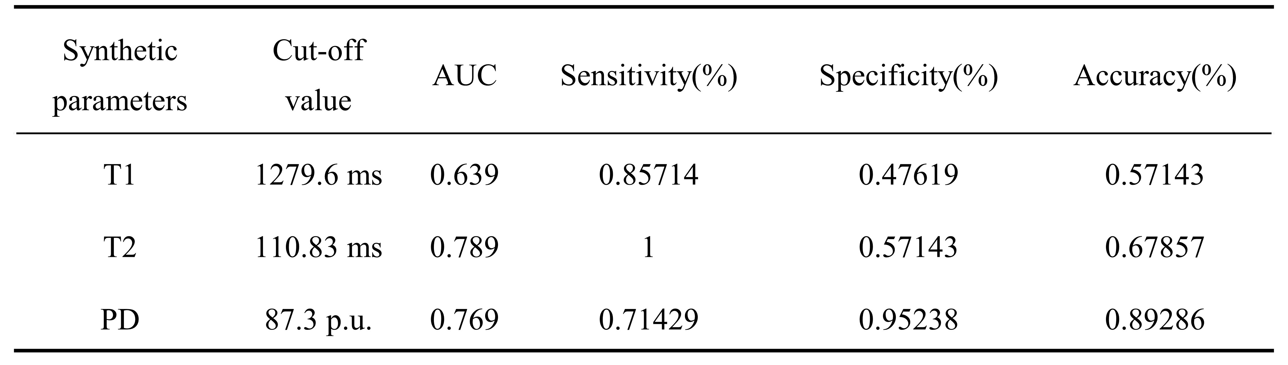
Results: Synthetic MRI-computed T2 and PD values were significantly higher in the soft pituitary tumor group than in the solid group (P<0.05) with cutoff values of 110.83 ms and 87.3 p.u., respectively.
Impact: Both T2 and PD value can assist surgeons in determination of surgical treatments (e.g. transsphenoidal resection or craniotomy) and prediction of surgical outcomes, such as resection completeness, indicating synthetic MRI could a strong imaging marker.
Introduction
Pituitary Adenoma is the third most common intracranial tumor, accounting for about 10%-15% of intracranial tumors[1]. Surgical resection is generally the first choice of treatment. Many factors such as tumor size, shape, texture and aggressiveness affect surgical outcomes and thus preoperative evaluation is essential[2]. It is difficult to completely remove solid pituitary tumors due to higher possibility of injuring peripheral blood vessels or nerves and remaining residuals after surgery. In contrast, the soft pituitary tumor highly likely invades the saddle itself and surrounding tissues, but it may gradually descend insides the saddle as brain blood pulsates and achieve a satisfactory resection. A study of diffusion weighted imaging in prediction of tumor texture and tumor resection degree has shown the soft pituitary tumor has higher ADC values and surgical resection rate[3]. MRI-based radiomics model using T1WI, T2WI, enhanced T1WI well predicts the texture of pituitary giant adenoma while machine learning T2WI-MRI radiomics model well classifies tumor texture with AUC, sensitivity and specificity of 93%, 100% and 87%[4]. However, synthetic MRI is an advanced and straightforward imaging is available in clinics and offer onsite quantitative information with good reliability and repeatability. Therefore, our study aims to investigate the classification performance of synthetic MRI for texture of pituitary tumor.Methods
A total of 28 patients with pituitary tumor prospectively underwent conventional MRI and quantitative MRI in our hospital from June to October 2023. Imaging features such as tumor shape, length diameter, and aggressiveness (Knosp grade) were evaluated using both plain and contrast enhanced MRI while longitudinal relaxation time (T1), transverse relaxation time (T2), and proton density (PD) maps were measured on corresponding parameter maps generated by Magnetic Resonance Imaging Compilation (MAGiC) sequence. According to tumor texture reported in surgical records, all patients were divided into solid and soft texture groups. Independent t test or Wilcoxon sign rank test was used to examine differences of imaging features, quantitative parameters, a surgical approach and resection degree of tumors according normality and variance equality assessed by Shapiro-Wilk and Levene test. Receiver operating characteristic curve (ROC) was plotted and the Area Under Curve (AUC) of each parameter was calculated to evaluate and compare the diagnostic efficacy of each parameter in predicting texture of pituitary tumors before surgery.Results
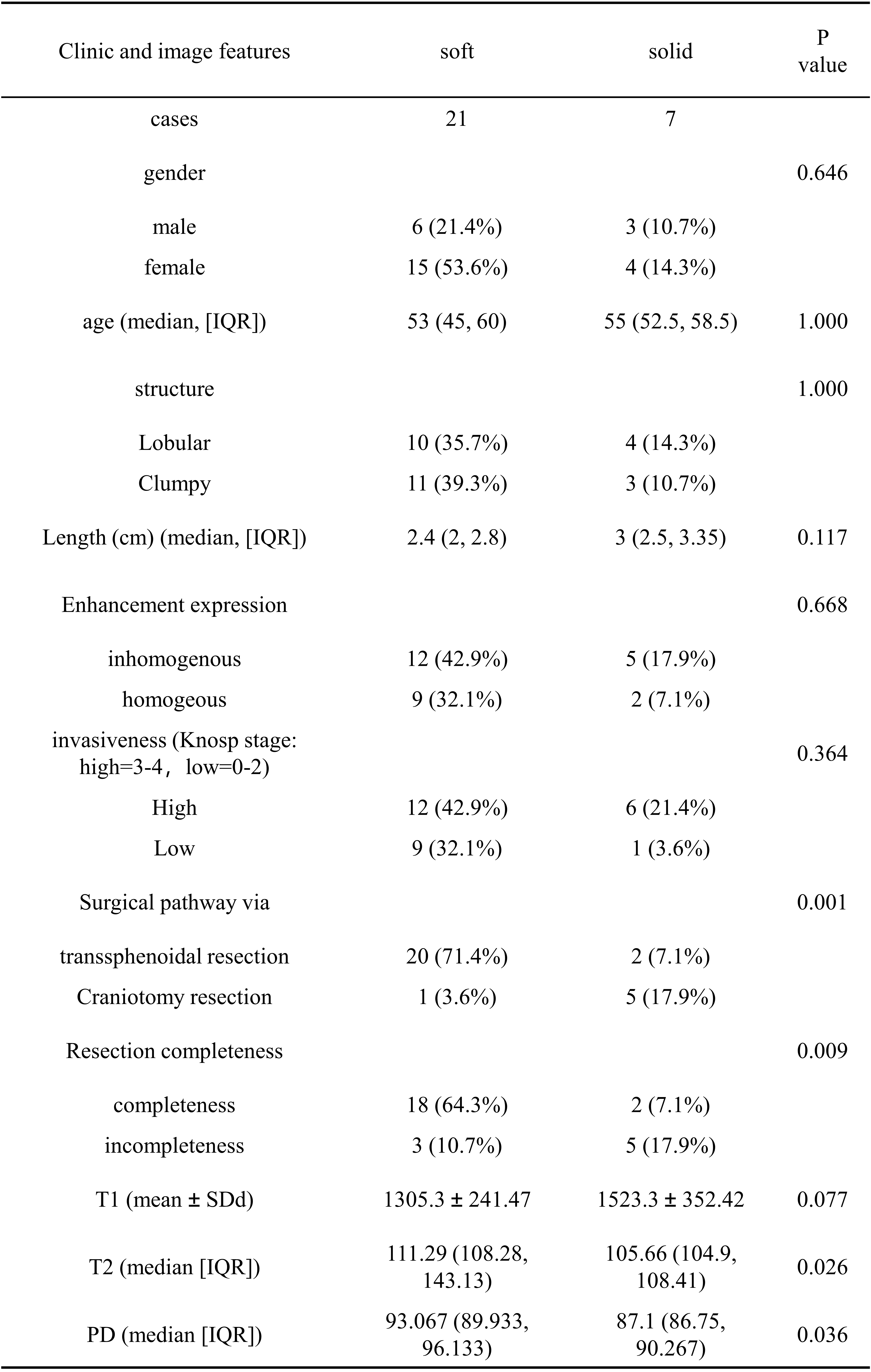
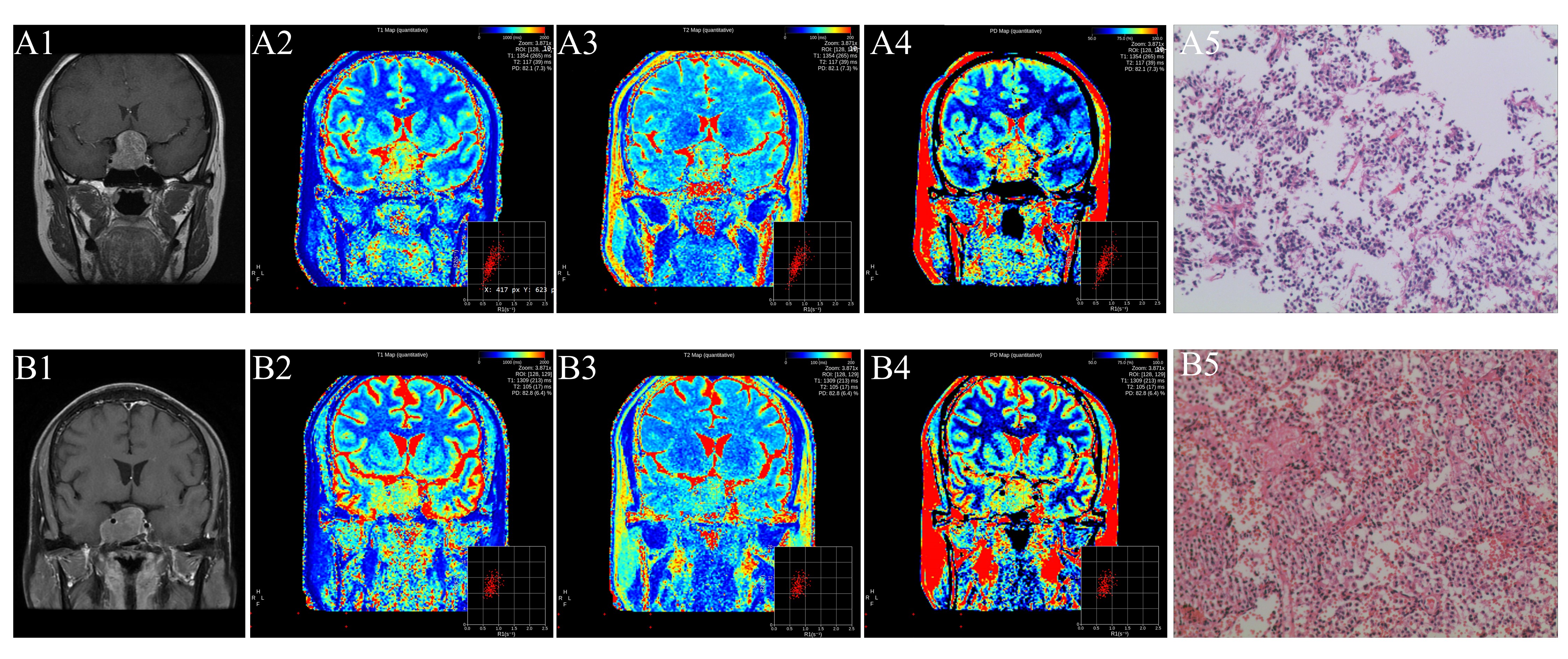
All demographics and clinical information of recruited patients were shown in Figure 1. The enhanced MRI sequences, MAGiC sequence, and pathological images of the solid and soft tumor groups are shown in Figures 2. The selection of surgical approach and the completeness of resection between texture groups were statistically significant. Compared to the solid texture group, transsphenoidal resection rather than craniotomy is more preferred to chosen in the soft texture group (P=0.001), and the completeness of resection was significantly higher in the soft texture group than that in the tough texture group (P=0.009). T2 and PD values were statistically higher in the soft tumor group than in the solid tumor group (P<0.05, Figure 3) with superior AUC of 0.789 and 0.769, sensitivity of 85% and 71%, specificity of 47% and 95%, and cutoff values of 110.83 ms and 87.3 proton density in unit (p.u.) to T1 values, respectively (Figure 4, 5).Discussion
T2 and PD values showed good classification performance on texture of pituitary tumors in our preliminary results. Collagen content is the main factor affecting the texture of pituitary adenomas[5],Some scholars have defined pituitary adenomas with collagen content exceeding 5% as fibrotic adenomas[6];in other words, the texture of the pituitary tumor is essentially the degree of fibrosis of the tumor. Previous studies have reported T1 and T2 values are closely related to aggravation of fibrosis degree in liver as well as human and animal kidney fibrosis[7, 8]. T2 value in combination with liver elasticity measured by magnetic resonance elastography (MRE) improve the diagnostic efficiency of liver fibrosis[9]. Therefore, our finding of higher T2 value instead of T1 value in soft pituitary tumor than solid ones encouraged us to enlarge more subjects to provide a more convening results and cutoff values for texture differentiation.Conclusion
Our finding of T2 and PD values in well preoperative prediction on texture of pituitary tumor, suggesting synthetic MRI provided a strong imaging basis for the selection of surgical approach or the preoperative evaluation of the resection degree.Acknowledgements
No acknowledgement found.References
[1] DALY A F, BECKERS A. The Epidemiology of Pituitary Adenomas [J]. Endocrinology and metabolism clinics of North America, 2020, 49(3): 347-55.
[2] RUTLAND J W, LOEWENSTERN J, RANTI D, et al. Analysis of 7-tesla diffusion-weighted imaging in the prediction of pituitary macroadenoma consistency [J]. Journal of neurosurgery, 2020, 134(3): 771-9.
[3] DING W, HUANG Z, ZHOU G, et al. Diffusion-weighted imaging for predicting tumor consistency and extent of resection in patients with pituitary adenoma [J]. Neurosurgical review, 2021, 44(5): 2933-41.
[4] CUOCOLO R, UGGA L, SOLARI D, et al. Prediction of pituitary adenoma surgical consistency: radiomic data mining and machine learning on T2-weighted MRI [J]. Neuroradiology, 2020, 62(12): 1649-56.
[5] LI P, ZHANG D, MA S, et al. Consistency of pituitary adenomas: Amounts of collagen types I and III and the predictive value of T2WI MRI [J]. Experimental and therapeutic medicine, 2021, 22(5): 1255.
[6] NAGANUMA H, SATOH E, NUKUI H. Technical considerations of transsphenoidal removal of fibrous pituitary adenomas and evaluation of collagen content and subtype in the adenomas [J]. Neurologia medico-chirurgica, 2002, 42(5): 202-12; discussion 13.
[7] HOAD C L, PALANIYAPPAN N, KAYE P, et al. A study of T1 relaxation time as a measure of liver fibrosis and the influence of confounding histological factors [J]. NMR in biomedicine, 2015, 28(6): 706-14.[8] FRIEDLI I, CROWE L A, BERCHTOLD L, et al. New Magnetic Resonance Imaging Index for Renal Fibrosis Assessment: A Comparison between Diffusion-Weighted Imaging and T1 Mapping with Histological Validation [J]. Scientific reports, 2016, 6: 30088.
[9] HOFFMAN D H, AYOOLA A, NICKEL D, et al. T1 mapping, T2 mapping and MR elastography of the liver for detection and staging of liver fibrosis [J]. Abdominal radiology (New York), 2020, 45(3): 692-700.
Figures