4985
Validation of U-Net models for direct EPI Segmentation of brain MRI: towards faster and accurate diffusion Analysis1National Taiwan University of Science and Technology, Taipei City, Taiwan
Synopsis
Keywords: Segmentation, Brain
Motivation: Brain sub-region segmentation from MRI scans aids in detailed structural analysis. We attempt to directly segment EPI to simplify diffusion metric analysis, potentially allowing for swift regional analysis of diffusion metrics.
Goal(s): Our primary goal is to develop deep learning U-Net models for EPI segmentation, aiming to circumvent the necessity for T1 images and to simplify the segmentation workflow.
Approach: We collected 3182 datasets from public MRI databases, enhancing ground-truth labels through distortion correction methods.
Results: The ASEG model achieves the highest Dice coefficient (0.709), reducing execution time significantly. Subsequent analyses show ASEG model's diffusion results correlate highly with conventional template registration.
Impact: The results enhanced speed and precision in EPI segmentation, promising substantial advancements in clinical and research domains through rapid acquisition of brain structural information. The anticipated open-source availability of this methodology stands to greatly facilitate clinical research involving regional brain analysis.
Purpose
We implemented a deep learning-based model for the direct segmentation of brain structures from echo-planar imaging (EPI). As the model solely depends on EPI, it circumvents the time-consuming registration with T1 images and is not affected by inaccuracies due to distorted EPI mismatching with T1 images. Our aim is to assess the feasibility of the direct EPI segmentation model on diffusion-weighted EPI (DWI) and to evaluate the precision of brain diffusion-related quantifications obtained by using the model.Material and Methods
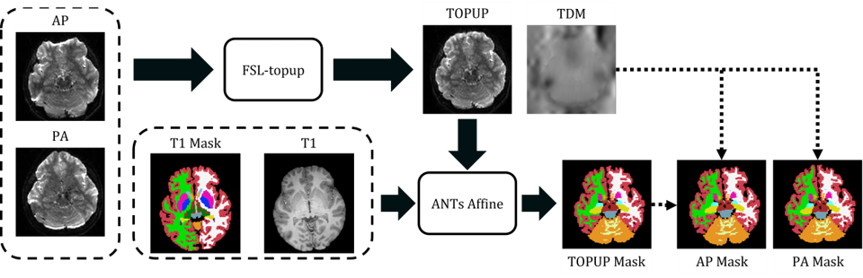
We collected 3182 brain MRI datasets from four databases: (1) HBN1, (2) LEMON2, (3) ADNI23, and (4) IXI4. Each dataset included DWI and 3D T1-weighted images (T1w). We randomly selected 2796 datasets from IXI, ADNI2, and HBN as the training set and 227 datasets as the validation set. For the testing set, we selected 159 datasets from LEMON. Figure 1 illustrates the preprocessing procedure5. For HBN and LEMON, each DWI dataset included anterior-to-posterior (AP) and posterior-to-anterior (PA) phase-encoded acquisitions, allowing for EPI distortion correction using the FSL TOPUP method6. For ADNI2 and IXI datasets, which lack reversed-gradient images, we employed the referenceless virtual-displacement mapping method (TigerEPI, https://github.com/htylab/tigerepi) to mitigate EPI distortion. Subsequently, we performed brain segmentation with FreeSurfer, producing brain masks7 that include automated segmentation (ASEG, 43 regions), white matter parcellation (WMP, 73 regions), and cortical parcellation (Desikan-Killiany-Tourville, DKT, 62 regions). The T1w and corrected DWI images were co-registered using ANTs Affine8 to generate brain masks aligned to the DWI images.We subsequently developed three 3D U-Net models; the input for each was the b0 image from the DWI, while the respective output ground truths (GT) were the corresponding masks for ASEG, DKT, and WMP. The training parameters for the models were as follows: optimizer: Adam, learning rate: 0.0005, loss function: Dice loss, epochs: 20, with no early stop condition implemented. After training, we evaluated the models' accuracy using the LEMON dataset, with the Dice coefficient serving as the metric9,10. Moreover, we computed the mean diffusivity (MD) and fractional anisotropy (FA) for brain regions using masks obtained from both the U-Net model and FreeSurfer. This was done to assess the viability of conducting direct regional DWI analysis using the 3D U-Net model.
Results
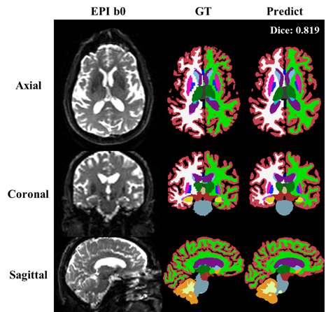
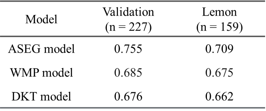
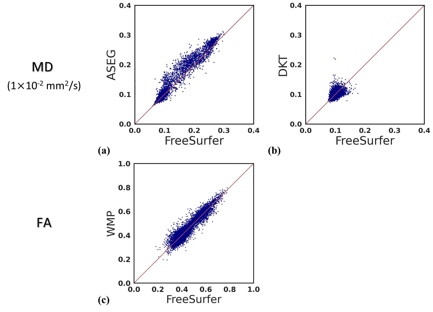
Figure 2 displays an example of brain segmentation from an ADNI2 dataset using the ASEG model (Dice = 0.819). Figure 3 shows a table listing the group averages of Dice coefficients across all validation (n = 227) and test datasets (n = 159). The accuracy metric utilized for evaluating the results of the three models is the average Dice coefficient, as presented in Table 1. Of the three models, the ASEG model produced the highest average Dice value in the test datasets (Dice = 0.709). We then used the masks derived from the three models and FreeSurfer to calculate the regional averages of MD and FA values corresponding to the subregions. Figure 4 displays scatter plots of all values, comparing U-Net with FreeSurfer: a) MDs of ASEG, b) MDs of DKT, and c) FAs of WMP.Discussions and Conclusions
Our study presents efficient U-Net models for direct echo-planar imaging (EPI) segmentation with cross-institutional validation. This method's elimination of T1 image registration mitigates inaccuracies due to EPI distortion, ensuring more reliable diffusion metric analysis. The DKT and WMP models showed relatively lower performance than ASEG, with Dice coefficients below 0.7. We anticipate that this could result from the fact that the ASEG model encompasses coarser brain parcellations, while DKT and WMP provide detailed cortical and white-matter parcellations. Further optimization for the three models merits future investigation. Our goal is to release the models in conjunction with the virtual displacement mapping in the open-source TigerEPI toolbox. In summary, our study provides a comprehensive evaluation of deep learning U-Net models for EPI segmentation across various brain subregions.Acknowledgements
We respectfully acknowledge the contributions of the participants and the investigators associated with the open-access datasets utilized in this work. Our team is grateful to the National Center for High-performance Computing for the provision of computational time and resources. The study was funded by the National Science and Technology Council, Taiwan, under grant 112-2314-B-011-002-MY2. We also extend our gratitude to OpenAI's ChatGPT-4 for its assistance in the grammatical refinement of our manuscript.
References
1. Ettehadi N, Kashyap P, Zhang XZ, et al. Automated Multiclass Artifact Detection in Diffusion MRI Volumes via 3D Residual Squeeze-and-Excitation Convolutional Neural Networks. Article. Front Hum Neurosci. Mar 2022;16:13. 877326.
2. Babayan A, Erbey M, Kumral D, et al. A mind-brain-body dataset of MRI, EEG, cognition, emotion, and peripheral physiology in young and old adults. Article; Data Paper. Sci Data. Feb 2019;6:21. 180308.
3. Mueller SG, Yushkevich PA, Das S, et al. Systematic comparison of different techniques to measure hippocampal subfield volumes in ADNI2. Article. NeuroImage-Clin. 2018;17:1006-1018.
4. Beheshti I, Maikusa N, Matsuda H. Effects of aging on brain volumes in healthy individuals across adulthood. Article. Neurol Sci. Jun 2019;40(6):1191-1198.
5. Kuo CC. Referenceless EPI Distortion Correction using Generative Adversarial Networks for Virtual Displacement Map Generation. National Taiwan University of Science and Technology; 2023.
6. Jenkinson M, Beckmann CF, Behrens TE, Woolrich MW, Smith SM. FSL. Review. Neuroimage. Aug 2012;62(2):782-790.
7. Fischl B. FreeSurfer. Review. Neuroimage. Aug 2012;62(2):774-781.
8. Avants BB, Tustison NJ, Song G, Cook PA, Klein A, Gee JC. A reproducible evaluation of ANTs similarity metric performance in brain image registration. Article. Neuroimage. Feb 2011;54(3):2033-2044.
9. AnbuDevi MKA, Suganthi K. Review of Semantic Segmentation of Medical Images Using Modified Architectures of UNET. Review. Diagnostics. Dec 2022;12(12):31. 3064.
10. Zhang L, Lan CF, Fu LR, Mao XH, Zhang M. Segmentation of brain tumor MRI image based on improved attention module Unet network. Article. Signal Image Video Process. Jul 2023;17(5):2277-2285.
Figures