4984
Pancreatic Fat Quantification in Human Diabetes and Its Relationship with β-Cell Reduction1Department of Endocrinology and Metabolism, Zhongshan Hospital, Shanghai, China, 2Department of Radiology, Zhongshan Hospital, Shanghai, China, 3Siemens Shenzhen Magnetic Resonance Ltd, Shenzhen, China
Synopsis
Keywords: Radiomics, Endocrine, Data acquisition, diabetes, metabolism, pancreas
Motivation: Patients with type 2 diabetes have more pancreatic fat, but its impact on β-cells is unclear.
Goal(s): Our goal is to explore the impact of pancreatic fat on β-cells.
Approach: We employed 6-point Dixon MRI to confirm the pancreatic fat fraction (PFF) in patients with type 2 diabetes. Histologic analysis was conducted to validate our findings and immunohistochemical staining was performed to observe changes in β-cells.
Results: We found a negative relationship between β-cell mass and PFF in patients with type 2 diabetes. This suggested that the increased pancreatic fat observed in type 2 diabetes might contribute to reducing the number of β-cells.
Impact: This study provided evidence that increased pancreatic fat in type 2 diabetes was associated with a decreased number of β-cells. These findings suggested targeting pancreatic fat accumulation could be a potential therapeutic approach for improving glycemic control in diabetic patients.
Introduction
Previous studies demonstrated an increase in pancreatic fat fraction (PFF) in patients with type 2 diabetes (T2DM)1-6, indicating that increased pancreatic fat might impair β-cell function in patients with and without type 2 diabetes1, 4-8. However, the assessment of β-cell function in these studies was limited to clinical indicators such as HOMA-β (Homeostatic Model Assessment of Beta Cell Function), lacking direct observations of β-cells. The direct measurement of PFF is challenging due to the deep retroperitoneal location of the pancreas. Therefore, imaging techniques have remarkable advantages in pancreatic fat quantification. Among these techniques, multi-echo Dixon MRI is currently the most developed technique9.Therefore, this study applied the advanced 6-point Dixon technique to explore the differences in PFF under various glucose metabolism conditions so as to elucidate the impact of pancreatic fat on β-cell function and its role in the pathogenesis and progression of diabetes. In addition, we also quantified the fat in donor pancreas using histologic methods and further explored the mechanisms by which pancreatic fat influenced β-cells.Methods
A total of 76 Chinese, aged 22-78 years, with (n = 38) and without (n = 38) type 2 diabetes (clinical characteristic are shown in Table 2), underwent abdominal MRI conducted on 1.5T MR (MAGNETOM Aera, Siemens Healthineers, Erlangen, Germany) and 3T MR systems (MAGNETOM Prisma, Siemens Healthineers, Erlangen, Germany) (57 and 19 participants, respectively). A 6-echo Dixon Volumetric Interpolated Breath-hold Examination (VIBE) sequence with fat quantification using a complex signal model10 for multiple lipid resonance with correction for relaxation effects was performed for PFF measurement on both systems. Pixel-wise fat fraction (FF) maps were inline generated after data acquisition. The scan parameters on both scanners are depicted in Table 1.Pancreatic tissue samples from organ donors (28 nondiabetic and 13 diabetic cases, clinical characteristic are shown in Table 3) were embedded in paraffin and cut into 5-μm-thick sections. Pancreatic FF (PFF) tissue samples were measured using histologic methods, and the changes in β-cells were observed through insulin immunohistochemical staining.Results
In our in vivo study, we observed a substantial increase in PFF measured using 6-point Dixon MRI in patients with diabetes compared with nondiabetic individuals (P <.001) (Fig. 1). Similarly, among donors with diabetes, PFF measured using histologic methods was considerably higher in patients with diabetes than in individuals without diabetes (P <.001). Insulin immunohistochemical staining revealed a correlation between an increase in PFF and a reduction in fractional β-cell area (r = –0.250, P =.004) and β-cell mass (r = –0.757, P =.003) in donors with diabetes. The detailed results are illustrated in Figure 2.Discussion
Our study demonstrated an elevated PFF measured by 6-point Dixon MRI in patients with type 2 diabetes. Histologic validation confirmed the presence of increased fat in the pancreas, accompanied by a reduction in β-cell mass. These findings emphasized the potential importance of pancreatic fat in the onset and progression of type 2 diabetes. Further research is needed to gain a deeper understanding of the underlying mechanisms and explore potential therapeutic interventions targeting pancreatic fat metabolism.Conclusions
Both MRI and histologic methods demonstrated a substantially higher PFF in patients with diabetes than in individuals without diabetes. PFF also displayed a remarkable negative correlation with the fractional β-cell area and β-cell mass.Acknowledgements
No acknowledgement found.References
- Tushuizen, M.E., et al., Pancreatic fat content and beta-cell function in men with and without type 2 diabetes. Diabetes care, 2007. 30(11): p. 2916-2921.
- Ou, H.-Y., et al., The association between nonalcoholic fatty pancreas disease and diabetes. PloS one, 2013. 8(5): p. e62561.
- Macauley, M., et al., Altered volume, morphology and composition of the pancreas in type 2 diabetes. PloS one, 2015. 10(5): p. e0126825.
- Lu, T., et al., Pancreatic fat content is associated with β-cell function and insulin resistance in Chinese type 2 diabetes subjects. Endocrine journal, 2019. 66(3): p. 265-270.
- Chin, S.O., et al., Pancreatic fat accumulation is associated with decreased β-cell function and deterioration in glucose tolerance in Korean adults. Diabetes/metabolism research and reviews, 2021. 37(7): p. e3425.
- Cao, M.-J., et al., Quantification of ectopic fat storage in the liver and pancreas using six-point Dixon MRI and its association with insulin sensitivity and β-cell function in patients with central obesity. European radiology, 2023.
- Heni, M., et al., Pancreatic fat is negatively associated with insulin secretion in individuals with impaired fasting glucose and/or impaired glucose tolerance: a nuclear magnetic resonance study. Diabetes/metabolism research and reviews, 2010. 26(3): p. 200-205.
- Wen, Y., et al., Pancreatic fat infiltration, β-cell function and insulin resistance: A study of the young patients with obesity. Diabetes research and clinical practice, 2022. 187: p. 109860.
- Wagner, R., et al., Metabolic implications of pancreatic fat accumulation. Nature reviews. Endocrinology, 2022. 18(1): p. 43-54.
- Zhong, X., et al., Liver fat quantification using a multi-step adaptive fitting approach with multi-echo GRE imaging. Magnetic resonance in medicine, 2014. 72(5): p. 1353-1365.
Figures
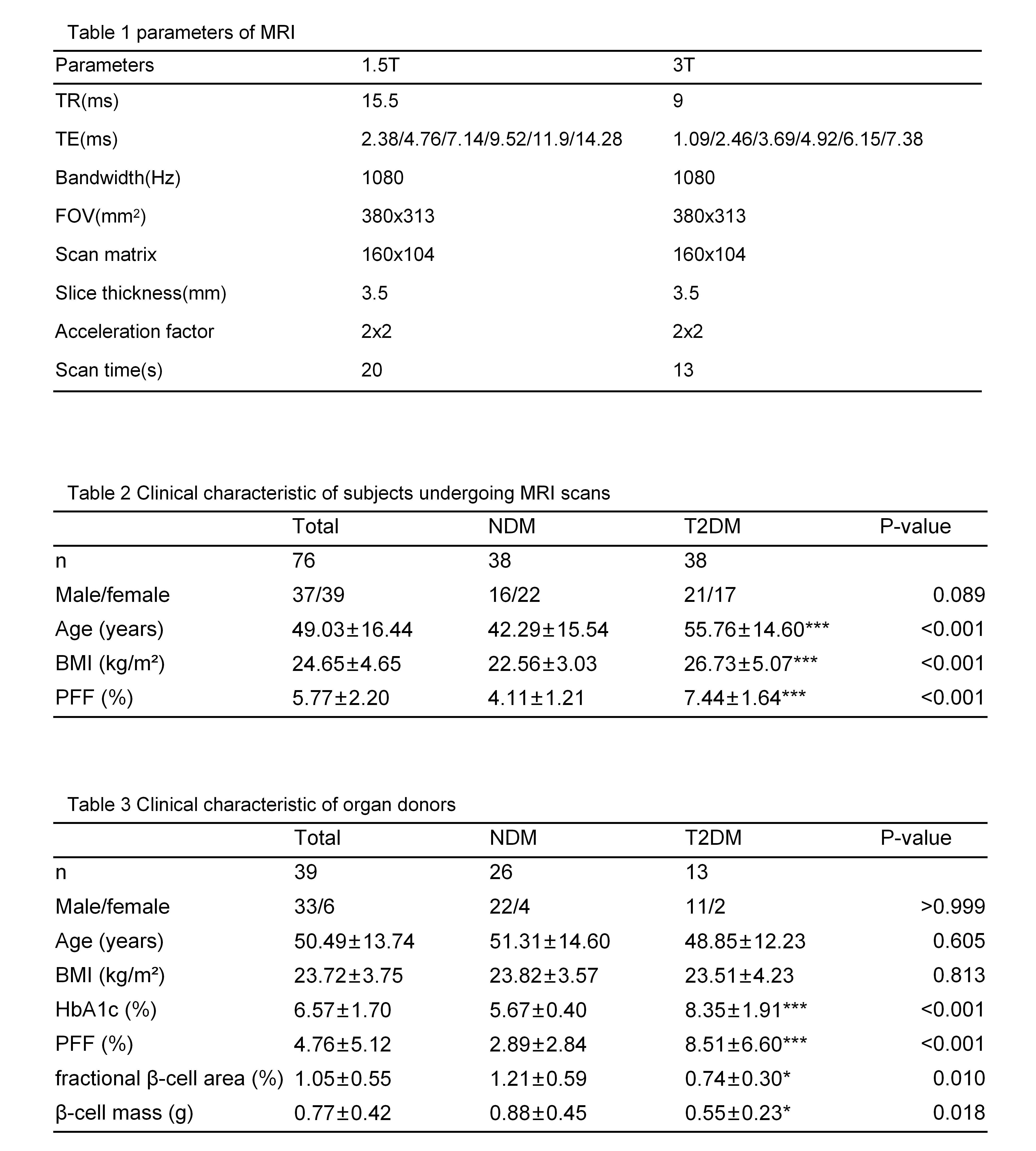
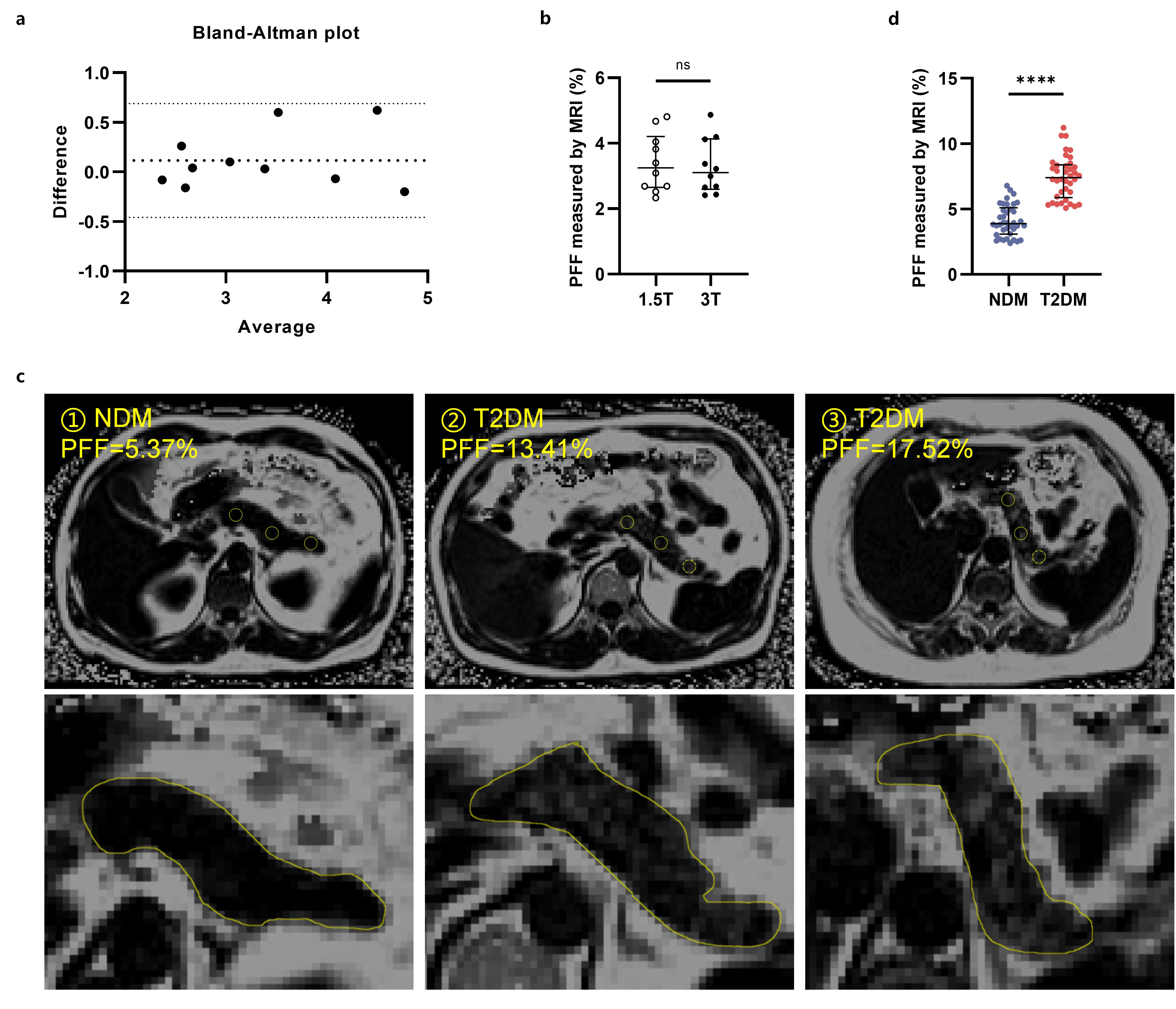
Figure 1. Increased PFF measured using 6-point Dixon MRI in patients with diabetes compared with those without diabetes.
(a and b) No difference in the measurement of PFF for the same individual between the 2 MRI systems. (a) Bland–Altman plot of PFF measurements taken from 10 nondiabetic individuals. (b) Paired-sample t test results. (c) Representative images of Dixon MRI. ①NDM, 42 years old, BMI 25.4; ②T2DM, 41 years old, BMI 28.0; ③T2DM, 67 years old, BMI 26.4. (d) PFF in patients with type 2 diabetes was considerably higher than that in the controls (4.11% ±1.21% vs 7.44% ±1.64%).
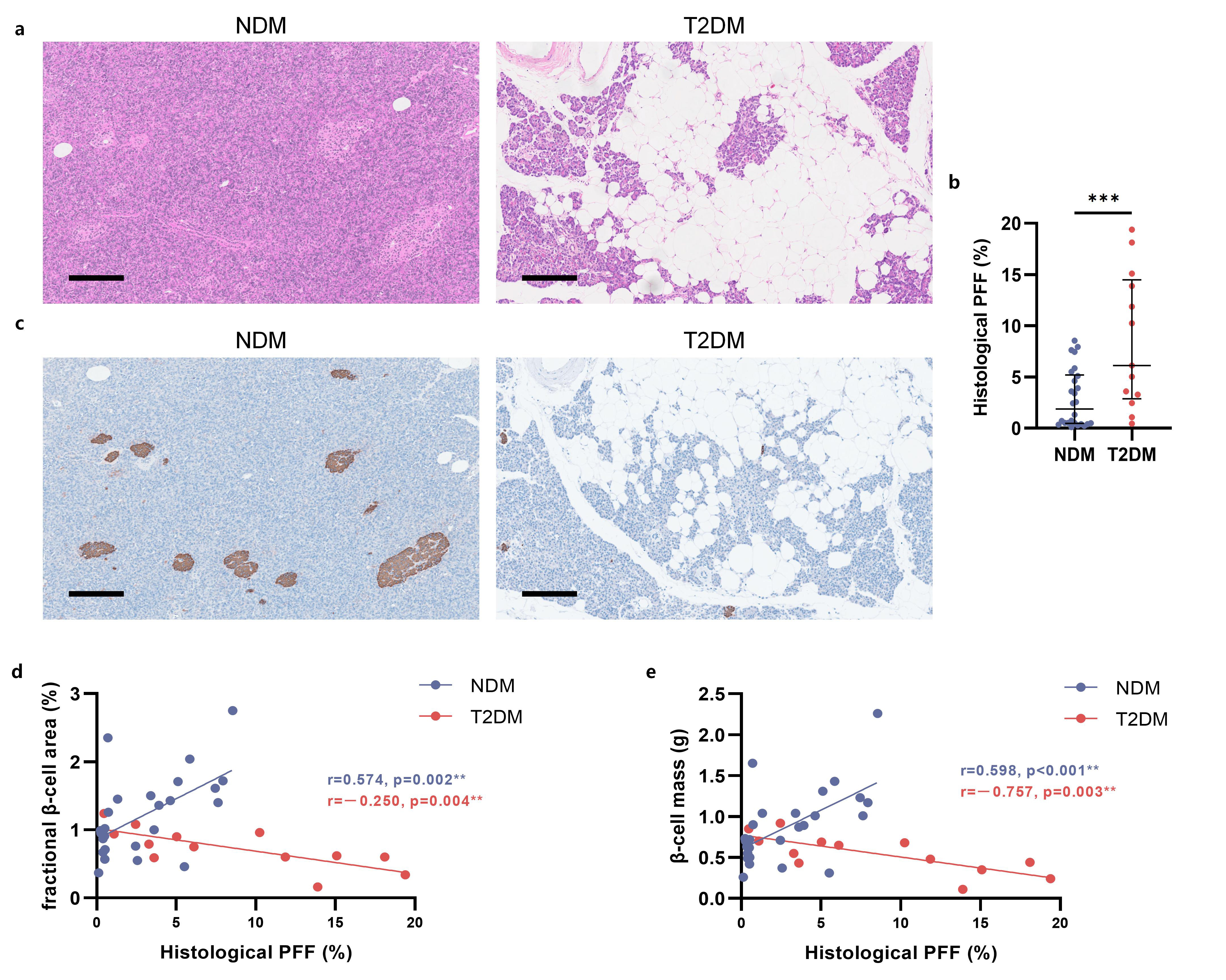
Figure 2. Increased PFF in donors with diabetes and its relationship with β-cell reduction.
(a) Representative images of pancreatic tissue stained with H&E from organ donors. NDM: male, 59 years old, PFF 3.26%; T2DM: male, 55 years old, PFF 15.08% (bar 200 μm). (b) Diabetic donors had an increased PFF compared with nondiabetic donors (2.89% ± 2.84% vs 8.81% ± 6.60%). (c) Representative images of insulin immunohistochemical staining on pancreatic tissue from organ donors. (d and e) Fractional β-cell area (d) and β-cell mass (e) were negatively correlated with PFF in donors with diabetes.