4983
Characterisation of brain T2* Values across the neonatal period at 7 Telsa1Guy's and St Thomas' NHS Foundation Trust, London, United Kingdom, 2London Collaborative Ultra high field System (LoCUS), London, United Kingdom, 3Centre for the Developing Brain, King's College London, London, United Kingdom, 4Centre for Neurodevelopmental Disorders, King's College London, London, United Kingdom
Synopsis
Keywords: Neonatal, Brain, High-field MRI, Neuro, Relaxometry
Motivation: Brain T2* values can provide information about tissue specific maturation, and can also provide knowledge important for optimising echo times for fMRI acquisition.
Goal(s): To characterise tissue specific T2* values in the neonatal brain at 7T.
Approach: Whole brain T2* maps from 14 neonates were generated using a 3D multi-echo acquisition.
Results: Median T2* values were: cortical grey matter: 58 ms, deep grey matter: 70 ms and white matter: 86 ms. Values differ markedly from those described in adults and measured at standard field strengths.
Impact: We describe tissue-specific T2* values in the neonatal brain at 7T, which may provide new information about brain development in health and disease, and provide a basis for optimising fMRI sequences for neonates at 7T.
Introduction
Brain T2* values are known to be significantly longer in early infancy due to marked differences in tissue composition1–3. They are also known to change significantly with static field strength, with values markedly reduced at ultra-high field4. T2* can provide information about tissue characteristics, including oxygenation, iron concentration and water content5–8 and can also help optimise functional MRI Blood Oxygen Level Dependent (BOLD) contrast by using an echo time matched to T2* values in the tissue of interest9. The increased signal to noise ratio (SNR) and higher resolution that can be achieved at 7T compared to lower field strengths is likely to be of particular value in neonates which have small but rapidly developing brain structures, during a key time for the establishment of their lifelong patterns of brain connectivity10. Although there are values in the literature for T2* in the adult brain at 7T4, they have not been characterised in neonates. In this study the aim was to establish the range of T2* in brain tissue at 7T across the neonatal period.Methods
We scanned 14 neonates of post menstrual age (PMA) 35.4-44.7 weeks with a variety of underlying clinical diagnoses (see Table 1). Images were acquired on a 7T scanner (MAGNETOM Terra, Siemens Healthcare, Erlangen, Germany) with a 1TX-32RX head coil (Nova Medical, Wilmington, MA, USA) in the LoCUS MRI Unit, St Thomas’ Hospital London. Ethical approval (NHS REC 19/LO/1384) was obtained following detailed safety evaluation11 and implementation of a local policy to mitigate risks of RF heating12.The sequence used for T2* mapping was a 3D 10 echo spoiled GRE sequence (TEs: 5 – 50 ms, spacing 5ms) with parameters 1 mm isotropic resolution, TR: 53 ms, flip angle=15°. An axial T2-weighted TSE sequence, 0.6 mm x 0.6 mm, 1.2 mm slice thickness, TR: 8640 ms, TE: 156 ms, echo train length 16, refocusing FA=120° was also acquired for tissue segmentation.
For T2* estimation non-linear unconstrained voxel-wise fitting was performed using MATLAB (2022b, The Mathworks, Natick MA) function fmincon, using equation
$$S(TE) = \mid S_{0}\exp\left(-\frac{TE}{T_2^*}\right)\mid$$
Automatic Segmentation of cortical grey matter, deep grey matter and white matter was performed on the axial TSE, using BOUNTI: Brain vOlumetry and aUtomated parcellatioN for 3D feTal MRI13. Binarised tissue segmentation masks were propagated from aligned T2 images to the T2* maps, which were resampled to 0.5 mm isotropic resolution. Regions of the T2* map with signal dropout were excluded using thresholding. The cortical grey matter mask was eroded to include only the middle cortex layer and thus limit the confounding effects of CSF and white matter partial voluming.
Median T2* was calculated for each tissue type in each infant. Median T2* across the cohort was then calculated and plotted against PMA. Linear regression was performed and Pearson’s rank correlation coefficients calculated.
Results
Example T2* maps are shown in Figure 1; Figure 2 shows examples of segmentations. Table 1 contains the T2* results for each subject. Across the population, median T2* values were: cortical grey matter: 58 ms, deep grey matter: 70 ms and white matter: 86 ms. Figure 3 shows scatter plots of the results and Table 2 shows the statistical analysis for correlation and regression. For all tissue types there was no clear relationship with age.Discussion
Neonatal T2* values are markedly longer than those reported in adults (average 7T T2* values across 6 adults were reported as 33.2 ms in grey matter and 26.8 ms in white matter4). This is consistent with findings at 1.5T 1–3. The difference to adult values was greatest in white matter. Generally white matter had larger inter-quartile ranges within each baby compared to grey matter, suggesting greater regional variability and in keeping with rapid maturation in the neonatal period as white matter fibre density increases, pre-myelination begins, and water content decreases. White matter had longer T2* compared to grey matter regions, consistent with results at 1.5T1.There was no significant trend of T2* with age. A previous study at 1.5T which showed a decrease in T2* from 33 weeks to 44 weeks PMA1. The reason for no strong correlation with PMA in this study may be due to the variety of underlying clinical diagnoses in our cohort.
Conclusion
We describe tissue specific T2* values in the neonatal brain and demonstrate that they are markedly longer than those described in adults. This suggests TEs in fMRI sequences at 7T for neonates should be adjusted to account for the longer T2* values.Acknowledgements
Wellcome Trust Collaboration in Science Grant [WT201526/Z/16/Z]References
- Rivkin MJ, Wolraich D, Als H, et al. Prolonged T2* values in newborn versus adult brain: Implications for fMRI studies of newborns. Magn Reson Med. 2004;51(6):1287-1291.2.
- Lee W, Donner EJ, Nossin-Manor R, Whyte HEA, Sled JG, Taylor MJ. Visual functional magnetic resonance imaging of preterm infants. Dev Med Child Neurol. 2012;54(8):724-729.
- Goksan S, Hartley C, Hurley SA, et al. Optimal echo time for functional MRI of the infant brain identified in response to noxious stimulation. Magn Reson Med. 2017;78(2):625-631.
- Peters AM, Brookes MJ, Hoogenraad FG, et al. T2* measurements in human brain at 1.5, 3 and 7 T. Magn Reson Imaging. 2007;25(6):748-753.
- Tamura H, Hatazawa J, Toyoshima H, Shimosegawa E, Okudera T. Detection of deoxygenation-related signal change in acute ischemic stroke patients by T2*-weighted magnetic resonance imaging. Stroke. 2002;33(4):967-971.
- Langkammer C, Krebs N, Goessler W, et al. Quantitative MR imaging of brain iron: A postmortem validation study. Radiology. 2010;257(2):455-462.
- Du YP, Chu R, Hwang D, et al. Fast multislice mapping of the myelin water fraction using multicompartment analysis of T2* decay at 3T: A preliminary postmortem study. Magn Reson Med. 2007;58(5):865-870.
- Kan H, Uchida Y, Ueki Y, et al. R2* relaxometry analysis for mapping of white matter alteration in Parkinson’s disease with mild cognitive impairment. Neuroimage Clin. 2022;33:102938.
- Bandettini PA, Wong EC, Jesmanowicz A, Hinks RS, Hyde JS. Spin‐echo and gradient‐echo epi of human brain activation using bold contrast: A comparative study at 1.5 T. NMR Biomed. 1994;7(1-2):12-20.
- Doria V, Beckmann CF, Arichi T, et al. Emergence of resting state networks in the preterm human brain. Proc Natl Acad Sci U S A. 2010;107(46):20015-20020.
- Malik SJ, Hand JW, Satnarine R, Price AN, Hajnal J V. Specific absorption rate and temperature in neonate models resulting from exposure to a 7T head coil. Magn Reson Med. 2021;86(3):1299-1313.
- Bridgen P, Tomi-Tricott R, Uus A, et al. Exploration of the potential of high resolution and contrast 7 Tesla MR brain imaging in neonates. medRxiv. Published online October 2, 2023:2023.09.28.23296232.
- Uus AU, Kyriakopoulou V, Makropoulos A, et al. BOUNTI: Brain vOlumetry and aUtomated parcellatioN for 3D feTal MRI. bioRxiv. Published online January 1, 2023:2023.04.18.537347.
Figures
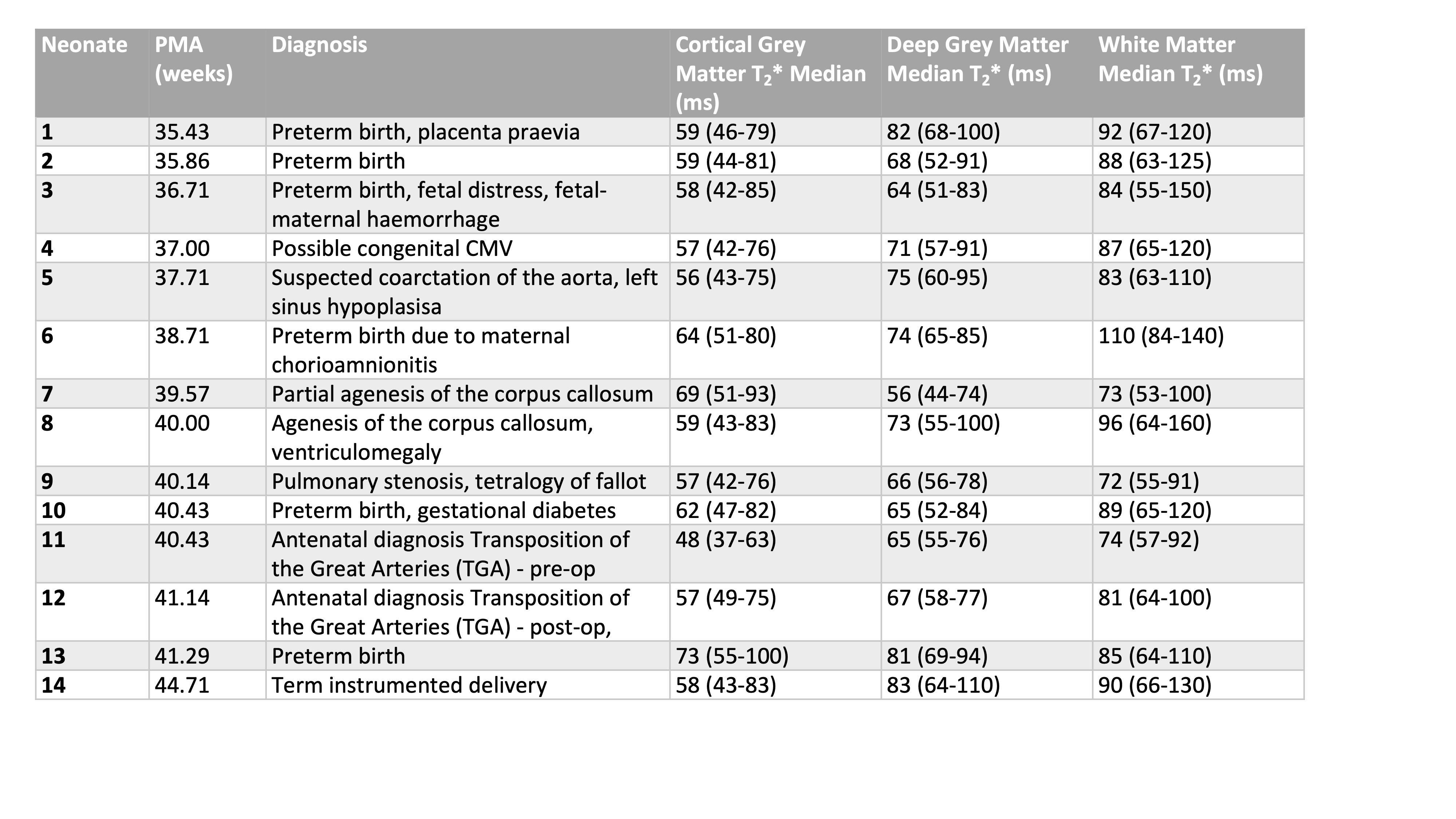
Table 1: Post menstrual age (PMA) of each neonate, and diagnosis, and measured T2* for cortical grey matter, deep grey matter and white matter, in order of PMA. T2* are medians of the segmentation (lower quartile to upper quartile)
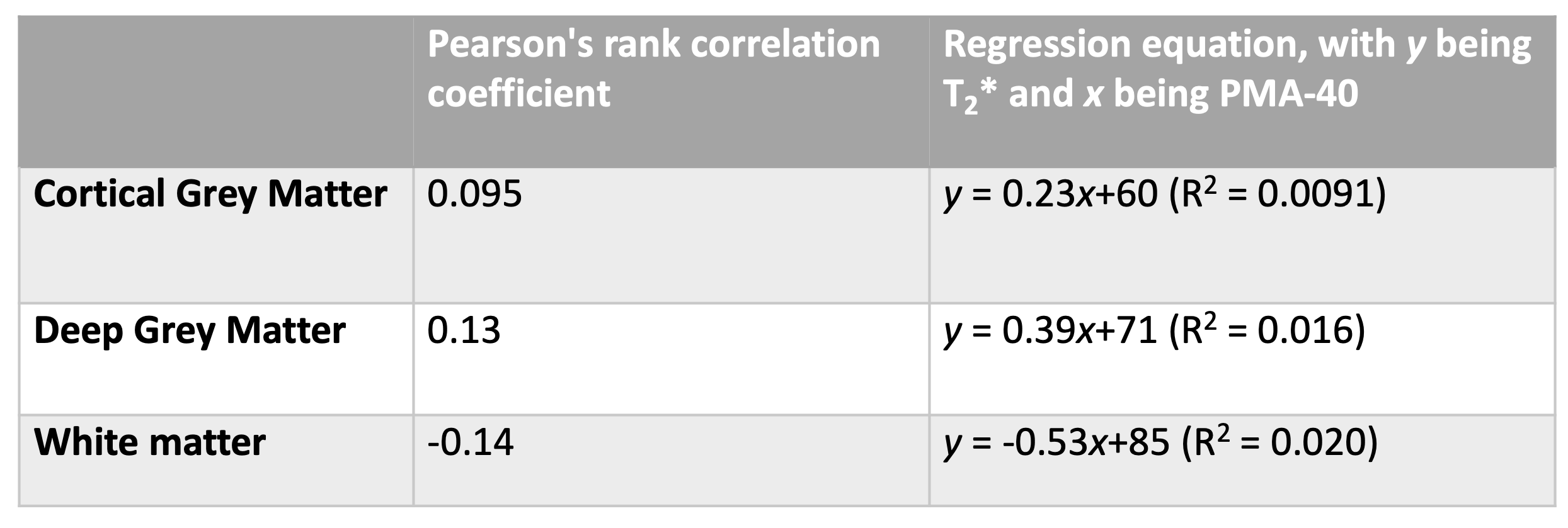
Table 2: Correlation coefficients and regression equations for tissue T2* compared with post menstrual age (PMA)In the regression equations y refers to T2* and x refers to PMA-40. Therefore, if y = Ax + B then B is the T2* at PMA=40 weeks.
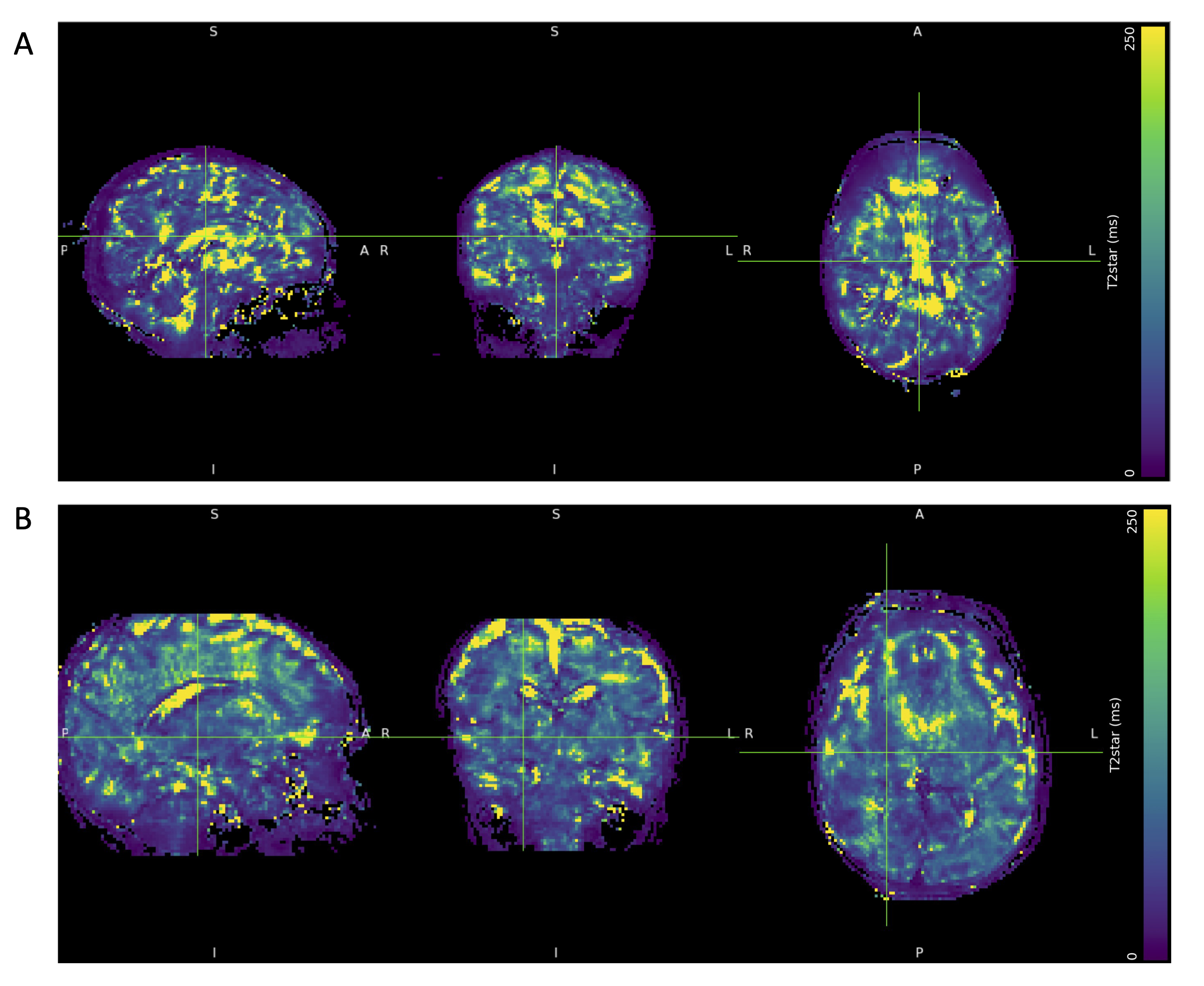
Figure 1: T2* maps from (A) Neonate 3 and (B) Neonate 14
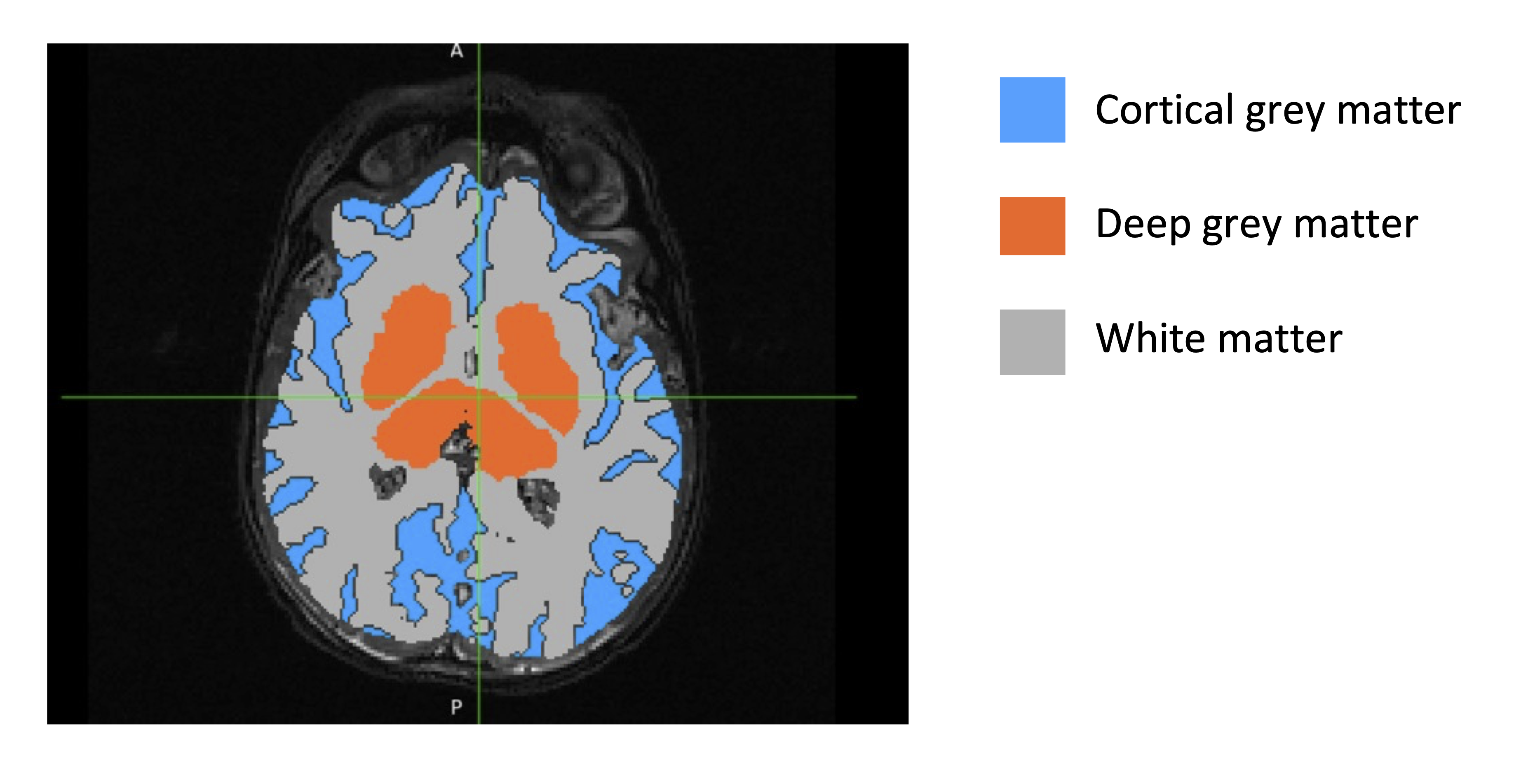
Figure 2: Example of final segmentation for Neonate 11 overlaid on Axial TSE image. Note: areas with signal drop out on the T2* map have been excluded in this mask.
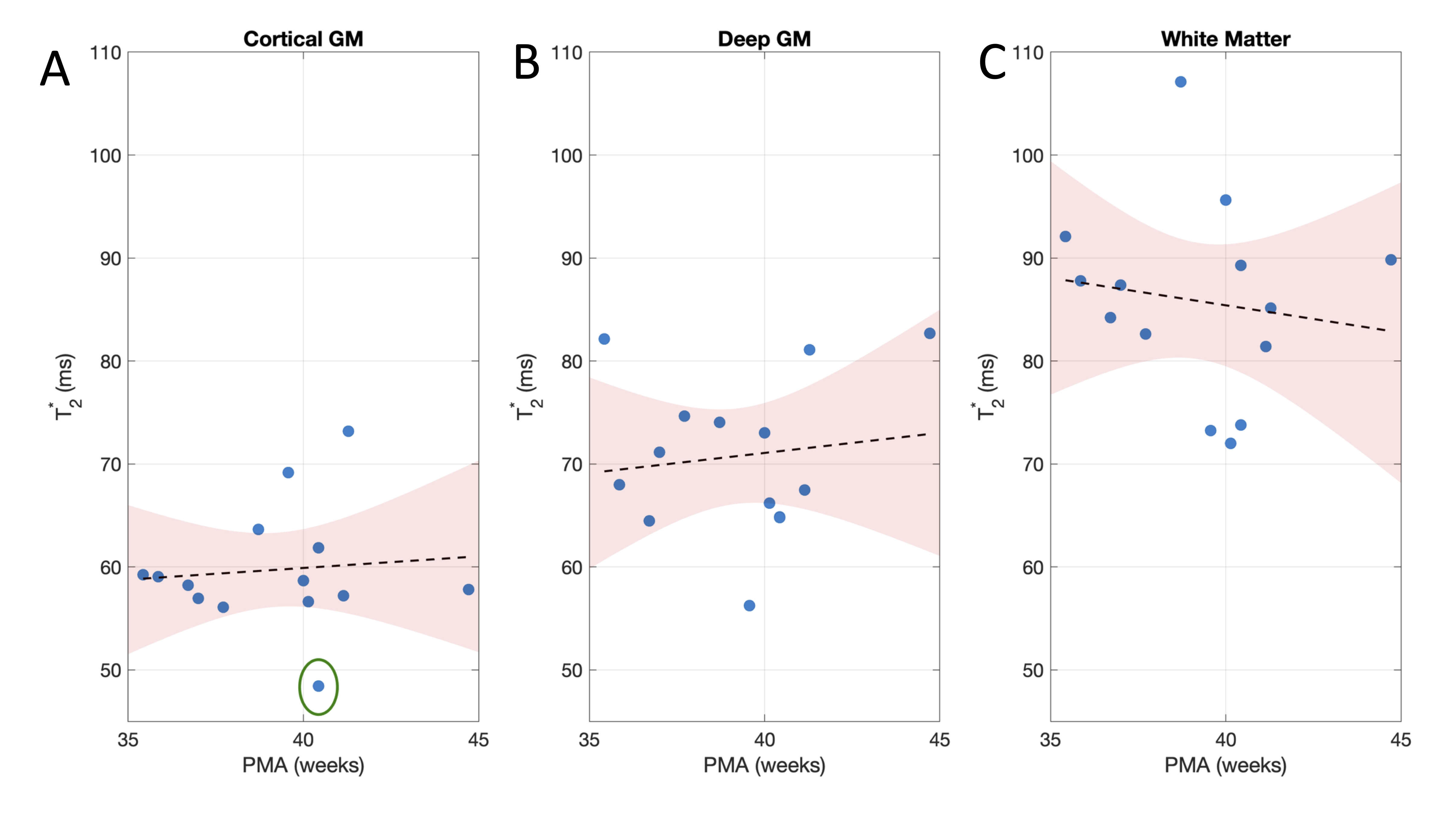
Figure 3: Measured T2* plotted against (A) cortical grey matter, (B) deep grey matter and (C) white matter, with linear regression lines and 95% confidence intervals. The lowest T2* was found in the cortical grey matter of neonate 11, circled in the figure. This infant had congenital cyanotic heart disease (TGA)) and the lower observed T2* was likely influenced by reduced tissue oxygenation related to this condition.