4982
Amide Proton Transfer Magnetic Resonance Imaging of Type 2 Diabetes Mellitus: A Preliminary Study
1Radiology department, The first affiliated hospital of Dalian medical university, Dalian City, China, 2Clinical & Technical Support, Philips Healthcare (Beijing), China, Beijing, China
Synopsis
Keywords: Other Neurodegeneration, Diabetes, APT T2DM
Motivation: APT imaging has been used to study neurodegenerative diseases,T2DM is also known to be associated with cognitive decline and brain structural changes,so, it is interesting to explore the cerebral metabolic alternation of T2DM.
Goal(s): This study investigated the feasibility of APT to monitor the condition of T2DM patients.
Approach: Twenty T2DM patients and Twenty age- and sex-matched healthy controls (HCs) underwent conventional MR and APT scanning,The image analysis of APT was performed by 3D Slicer software.
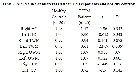
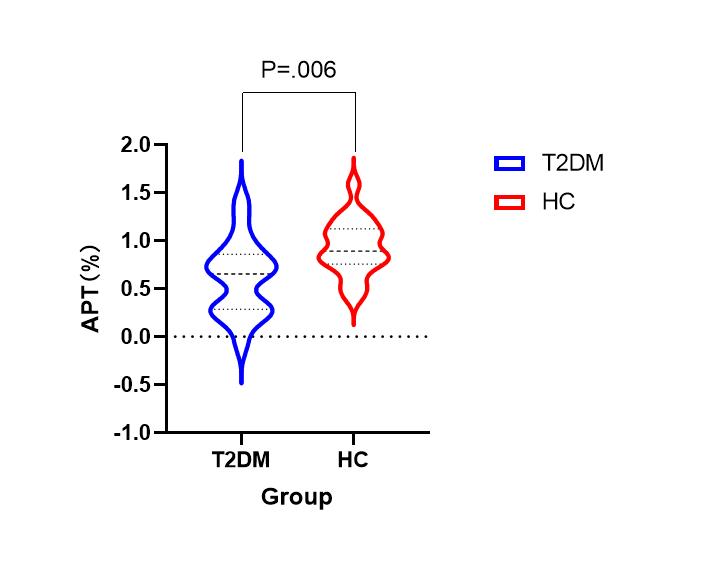
Results: The results showed that the APT values in the left temporal white matter of T2DM patients were lower and significantly different from healthy controls.
Impact: By APT technique, temporal lobe white matter APT signal values were found to be lower in T2DM patients than in healthy controls, which can provide more information on cerebral metabolic changes in T2DM patients.
Purpose
Amide proton transfer magnetic resonance imaging (APT) is a novel method that detects low-concentration endogenous mobile proteins and peptides in tissue non-invasively. The purpose of this work is to initially explore the clinical application of APT on cerebral metabolic changes in Type 2 Diabetes Mellitus.Introduction
Type 2 diabetes mellitus (T2DM) is a common, chronic, and progressive metabolic disorder characterized by abnormally high blood glucose levels for a prolonged period, termed hyperglycemia, due to insulin resistance and decreased production of insulin. Typical T2DM complications include retinopathy, kidney failure, and peripheral neuropathy. T2DM is also known to be associated with cognitive decline and brain structural changes[1,2]. Recently, APT imaging has been used to study neurological diseases, mainly for neurodegenerative diseases[3]. Therefore, it is interesting to explore the cerebral metabolic alternation of T2DM. So far, there is no study published about APT in T2DM patients, and whether APT imaging can provide unique information for T2DM is still unknown. This study aimed to investigate the feasibility of using APT imaging to detect cerebral abnormality in patients with T2DM.Methods
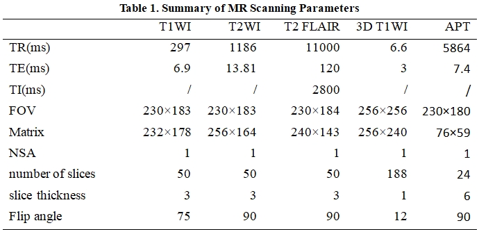
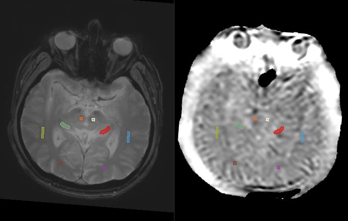
Twenty T2DM patients and Twenty age- and sex-matched healthy controls (HCs) underwent conventional MR and APT scanning using a 3.0 T MR scanner (Ingenia CX, Philips Healthcare, the Netherlands) with a 16-channel head phase-array breast coil. Clinical data and laboratory indicators of the subjects were recorded within one week before or after the MRI examination. The image analysis of APT was performed by 3D Slicer software (https://www.slicer.org/). The T2-weighted image was used as the anatomical reference to draw regions of interest (ROIs) of the bilateral hippocampus (HC), temporal white matter (TWM), occipital white matter (OWM), and cerebral peduncles (CPs). APT values of above ROIs were measured on APT images, as shown in Figure 1. The ROIs were drawn to include as much of the measured tissues as possible while avoiding the adjacent cerebrospinal fluid (CSF) containing spaces to decrease partial volume effects from the fluid. All data were analyzed using the statistical package SPSS (Version 27). Two independent sample t-tests were used to compare the APT values of ROIs between the T2DM and healthy groups. P <0.05 was considered statistically significant.Results
APT values of left TWM were significantly lower in T2DM patients than in healthy controls. However, no significant differences in APT values of other brain structures were observed between T2DM patients and healthy controls. Detailed results were summarised in Table 2.Discussion and Conclusion
In this study, the APT value of left temporal white matter was lower in T2DM patients than in healthy controls. In contrast, there were no significant statistical differences in APT values of other cerebral structures between T2DM patients and healthy controls. There are two possible reasons for the findings: (1) The temporal white matter was affected the earliest and most severely in T2DM patients; (2) Lower APT values of the temporal white matter in T2DM patients may be associated with decreased cytosolic proteins and peptides. The metabolimic alternation may be related to reduced cognitive function in T2DM patients. This study indicated that patients with T2DM do have cerebral metabolic changes, which might lead to structural damage to the temporal white matter. Further studies are attractive to explore the clinical value of APT in monitoring the condition of patients with T2DM.Acknowledgements
No acknowledgement found.References
[1]den Heijer T, Vermeer SE, van Dijk EJ, Prins ND, Koudstaal PJ, Hofman A, Breteler MM. Type 2 diabetes and atrophy of medial temporal lobe structures on brain MRI. Diabetologia. 2003;46:1604–1610. [2] Cukierman T, Gerstein HC, Williamson JD. Cognitive decline and dementia in diabetes: systematic overview of prospective observational studies. Diabetologia. 2005;48:2460–2469. [3] Dou W, Lin CE, Ding H, Shen Y, Dou C, Qian L, Wen B, Wu B. Chemical exchange saturation transfer magnetic resonance imaging and its main and potential applications in pre-clinical and clinical studies. Quant Imaging Med Surg. 2019 Oct;9(10):1747-1766.Figures