4980
Detection of intracranial hemorrhage in healthy neonatal mice by T2*-weighted 7T MRI1Graduate School of Pharmaceutical Sciences, The University of Tokyo, Tokyo, Japan, 2Institute for Quantum Life Science, National Institutes for Quantum Science and Technology, Chiba, Japan, 3Sydney Imaging Core Research Facility, The University of Sydney, Sydney, Australia
Synopsis
Keywords: Blood Vessels, Normal development, high-field MRI
Motivation: Neonatal brain hemorrhage, which are observed in premature and even in full-term infants, could cause adverse neurological damage, however, the mechanism of hemorrhages is unknown and the validated information for neonatal hemorrhages, especially in asymptomatic neonates, is lacking.
Goal(s): Our goal was to detect hemorrhages in the brains of untreated postnatal mice and evaluate the spatiotemporal information.
Approach: T2*-weighted MR images were obtained from records of C57BL/6J mice at P0-1 and acquired hemorrhages were cross-validated by histological analysis.
Results: We found that hemorrhages occur throughout the brain of healthy neonatal mice.
Impact: In vivo MRI of untreated postnatal mice may serve as a useful tool to further investigate asymptomatic hemorrhages in full-term infants and will deepen our understanding of not only the mechanism of neonatal hemorrhages but also the brain development.
Introduction
The neonatal brain hemorrhage or the intraventricular hemorrhage (IVH) due to blood vessel rupture in newborns is the most common complication that could result in adverse neurological outcomes and death1. Resulting long-term neurological deficits could be the expansion of the cerebroventricular system and subsequent mechanical compression of brain tissue2. Thus, IVH continues to be a major problem of premature infant in modern neonatal intensive care units worldwide3. However, the mechanisms and early detection of brain hemorrhage are poorly understood. Histological and ultrasonic studies were made to explore intracranial hemorrhage in vivo 4,5, but the histological studies are impractical in living bodies while the ultrasonic imaging could not be applicable in newborns and may deliver misleading information. Magnetic resonance imaging (MRI) provides excellent detail of the immature brain such as for the visualization of germinal matrix and the cerebral maturation 6–8. Recent studies shows that the role of tissue iron deposition in pathogenesis of the brain injury could be related to the cause of intracerebral hemorrhage (ICH) 9,10. MRI has the potential to quantify iron overload in the tissue using T2* relaxation 11,12. T2*-based application in MRI are well-established application such as for susceptibility-weighted imaging, functional MR imaging and iron overload imaging13. In this study, owing to the advantages of high field MRI, we performed postnatal imaging of the micro bleeding on newly born mice. The T2*-weighted MR images obtained were compared and analyzed with the histological images of the mice brain for the cross validation of micro bleeding spots in mice brain.Methods
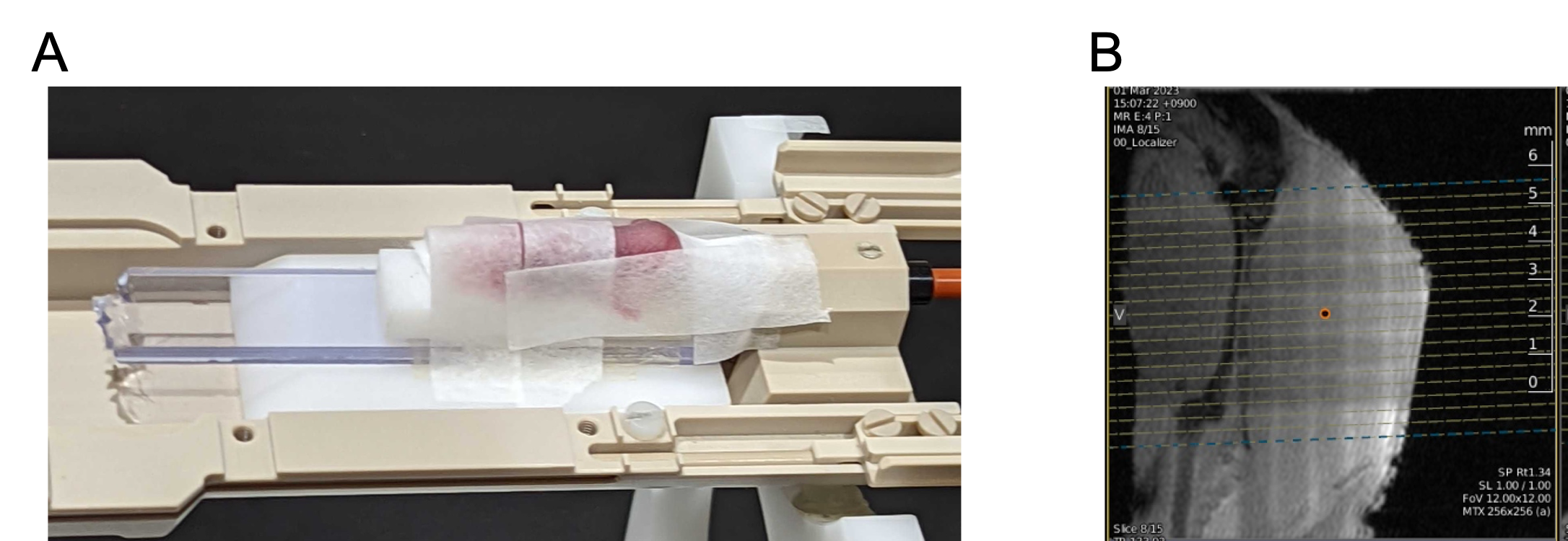
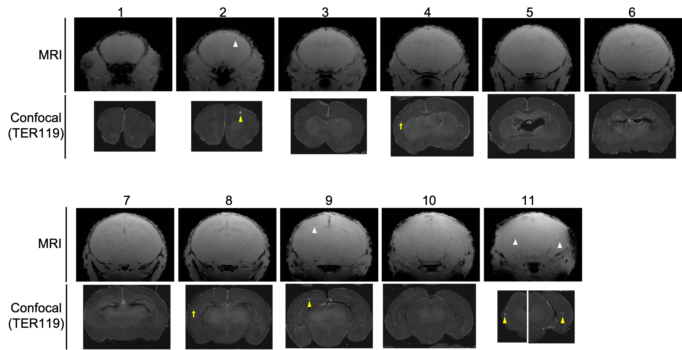
Newly born C57BL/6J mice (1-2 days after birth) were subjected for the experiments. Pregnant mice were habituated under climate-controlled breeding house, 2-3 weeks prior giving birth to new mice. The Institutional Animal Care and Use Committee of the Institute approved all animal experiments.During the MRI measurement, mice were lightly anesthetized by isoflurane (1.0 %) to avoid head movement during scanning (Fig.1A). T2-weighted and T2*-weighted measurements were performed using a horizontal 7T Bruker BioSpec 70/40 MRI system with 86 mm volume transmit and a 4-channel phased array receiving cryoprobe (Bruker Biospin, Ettlingen, Germany). The MRI system uses ParaVision 360 software for MR acquisition and image processing. Figure 1A shows the experimental setup of mice for MR measurement and figure 1B shows the sagittal localizer MR image for the respective setup. The T2-weighted images are acquired for visualization of the mouse brain using 2D-FLASH sequence with TR/TE = 3000/36ms, slice thickness = 0.750mm, slice gap = 0mm, average = 2, Rare Factor 8, resolution = 0.075*0.075 mm2 and total scan time was 1 min 30 sec. Figure 2 shows the representative T2*-weighted images aligned with the confocal (TER 119 images). T2*-weighted images were acquired at TR/TE = 600/8 ms, slice thickness = 0.35 mm, slice gap = 0 mm, flip angle = 60-degree, average = 4, Field of View 12.8*12.8mm, resolution = 0.025*0.025 mm2 and total scan time was 13 min 40 sec. Quantification of hemorrhage spots was conducted using brain sections immunostained with TER119 (MAB1125; R&D systems) and Isolectin GS-IB4 From Griffonia simplicifolia, biotin-XX Conjugate (I21414; Thermo Fisher Scientific). The spot with more than two erythrocytes outside of vessels was defined as a hemorrhage.Results/Discussion
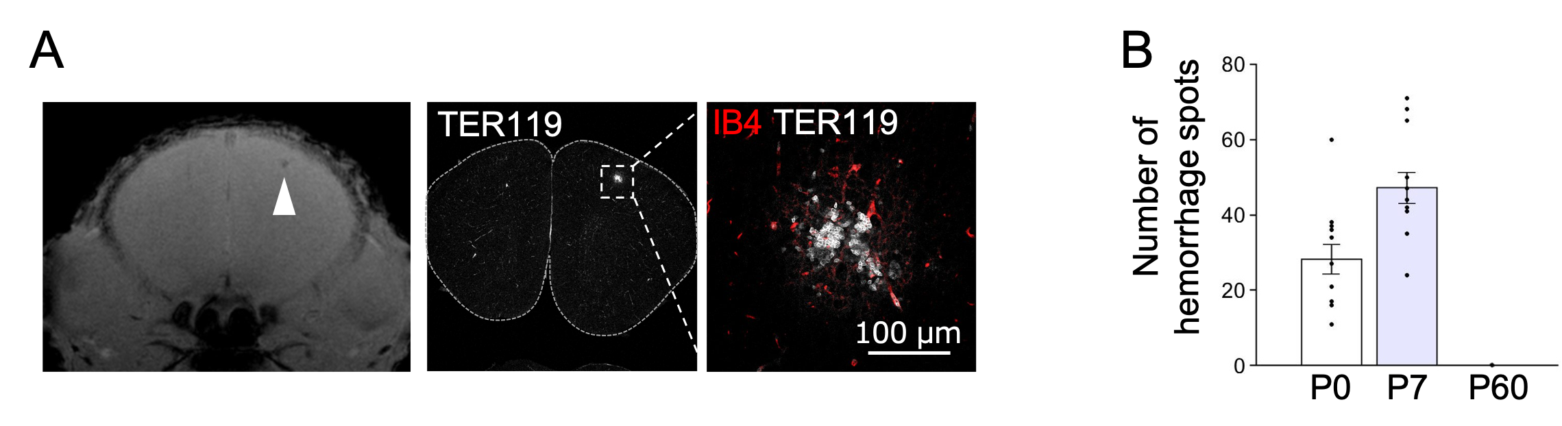
C57BL/6J untreated mice at P1 were anesthetized with isoflurane and applied T2*-weighted MR imaging, the method that can detect microbleeds with high sensitivity (Fig. 1A). The sagittal localizer image for the T2*-weighted MR imaging is shown in Fig.1B illustrates the suitable alignment of the mouse brain for the acquisition of T2-weighted and T2*-weighted measurements. We obtained coronal T2*-weighted MR images (Fig. 2) and 12 MR images that have been corroborated by histological analysis are presented in Fig. 2. We found some putative hemorrhages throughout the brain, including cortex (Fig. 2, upper). Immunostaining against TER119 (erythrocyte marker) and Isolectin B4 (blood vessel marker) following observation with MRI corroborated that hemorrhages occur with healthy development (Fig. 2, lower and Fig. 3A). We confirmed that some line-shaped signal of TER119 (Fig. 2, yellow allows) was restricted inside vessels and not hemorrhages. We also found hemorrhages at P7 but there were no hemorrhages in adult brains (Fig. 3B).Conclusion
Using high field MRI and histological analysis, we demonstrated that hemorrhages occur in the brains of healthy neonatal mice. These finding are consistent with previous human clinical studies14. In vivo MRI of healthy postnatal mice may serve as a useful tool to further investigate asymptomatic ICH during development and will deepen our understanding of healthy brain development.Acknowledgements
No acknowledgement found.References
- P. Ballabh, Intraventricular hemorrhage in premature infants: mechanism of disease. Pediatr Res. 67, 1–8 (2010).
- K. Aquilina, E. Chakkarapani, S. Love, M. Thoresen, Neonatal rat model of intraventricular haemorrhage and post haemorrhagic ventricular dilation with long-term survival into adulthood, Neuropathol Appl Neurobiol. 37, 156–165 (2011).
- D. Wilson-Costello, H. Friedman, N. Minich, A. Fanariff, M. Hack, Improved survival rates with increased neurodevelopmental diasibility for extremely low birth weight infants in the 1990s. Pediatrics. 115, 997–1003 (2005).
- K. Funamoto, I. Takuya, K. Funmaoto, C.L. Velayo, Y. Kimura, Ultrasound imaging of mouse fetal intracranial hemorrhage due to ischema/reperfusion. Front Physiol. 24, 340 (2017).
- B. Piccolo, M. Marchignoli, F. Pisani, Intraventricular hemorrhage in preterm newborn’ Predictors of mortality. Acta Biomed. 93, e2022041 (2022).
- MR. Battin, EF. Maalouf, SJ. Counsell, et al., Magnetic resonance imaging of the brain in verypretem infants: visualization of gminal matrix, early myelination and cortical folding. Pediatrics. 101, 957–962 (1998).
- AM. Childs, LA. Ramenghi, L. Cornette, et al., Cerebral Maturation in premature infants: quantative assessment using MR imaging. Pam J Neuroradiol. 22, 1577–1582 (2001).8.
- SJ. Counsell, MA. Rutherford, FM. Cowan, et al., Magnetic resonance imaging of preterm brain injury. Archives of Disease in Childhood – Fetal and Neonatal Edition. 88, F269–F274 (2003).
- J. Wu, Y. Hua, RF. Keep, et al., Iron and iron-handling proteins in the brain after after intracerebral hemorrhage. Stroke. 34, 2964–2969 (2003).
- Y. Hua, T. Nakamura, et al., Long-term effect of experimental intracerebral hemorrhage: the role of iron. J. Neurosurg. 104, 305–312 (2006).
- G. Wu, G. Xi, Y. Hua, O. Sagher, T2* magnetic resonance imaging sequences reflect brain tissue iron deposition following intracerebral hemorrhage. Transl Stroke Res. 1, 31–34 (2010).
- JC. Wood, Use of magnetic Resonance Imaging to monitor iron overload. Hematol Oncol Clin North Am. 28, 747–764 (2014).
- GB. Chavhan, PS. Babyn, B. Thomas, et al., Principles, techniques and applications of T2*-based MR imaging and its special applications. RadioGraphics. 29, 1433–1449 (2009).
- V. Kumpulainen, SJ. Lehtola, JJ. Tuulari, et al., Prevalence and Risk Factors of Incidental Findings in Brain MRIs of Healthy Neonates-The FinnBrain Birth Cohort Study. Front Neurol. 10, 1347 (2020).
Figures