4977
Hybrid dynamic bright and black blood angiography by vessel-selective saturation angiography1Neuroradiology, Universitätsklinikum Hamburg-Eppendorf, Hamburg, Germany, 2Radiology, UKSH Kiel, Kiel, Germany
Synopsis
Keywords: Blood Vessels, Vessels
Motivation: Examining a simultaneous bright and black blood sequence.
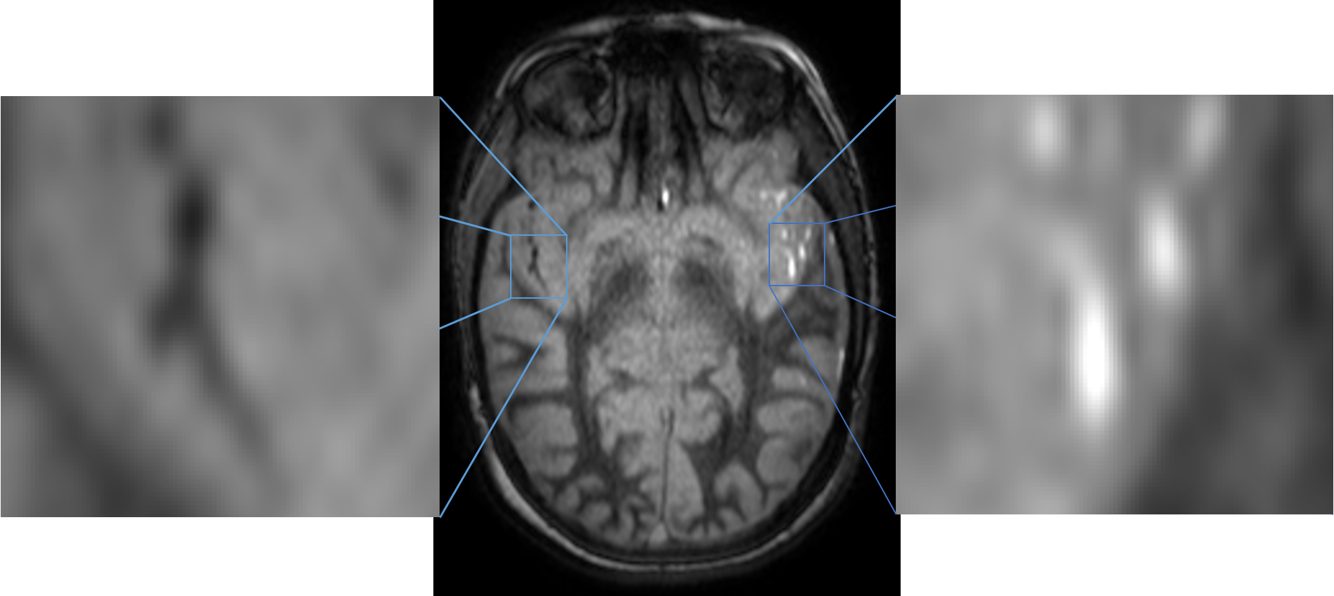
Goal(s): Demonstrate ability to simultaneously acquire bright and black blood contrast in one image, providing simultaneous information on time-resolved artery-selective blood flow as well as the vessel walls integrity.
Approach: Feasibility was evaluated by comparing calculated magnetization from a Bloch simulation and measured signal values from volunteer.
Results: Simulated and measured image data showed a largely similar course over time points. An outlier time point has to be further investigated. Best black blood contrast to surrounding tissue was not found at the time point with the lowest blood signal.
Impact: The study demonstrates the ability to simultaneously acquire bright and black blood contrast in a single image set and resulting simultaneous information on time-resolved artery-selective blood flow as well as the vessel walls integrity.
Introduction
The integrity of vessel walls and changes in blood flow are involved in many diseases. This study demonstrates the ability to simultaneously acquire bright and black blood contrast in one image and resulting simultaneous information on time-resolved artery-selective blood flow as well as the condition of vessel walls. It is achieved by comparing calculated magnetization from a Bloch simulation and measured signal values from volunteer measurements.Materials and Methods
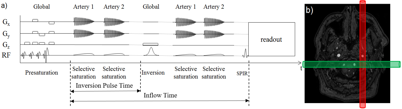
Six healthy volunteers were enrolled to acquire images of both carotid arteries and the posterior circulation using a 3 T Philips Achieva MRI scanner (Philips Healthcare, Best, The Netherlands), resulting in three datasets from each volunteer.The imaging sequence used consisted of four initial 90° saturation pulses to null out the signal from the brain tissue and intracerebral arterial and venous blood. During the inflow period, two two-dimensional 90° cylindrical pulses, positioned over two of the three major brain-feeding arteries are sequentially applied caudally to the imaging volume to saturate the inflowing blood. As a result, only blood within non-saturated vessels provides a high signal in the acquired image and the low signal provided by the saturated vessels allows for the output of a black-blood contrast.
After selective saturation, a non-selective 180° inversion pulse inverted the magnetization of static tissue and blood in the FOV. Further selective pulses to saturate the later incoming blood followed this. Each scan contains six frames with increasing inflow and inversion pulse times for time-resolved acquisition.
In addition to the imaging, a Bloch simulation considering the different inflow and inversion pulse times (Fig. 1) was performed to calculate the magnetization of saturated, non-saturated blood and static tissues such as gray matter, white matter, and cerebrospinal fluid.
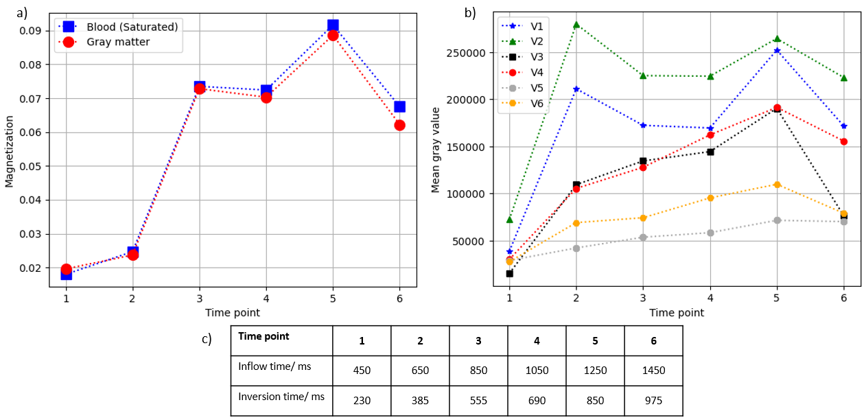
During the evaluation, the image signal of the vessels with saturated blood and the gray matter were determined. To do this, non-saturated vessels in each dataset were segmented and the resulting masks were applied to the other two datasets. The mean of the vessels from the different images of each volunteer was calculated. Good visualization of the vessels requires a high contrast to the surrounding tissue. For this reason, the contrast between blood and gray matter was determined.
This was followed by a qualitative comparison of the expected magnetization values resulting from the Bloch simulation with the actual signal values of the saturated blood and gray matter from the volunteer measurements and an assessment of the contrast between saturated blood and gray matter.
Results and Discussion
The evaluation and comparison of the data from the simulation and the measured images showed a similar course of the signal from time point 3 to time point 6. The lowest signal was measured at time point 1. On the one hand, this can be explained by the expected low magnetization from the Bloch simulation and, on the other hand, the effect of the presaturation-pulses has probably not yet decayed completely at this time point. At time point 2, an unexpectedly high signal was measured in both gray matter and blood in two volunteers. Reasons for this outlier need to be further investigated. Despite the small difference in magnetization, a visually sufficient contrast between blood and mass can be seen at all times. The highest contrast between unsaturated blood and gray matter was calculated in time point 5. In further studies, it would be recommended to optimize the inflow and inversion time using the Bloch simulation to reach the time point of highest possible contrast.Conclusion
The analysis showed that the signal progressed as expected from the Bloch simulation, except for one time point that needs to be investigated further. This technique enabled black and bright blood contrast to be acquired simultaneously in selected vessels without using contrast agents. With image post-processing, it is possible to create a holistic image of the entire cerebral vasculature.Acknowledgements
No acknowledgement found.References
Lindner T, Larsen N, Jansen O, Helle M: Selective arterial spin labeling in conjunction with phase-contrast acquisition for the simultaneous visualization of morphology, flow direction, and velocity of individual arteries in the cerebrovascular system. MRM. 2016; 78(4):1469-1475Figures