4976
The causal effect of insomnia on the hippocampal volume and hippocampal plasticity1Department of Psychology, Hangzhou Normal University, Hangzhou, China, 2Faculty of Psychology, Beijing Normal University, Beijing, China
Synopsis
Keywords: Gray Matter, Brain, Mendelian randomization; sleep;hippocampus
Motivation: The causal relationship between the sleep-related traits and the plasticity of subcortical brain volumes remains unclear.
Goal(s): This study aims to explore the causal relationship between two sleep-related traits (i.e., sleep duration and insomnia) and subcortical volumes.
Approach: Two-sample Mendelian randomization (MR) analysis
Results: We found a significant causal effect of insomnia but not sleep duration on the hippocampal volume. Moreover, insomnia showed significant causal influence on the structural plasticity of the hippocampus, which may associated with the rates of hippocampal atrophy.
Impact: The causal effect of insomnia on the hippocampal volume and plasticity may explain the adverse effect of insomnia on memory and may offer new evidence which could push the exploration of sleep management to delay the course of neurodegenerative diseases.
Introduction
It is widely perceived that shorter sleep could be a pervasive negative factor for physical, mental, and cognitive health1-3, yielding an increased risk of Alzheimer’s disease (AD) and other dementias4-6. The question of whether a causal relationship between sleep and brain health has recently garnered the attention of researchers, and only a few studies have tested this causal relationship7-8. One study suggested that sleep duration may not affect brain volume7, while the other study illustrated that sleep disorder had a significant causal influence on cortical brain volume8. However, the later study only tested the causal relationship between sleep disorder and cortical regions but not subcortical areas such as the hippocampus, which is an important brain structure for memory and AD. Moreover, whether sleep duration or sleep disorder has a causal influence on the plasticity of the subcortical structure remains unclear. Hence, we performed a two-sample Mendelian randomization (MR) analysis to reveal the causal effect of both sleep duration and sleep disorder on subcortical volumes and plasticity.Method
Datasets: We used the GWAS summary statistics of the traits about sleep 9, subcortical volume 10, and the plasticity of the subcortical volume 11 in the present study. The sleep-related traits included the sleep duration (n = 453532) and the insomnia (n = 455744). The subcortical volume included the bilateral thalamus, caudate, putamen, pallidum, hippocampus, amygdala, and accumbens (n = 33224). The plasticity of subcortical volume (e.g. longitudinal change) included the same regions except that the original study averaged the volume across the hemispheres for each nuclei (n = 15100). All data were based on cohorts of European ancestry, and there was a small sample overlap between sleep-related traits and the subcortical volume (~ 5%) or the plasticity of subcortical volume (~ 0.5%).Two-sample Mendelian randomization analysis: We selected the instrumental SNPs using the clump function in PLINK software and took the genomic data of the European superpopulation in the 1000 Genomes Project as the LD reference (MAF > 0.1, LD pruning r2 = 0.001, window size = 10kb, p-value < 5e-8). We then removed the SNPs that showed significant association with outcomes (p < 5e-8) and harmonized the data using the TwoSmpleMR R package to ensure that the same allele was used to estimate the genetic variant association. Inverse variance-weighted (IVW) regression 12 with multiplicative random effects was conducted as the primary analysis method. For the significant results with the IVW method, we would conduct four other methods to assess the robustness of the results. All of these methods were conducted in the TwoSmpleMR package. In addition, we performed the MR-Egger regression to examine the potential bias of directional pleiotropy 13 and a leave-one-out analysis to check whether the causal association was driven by a single SNP.
Results
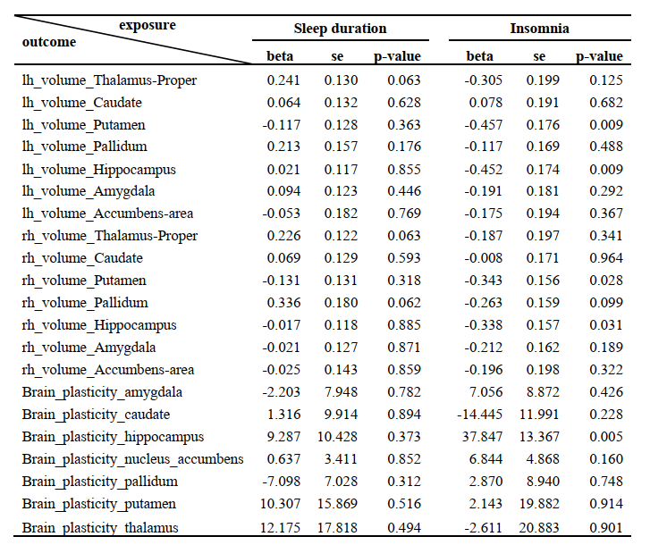
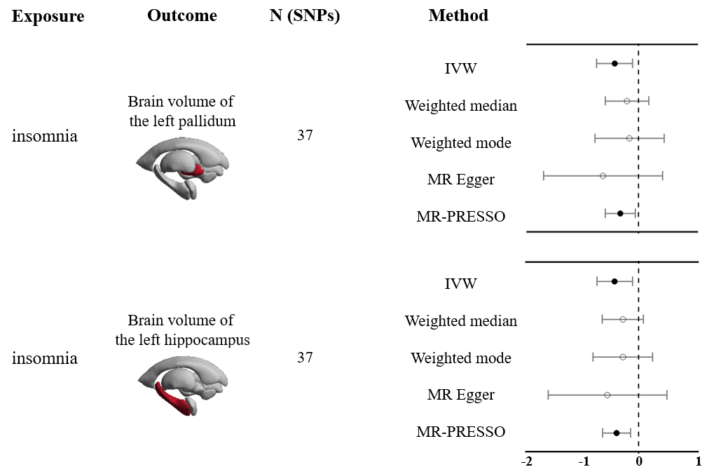
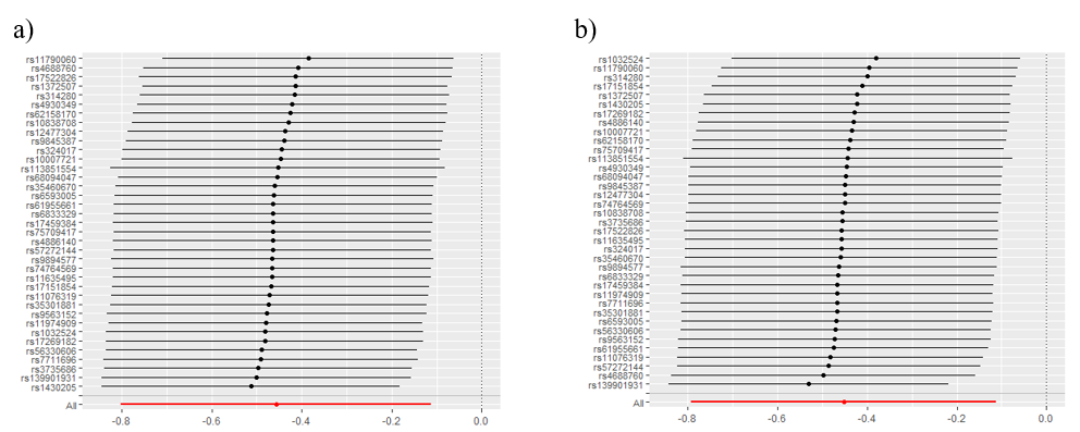
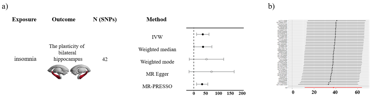
We first investigated the causal relationship between sleep duration and subcortical brain volumes, but no significant results were found (p's > 0.05, Table 1). On the contrary, insomnia showed significant causal associations with the brain volume of the left putamen (IVW method, b = -0.46, 95% CI of -0.80 to -0.11, p < 0.01) and the left hippocampus (b = -0.45, 95% CI of -0.79 to -0.11, p < 0.01, Fig 1). MR-PRESSO method yielded similar estimates (Fig 1). Sensitivity analyses showed no evidence of directional pleiotropy (p > 0.6) with the MR-Egger intercept test and no bias in the leave-one-out plot (Fig 2a-b). Suggesting the causal effect of insomnia on the atrophy of the left putamen and hippocampus. Furthermore, we investigate the causal relationship between the two sleep-related traits and the plasticity of subcortical brain regions. We also did not find a significant effect on sleep duration but found a significant causal effect for insomnia on the plasticity of the hippocampus (IVW, b = 37.85, 95% CI of 11.65 to 64.04, p < 0.005, Fig 3a). Other methods including weighted median and MR-PRESSO also showed significant effects. Sensitivity analyses showed no evidence of directional pleiotropy (p > 0.4) with the MR-Egger intercept test and no bias in the leave-one-out plot (Fig 3b).Discussion and Conclusion
Our study employed MR analysis to investigate the causal impact of sleep duration and insomnia on subcortical brain volume and plasticity. We did not find significant relationship between sleep duration and subcortical brain volumes but found evidence for causal impacts of sleep disorders (i.e., insomnia) on the hippocampal volume and plasticity. The causality supports the hypothesis that sleep disorder may be a vital element on the causal pathway to neurodegenerative diseases by affecting the structure of human brain.Acknowledgements
This work was supported by Zhejiang Provincial Natural Science Foundation of China under Grant No. Q23C090024.References
1. Lyon, L. Is an epidemic of sleeplessness increasing the incidence of Alzheimer’s disease? Brain 142, e30 (2019).
2. Walker, M. P. A societal sleep prescription. Neuron 103, 559–562 (2019).
3. Krause, A. J. et al. The sleep-deprived human brain. Nat. Rev. Neurosci. 18, 404–418 (2017)
4. Shi, L. et al. Sleep disturbances increase the risk of dementia: a systematic review and meta-analysis. Sleep Med. Rev. 40, 4–16 (2018).
5. Hatfield, C. F. et al. Disrupted daily activity/rest cycles in relation to daily cortisol rhythms of home-dwelling patients with early Alzheimer’s dementia. Brain 127, 1061–1074 (2004).
6. Videnovic, A. et al. “The clocks that time us”—circadian rhythms in neurodegenerative disorders. Nat. Rev. Neurol. 10, 683–693 (2014)
7. Fjell, A. M. et al. No phenotypic or genotypic evidence for a link between sleep duration and brain atrophy. Nature Human Behaviour, 1-15 (2023).
8. Gao, X. et al. Sleep disorders causally affect the brain cortical structure: A Mendelian randomization study. Sleep Medicine, 110, 243-253 (2023).
9. Jiang, L. et al. A resource-efficient tool for mixed model association analysis of large-scale data. Nat. Genet. 51, (2019).
10. Smith, S. M. et al. An expanded set of genome-wide association studies of brain imaging phenotypes in UK Biobank. Nat. Neurosci. 24, 737–745 (2021).
11. Brouwer, R. M. et al. Genetic variants associated with longitudinal changes in brain structure across the lifespan. Nat. Neurosci. 25, 421–432 (2022).
12. Burgess, S., Butterworth, A. & Thompson, S. G. Mendelian randomization analysis with multiple genetic variants using summarized data. Genet. Epidemiol. 37, 658–665 (2013).
13. Bowden, J., Smith, G. D. & Burgess, S. Mendelian randomization with invalid instruments: Effect estimation and bias detection through Egger regression. Int. J. Epidemiol. 44, 512–525 (2015).
Figures