4974
Accelerated Cortical Thickness Mapping Using Deep Learning1National Taiwan University of Science and Technology, Taipei, Taiwan
Synopsis
Keywords: Gray Matter, Data Analysis, Deep learning,Cortical thickness,ADNI
Motivation: The long processing time of current CT mapping methods hampers their use in clinical research. A faster and reliable CT mapping alternative is needed.
Goal(s): To create a deep-learning model that reduces CT mapping time without compromising accuracy or the ability to classify Alzheimer's disease.
Approach: We trained a 3D U-Net-based model on T1-weighted MRI datasets to produce CT maps, generating two model variants—one using skull-stripped and the other using both whole-brain and skull-stripped images. Performance was benchmarked against FreeSurfer.
Results: The complete Unet-based CT mapping workflow, inclusive of preprocessing, was executed in under a minute without relying on GPU acceleration.
Impact: The developed deep-learning-based method, executed within a minute, could accelerate neurological research related to CT values by providing fast and reliable procedure for CT mapping.
Introduction
Cortical thickness (CT) mapping, conducted with FreeSurfer1 and ANTS via high-resolution 3D T1-weighted (T1w) whole-brain images, is of significant importance in neurological research3,4. However, the extensive computation time required by FreeSurfer and ANTS, which can last several hours, presents a substantial challenge for applying CT mapping in clinical research settings. In this study, we have developed a deep-learning-based generative model that achieves CT mapping in under a minute. We compared this model's performance with FreeSurfer, examining estimation accuracy, reproducibility, and its efficacy in classifying Alzheimer’s disease.Material and Methods
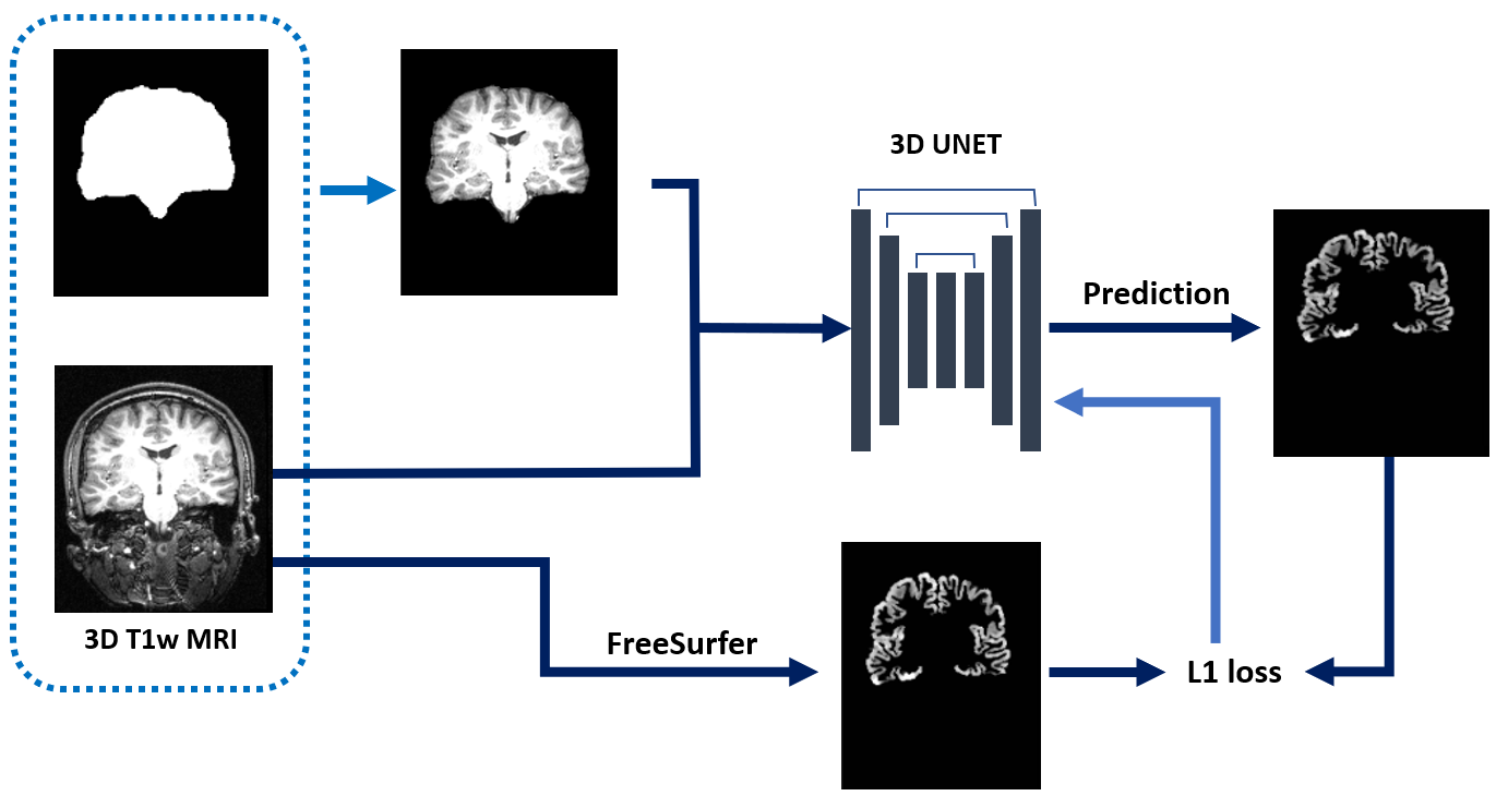
We curated T1-weighted datasets from open-access repositories, assembling 14,504 datasets for training5-8 and 1877 datasets for testing5, 9-12. These datasets were processed to create suitable inputs and outputs for the deep learning model. The input preparation involved min-max normalization and transforming to isotropic voxel resolution (1x1x1 mm³). We generated two forms of input data: the full-head T1w volumes and the skull-stripped brain volumes using TigerBx13. For the outputs, we utilized the FreeSurfer's recon-all pipeline coupled with volume resampling to craft CT maps for each dataset. These maps constituted the "ground truth" employed in both the training phase and subsequent evaluation of the model.Figure 1 illustrates the training process of our model, which employs a 3D U-Net architecture to generate CT maps from T1w volumes. The training was conducted with the following parameters: an initial learning rate set at 0.0001, a constant annealing scheduler, L1 loss function, and an Adam optimizer 20 epochs. We developed two separate models based on the training data composition. One utilized skull-stripped brain volumes exclusively, while the other was trained on both variations of input datasets, i.e., with and without skull-stripping. These models were designated as the Unet and Augmented Unet (Aug-Unet), correspondingly. We employed augmentation techniques to train an additional model, where during the input step, we randomly selected either skull-stripped images or whole brain images to train.
We evaluated the cortical thickness maps produced by FreeSurfer, Unet, and Aug-Unet using three distinct subsets within the test datasets. The 'balance-set' consisted of 154 datasets, selected to represent an approximately balanced age distribution ranging from 4 to 91 years. The 'SIMON' subset included 95 datasets from the SIMON database11, capturing multiple imaging sessions of a single healthy male subject. Lastly, the 'ADNI' subset contained 1628 datasets sourced from the Alzheimer's Disease Neuroimaging Initiative (ADNI)12, evenly divided among three cognitive states: cognitively normal (CN) individuals, mild cognitive impairment (MCI), and Alzheimer’s disease (AD).
Results
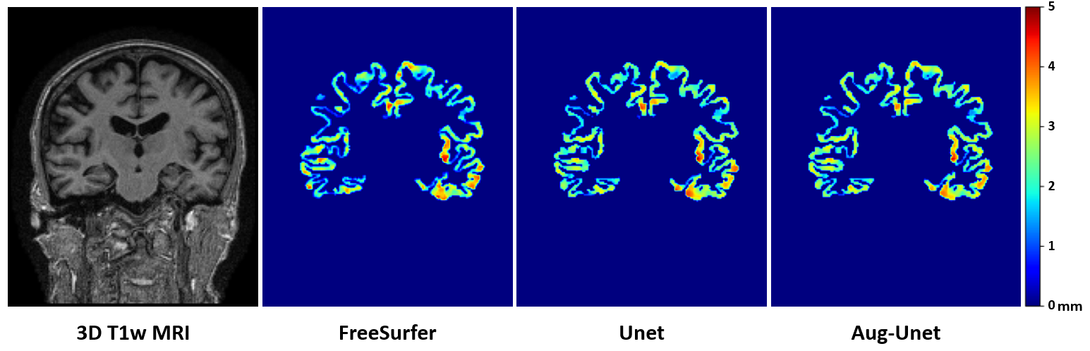
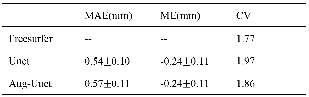
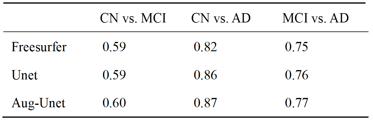
Figure 2 showcases the original T1w MRI scans alongside the CT maps predicted by FreeSurfer, Unet, and Aug-Unet. The comparative performance results, namely mean absolute errors (MAE), mean errors (ME), and coefficient of variation (CV), of these methods are encapsulated in Figure 3. When using the balance-set for a comparative analysis with FreeSurfer, Unet and Aug-Unet demonstrated MAEs of 0.54 mm and 0.57 mm, respectively. With the SIMON dataset, we computed the CV values for the whole-brain CT values averaged across the three methods. For the ADNI datasets, we computed the average CT values across 62 brain subregions using the Desikan-Killiany-Tourville (DKT) parcellation, implemented via TigerBx or FreeSurfer. Subsequently, we conducted receiver operating characteristic analysis for classifying CN, MCI and AD states using these CT values. Within the various regions defined by the DKT regions, the CT measurement of the lh-entorhinal region stands out with the highest area under the curve (AUC) in the classification of Alzheimer's Disease (AD) using the FreeSurfer analysis. Figure 4 presents the area under the curve (AUC) metrics for these classification tasks, specifically highlighting the CT values from the left hemisphere entorhinal (lh-entorhinal) region. The other regions are not shown due to limited space of this abstract.Discussions and Conclusions
Our Unet-based approach for CT mapping was developed and assessed against large-scale training databases. The model produced CT estimates with an average MAE of approximately 0.5 mm and demonstrated reproducibility and AD classification performance on par with FreeSurfer. Notably, Aug-Unet had a slight edge over Unet, hinting that the augmentation step enhanced the model's ability to generalize. The complete Unet-based CT mapping workflow, inclusive of preprocessing, was executed in under a minute without relying on GPU acceleration. We are preparing to make the model openly available for community use in CT-related research. In summary, our deep-learning-based CT mapping technique has been validated and may serve as an efficient tool for clinical scenarios involving brain cortical thickness assessment.Acknowledgements
We are grateful to the National Center for High-performance Computing for computer time and facilities. This study was supported by the National Science and Technology Council, Taiwan (112-2314-B-011-002-MY2). We respectfully acknowledge the participants and the investigators of the open-access datasets that were adopted in this work. The assistance of OpenAI's ChatGPT-4 in refining the grammatical structure of this manuscript is hereby acknowledged.References
1. Fischl B. FreeSurfer. NeuroImage. 2012;62(2):774-81.2. Tustison NJ, Cook PA, Klein A, Song G, Das SR, Duda JT, et al. Large-scale evaluation of ANTs and FreeSurfer cortical thickness measurements. NeuroImage. 2014;99:166-79.
3. Thorns J, Jansma H, Peschel T, Grosskreutz J, Mohammadi B, Dengler R, et al. Extent of cortical involvement in amyotrophic lateral sclerosis - an analysis based on cortical thickness. Bmc Neurology. 2013;13.
4. Schultz CC, Koch K, Wagner G, Roebel M, Schachtzabel C, Gaser C, et al. Reduced cortical thickness in first episode schizophrenia. Schizophrenia Research. 2010;116(2-3):204-9.
5. Kennedy DN, Haselgrove C, Riehl J, Preuss N, Buccigrossi R. The NITRC image repository. Neuroimage. 2016;124(Pt B):1069-73.
6. Markiewicz CJ, Gorgolewski KJ, Feingold F, Blair R, Halchenko YO, Miller E, et al. The OpenNeuro resource for sharing of neuroscience data. Elife. 2021;10.
7. Souza R, Lucena O, Garrafa J, Gobbi D, Saluzzi M, Appenzeller S, et al. An open, multi-vendor, multi-field-strength brain MR dataset and analysis of publicly available skull stripping methods agreement. NeuroImage. 2018;170:482-94.
8. Di Martino A, Yan CG, Li Q, Denio E, Castellanos FX, Alaerts K, et al. The autism brain imaging data exchange: towards a large-scale evaluation of the intrinsic brain architecture in autism. Molecular Psychiatry. 2014;19(6):659-67.
9. Kennedy DN, Haselgrove C, Hodge SM, Rane PS, Makris N, Frazier JA. CANDIShare: a resource for pediatric neuroimaging data. Neuroinformatics. 2012;10(3):319-22.
10. Marcus DS, Wang TH, Parker J, Csernansky JG, Morris JC, Buckner RL. Open Access Series of Imaging Studies (OASIS): cross-sectional MRI data in young, middle aged, nondemented, and demented older adults. J Cogn Neurosci. 2007;19(9):1498-507.
11. Duchesne S, Chouinard I, Potvin O, Fonov VS, Khademi A, Bartha R, et al. The Canadian Dementia Imaging Protocol: Harmonizing National Cohorts. J Magn Reson Imaging. 2019;49(2):456-65.
12. Petersen RC, Aisen PS, Beckett LA, Donohue MC, Gamst AC, Harvey DJ, et al. Alzheimer's Disease Neuroimaging Initiative (ADNI): clinical characterization. Neurology. 2010;74(3):201-9.
13. Weng JS, Huang TY. Deriving a robust deep-learning model for subcortical brain segmentation by using a large-scale database: Preprocessing, reproducibility, and accuracy of volume estimation. NMR Biomed. 2023;36(5):e4880.
Figures