4973
Noninvasive quantification of ketamine-induced structural plasticity in mice using multishell diffusion weighted imaging1Department of Radiology, Stanford University, Stanford, CA, United States, 2Department of Anesthesiology, Perioperative and Pain Medicine, Stanford University, Stanford, CA, United States
Synopsis
Keywords: Pharmacology, Diffusion/other diffusion imaging techniques, Pharmacology
Motivation: To monitor treatment, we need noninvasive quantitation of the microstructural changes known to accompany rapid-acting antidepressant therapy.
Goal(s): Our goal was to develop a noninvasive biomarker of the structural plasticity induced by rapid-acting antidepressants.
Approach: Neurite orientation dispersion and density imaging (NODDI) was assessed before and after treating male and female mice with ketamine, with or without opioid blockade.
Results: Ketamine induced alterations of orientation dispersion index (ODI) and neurite density index (NDI) in brain regions involved in mood regulation and reward processing, particularly in females. Curiously, opioid blockade induced trendwise increases in NDI and ODI across multiple brain regions, independent of ketamine.
Impact: Our preliminary results indicate that multishell diffusion MRI tracks the brain microstructural changes known to be induced by ketamine and related rapid-acting antidepressants, enabling a noninvasive quantitative biomarker that could be used to track individual patient response to antidepressant therapy.
INTRODUCTION
Psychedelic and dissociative anesthetic drugs such as psilocybin and ketamine, respectively, constitute a promising new class of antidepressant therapy which produces lasting therapeutic effects rapidly in patients who have not responded to other antidepressants. A proposed mechanism of action of these drugs is structural plasticity: in rodents, stress decreases dendrite length and spine number in cortical neurons1,2 whereas ketamine and psilocybin induce dendrite growth and spinogenesis3,4, measured by histopathology. Currently, there is no noninvasive, translational method for measuring this structural plasticity in living subjects and, more broadly, there is a critical paucity of biomarkers for assessing at an early stage whether an antidepressant is effective for a given patient. For this study, we hypothesized that ketamine’s neuroplasticity effects could be measured via advanced multishell diffusion MRI-based models such as neurite orientation dispersion and density imaging (NODDI)5. The neurite density parameter from NODDI is hypothesized to provide sensitivity to the expected increased density of dendrites, which tend to be oriented less coherently than axons in white matter. In prior work, we showed that the structural plasticity effects of ketamine are sex- and opioid receptor-dependent6; therefore,we also sought to characterize the effects of sex and naltrexone (an opioid receptor blocker) pretreatment on ketamine’s effects on the NODDI neurite density index (NDI) and orientation dispersion index (ODI).METHODS
In C57BL/6 mice, we acquired in vivo multishell diffusion weighted images and T2-weighted structural images at time points 30 minutes prior to and 24 hours after treatment with one of the following: intraperitoneal (i.p.) saline (n=5 males, 4 females), 10 mg/kg i.p. ketamine (n=7 males, 3 females), 10 mg/kg subcutaneous (s.c.) naltrexone followed by i.p. saline (n=7 males), or 10 mg/kg s.c. naltrexone followed by i.p. ketamine (n=7 males). Four subjects were excluded due to motion artifacts or incomplete scan protocols. Scanned animals were perfused immediately after the second timepoint for histology. Images were acquired on a Bruker 7T BioSpec with a CryoProbe RF coil with the following sequence parameters: b=1000, 2500 s/mm2, 40 diffusion directions, TE / TR = 21.7 / 4000 ms. For processing the data, we corrected for eddy currents and performed diffusion tensor fitting with FSL7, fit NODDI parameters with the Microstructure Diffusion Toolbox8, skull-stripped images with the FSL brain extraction tool, registered to a high-resolution fractional anisotropy atlas with FLIRT and 3dqWarp, and compared ODI and NDI across groups via custom scripts for voxel-based and region of interest analyses.RESULTS
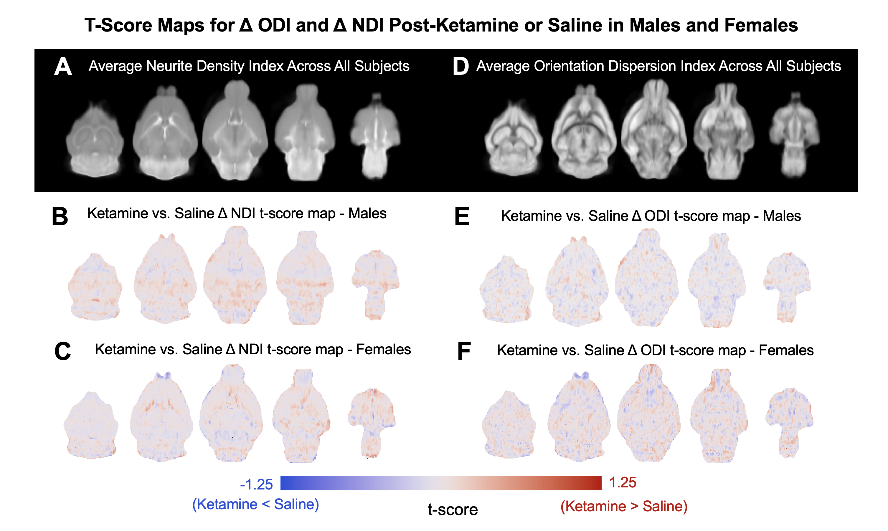
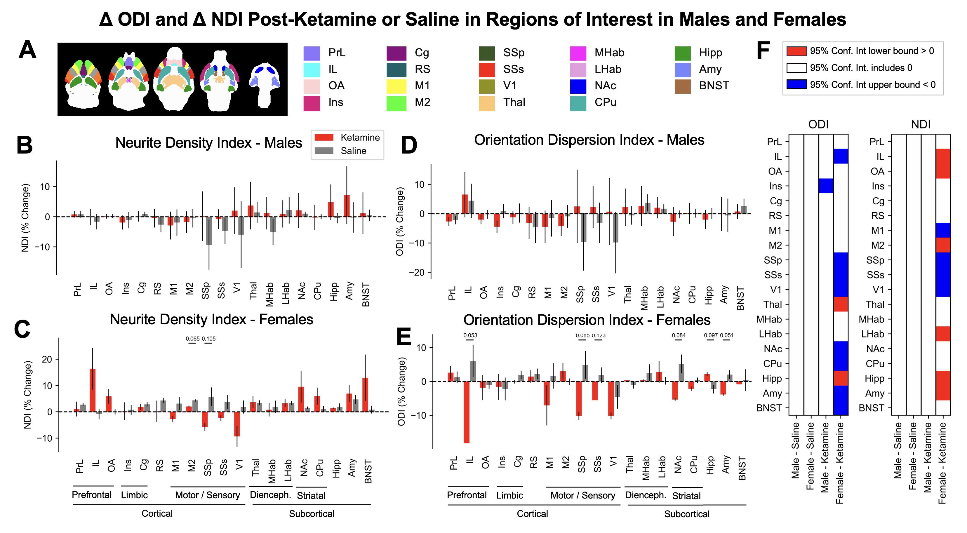
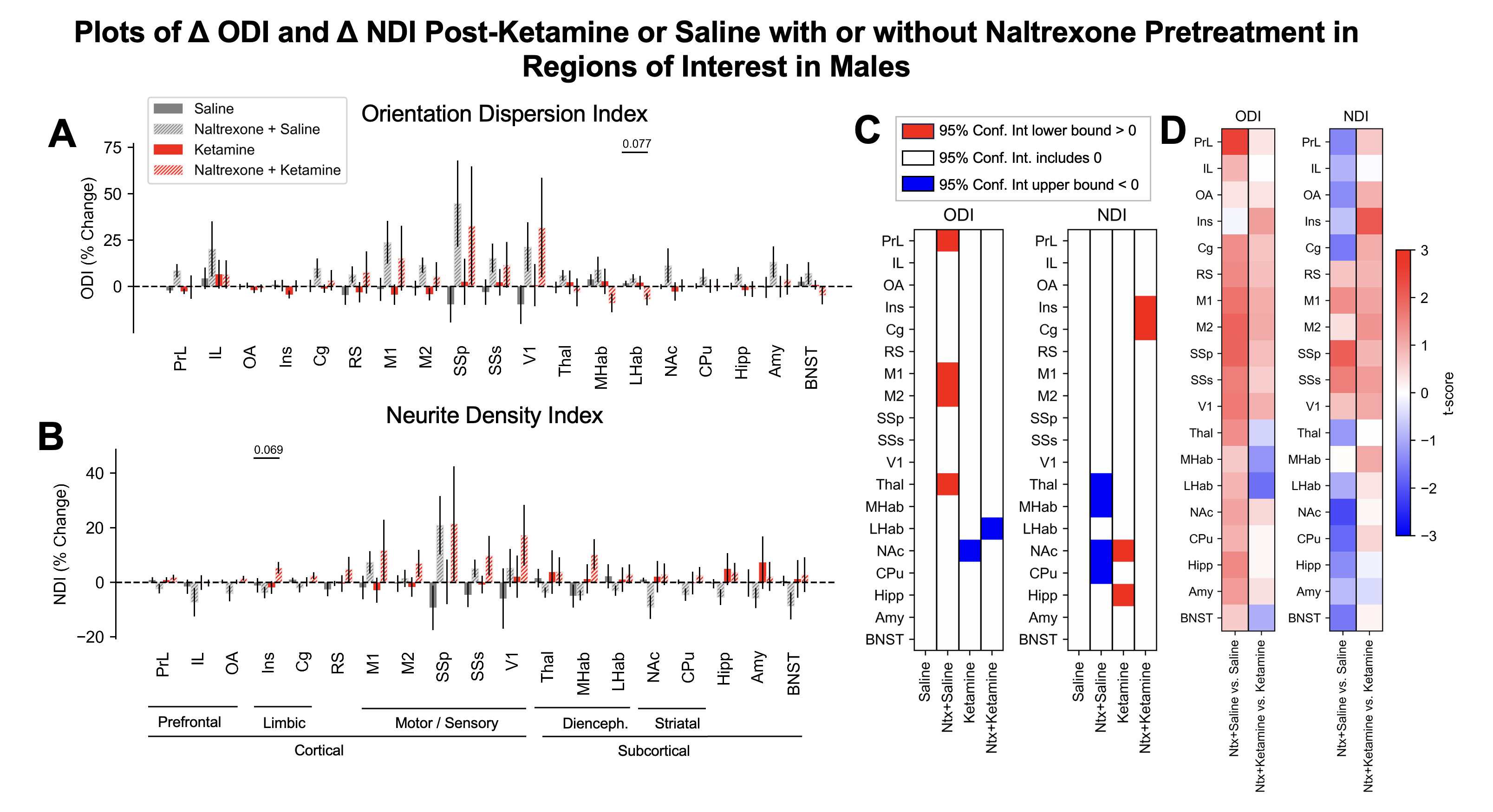
In female mice, we observed changes in ODI and NDI in infralimbic cortex, a brain region where drug-induced structural plasticity effects have repeatedly been reported using histopathology (ΔODI confidence interval: [-18.3%, -18.2%]; ΔODI Welch’s p-value vs. sham: 0.049; ΔNDI confidence interval: [3.4%, 29.4%]; ΔNDI Welch’s p-value vs. sham: 0.15), as well as other brain regions involved in mood regulation and reward processing, such as the nucleus accumbens (ΔODI confidence interval: [-6.2%, -4.5%]; ΔODI Welch’s p-value vs. sham: 0.10) (Fig. 1C,F; 2C-F). We did not observe significant changes in ODI and NDI in male mice in any regions of interest following ketamine treatment, although trendwise effects in similar brain regions were noted (Fig. 1B,E; 2B,D,F). We found that naltrexone pretreatment seemed to both modulate the effects of ketamine on NDI and ODI and induce changes in NDI and ODI, but independent of ketamine treatment (Fig. 3A-D).DISCUSSION
Our results suggest that the effects of ketamine brain microstructural plasticity in cortical and subcortical areas can indeed be quantified using NODDI. In particular, ODI tended to be decreased and NDI tended to be increased in relevant brain regions in female mice following ketamine treatment. As a next step, we intend to use NODDI to quantify the structural plasticity effects of psilocybin, a serotonergic rapid-acting antidepressant and psychedelic drug. Additionally, we are currently preparing to immunostain and image postsynaptic density protein 95, a marker of synapses, in whole-mounted, iDISCO-cleared9 tissue of the mice imaged in the study, to correlate multishell diffusion imaging and NODDI parameters to a validated marker of structural plasticity3,4.CONCLUSION
Multishell diffusion imaging parameters show great promise for quantifying changes in brain microstructural plasticity, providing a noninvasive biomarker of the therapeutic effects of rapid-acting antidepressants. Future studies will translate these results to determine whether multishell diffusion imaging metrics are altered in disease-relevant brain regions following systemic or targeted treatment with ketamine or other rapid-acting antidepressants in psychiatric patients.Acknowledgements
We acknowledge the Stanford University Wu Tsai Institute Neurosciences Preclinical Imaging Laboratory (NPIL) for funding via the NPIL Pilot Award and for technical support from NPIL Director Dr. Jieun Kim. We also acknowledge funding from the National Science Foundation Graduate Research Fellowship Program for SNE.References
1. Silva-Gómez, A. B., Rojas, D., Juárez, I. & Flores, G. Decreased dendritic spine density on prefrontal cortical and hippocampal pyramidal neurons in postweaning social isolation rats. Brain Research 983, 128–136 (2003).
2. Cook, S. C. & Wellman, C. L. Chronic stress alters dendritic morphology in rat medial prefrontal cortex. Journal of Neurobiology 60, 236–248 (2004).
3. Li, N. et al. mTOR-Dependent Synapse Formation Underlies the Rapid Antidepressant Effects of NMDA Antagonists. Science (2010) doi:10.1126/science.1190287.
4. Ly, C. et al. Psychedelics Promote Structural and Functional Neural Plasticity. Cell Reports 23, 3170–3182 (2018).
5. Zhang, H., Schneider, T., Wheeler-Kingshott, C. A. & Alexander, D. C. NODDI: Practical in vivo neurite orientation dispersion and density imaging of the human brain. NeuroImage 61, 1000–1016 (2012).
6. Ianni, T. D., Azadian, M. M., Ewbank, S. N., Michaelides, M. & Airan, R. D. Sex dependence of opioid-mediated responses to subanesthetic ketamine. 2022.09.06.506854 Preprint at https://doi.org/10.1101/2022.09.06.506854 (2022).
7. Woolrich, M. W. et al. Bayesian analysis of neuroimaging data in FSL. NeuroImage 45, S173–S186 (2009).
8. Harms, R. L., Fritz, F. J., Tobisch, A., Goebel, R. & Roebroeck, A. Robust and fast nonlinear optimization of diffusion MRI microstructure models. NeuroImage 155, 82–96 (2017).
9. Rijsketic, D. R. et al. UNRAVELing the synergistic effects of psilocybin and environment on brain-wide immediate early gene expression in mice. Neuropsychopharmacology 48, 1798–1807 (2023).
Figures