4971
Language Lateralization on resting state fMRI Vs task fMRI, in neurosurgical cases1MRI, P.D.Hinduja Hospital, Mumbai, India
Synopsis
Keywords: Functional Connectivity, fMRI (resting state), fMRI (task)
Motivation: Understanding hemispheric language dominance is crucial for surgery, yet task-based fMRI has many challenges. Resting-state fMRI shows promise, but its ability to depict language lateralization is still evolving, with varied results in literature.
Goal(s): To assess language dominance on rest fMRI in neurosurgical patients by different methods and seeing concordance with task-based fMRI.
Approach: We explored three calculation techniques for lateralization: first using individual seed volumes, second using voxel activation in ipsilateral hemisphere, and third using net voxel activation generated in bilateral hemispheres
Results: Maximum of 54.55% concordance of rest-fMRI with task-based fMRI was seen for language dominance, with one of the methods.
Impact: Our results emphasize that rest fMRI for language dominance should be used with caution and as an adjunct to task fMRI in neurosurgical patients.
ABSTRACT
INTRODUCTION: While task-based functional magnetic resonance imaging (fMRI) is being routinely performed for preoperative planning and language dominance, it has limitations due to dependency on patient’s cooperation in clinical settings and challenges in cases of children and neurocognitive impairment. Resting-state fMRI (rs-fMRI) provides an alternative approach by analyzing spontaneous brain activity during rest, eliminating the need for explicit tasks and enabling the mapping of multiple networks, making it a promising method. However, rs-fMRI is still evolving and its effectiveness compared to task-based fMRI in language lateralization remains uncertain due to diverse study results (1)(2).The majority of studies utilize Independent Component Analysis (ICA) for processing rs-fMRI (3), with fewer using seed-based analysis. There are no standard methods for calculating the Laterality Index (LI) in rs-fMRI to determine language dominance, with published research trying multiple methods with varied results (4)(5). To add to it, there is limited research in clinical settings in neurosurgical patients, which is what we have attempted.
METHODS: 22 patients, 9 to 65 Years (M: F = 13 : 9), underwent both language task-based fMRI and rest- fMRI, on 3.0T Philips Ingenia and post processing done on FDA-approved Brainance MD software by Advantis Medical Imaging. We employed seed-based technique to analyze rs-fMRI data and compared language lateralization on rest versus task fMRI.
For rs-fMRI, we explored three methods to calculate Laterality Index (LI):
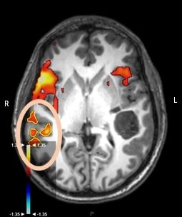
1. Individual Seed Volumes Method: We used the Broca seed volume, chosen for its role in expressive language function and temporal reliability (6) (7), with the formula (LS - RS) / (LS + RS), where LS and RS represent volumes of the Broca's seeds.
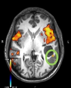
2. Intra-hemispheric Calculation: This method calculates functional connectivity in the ipsilateral hemisphere generated by the Broca's seed, with the formula (LSLH - RSRH) / (LSLH + RSRH), where LH and RH represent voxel counts in the left and right hemispheres respectively (5).
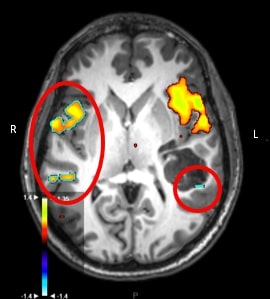
3. Cross hemispheric Calculation: This formula considered voxel activation in both hemispheres produced by the individual Broca’s seed. The formula used was ((LSLH - LSRH) / (LSLH + LSRH)) - ((RSRH - RSLH) / (RSLH + RSRH)), where LSLH and LSRH are voxel counts in the left & right hemisphere due to the left seed, and RSRH and RSLH are similar voxel counts due to the right seed (5).
We used an LI value less than or equal to -0.1 for right dominance, while a value greater than or equal to 0.1 as left dominance and LI values between 0.1 and -0.1 were classified as bilateral, based on previous research studies (8)(9).
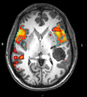
RESULTS: While most patients demonstrated left lateralization during task fMRI, rs-fMRI results were more diverse. The individual seed volumes (method 1) and a cross-hemispheric approach (method 3) both yielded concordance value of 45.45 % with task-based outcomes. The intra-hemispheric calculation (method 2) showed a higher concordance at 54.55%.
DISCUSSION: Our study investigated task-based and resting-state fMRI concordance in language lateralization using a seed-based technique in neurosurgical patients. We employed 3 different methods on rs-fMRI, to calculate LI. But the concordance of each of these methods with task fMRI, showed a maximum of 54.55% , which was demonstrated by the method using intra-hemispheric calculation.
A similar study in literature (5) using a seed based technique on rs-fMRI, with above methods 2 and 3, showed concordance of dominance of 20- 30% for the intra-hemispheric resting state LI technique (method 2) and 50 - 63% for the resting state LI intra- minus interhemispheric difference technique (method 3).
Rest-fMRI has been consistently found to have good results in most studies for localization of eloquent areas (10)(11). However, its role in depicting language lateralization seems less reliable. A possible explanation for our findings could be that these methods are not being able to accurately represent the complex neural processes involved in language lateralization. In hindsight, these new techniques require further refinement and validation. This observation emphasizes the need for methodological rigor in neuroimaging studies. Continuous research is crucial to improve the accuracy of rest-fMRI analysis techniques, emphasizing the complexity of understanding resting-state language networks, especially in clinical cases.
CONCLUSION: Our study provides valuable insights into the challenges of resting-state fMRI analysis in language lateralization, with the results emphasizing that rest fMRI for language dominance should be used with caution and as an adjunct to task fMRI in neurosurgical patients. While our initial results did not show good concordance with task-based fMRI, the discrepancies observed offer crucial cues for future research.
Acknowledgements
Ms Aditi Erande, to help us in statistics
References
1. Sair HI, Yahyavi-Firouz-Abadi N, Calhoun VD, Airan RD, Agarwal S, Intrapiromkul J, Choe AS, Gujar SK, Caffo B, Lindquist MA, Pillai JJ. Presurgical brain mapping of the language network in patients with brain tumors using resting-state fMRI: Comparison with task fMRI. Hum Brain Mapp. 2016 Mar;37(3):913-23.
2. Lemée JM, Berro DH, Bernard F, Chinier E, Leiber LM, Menei P, Ter Minassian A. Resting-state functional magnetic resonance imaging versus task-based activity for language mapping and correlation with perioperative cortical mapping. Brain Behav. 2019 Oct;9(10):e01362.
3. Branco P, Seixas D, Deprez S, Kovacs S, Peeters R, Castro SL, Sunaert S. Resting-State Functional Magnetic Resonance Imaging for Language Preoperative Planning. Front Hum Neurosci. 2016 Feb 1;10:11.
4. Singh S, Alhasan MSM, Wang Z, Clarke R, Xi Y, Maldjian JA, Wagner B, Booth T. Establishing language laterality: does resting-state functional MRI help?. J Neurosurg Pediatr. 2023 May;31(5):496-502.
5. Rolinski R, You X, Gonzalez-Castillo J, Norato G, Reynolds RC, Inati SK, Theodore WH. Language lateralization from task-based and resting state functional MRI in patients with epilepsy. Hum Brain Mapp. 2020 Aug 1;41(11):3133-3146.
6. Tomasi D, Volkow ND. Resting functional connectivity of language networks: Characterization and reproducibility. Mol Psychiatry. 2012;17:841-854.
7. Zhu L, Fan Y, Zou Q, Wang J, Gao JH, Niu Z. Temporal reliability and lateralization of the resting‐state language network. PLoS One. 2014;9:e85880.
8. Nath A, Robinson M, Magnotti J, Karas P, Curry D, Paldino M. Determination of Differences in Seed-Based Resting State Functional Magnetic Resonance Imaging Language Networks in Pediatric Patients with Left- and Right-Lateralized Language: A Pilot Study. J Epilepsy Res. 2019 Dec 31;9(2):93-102.
9. Brumer I, De Vita E, Ashmore J, Jarosz J, Borri M. Implementation of clinically relevant and robust fMRI-based language lateralization: Choosing the laterality index calculation method. PLoS One. 2020 Mar 12;15(3):e0230129.
10. Tie Y, Rigolo L, Norton IH, Huang RY, Wu W, Orringer D, Mukundan S Jr, Golby AJ. Defining language networks from resting-state fMRI for surgical planning--a feasibility study. Hum Brain Mapp. 2014 Mar;35(3):1018-30.
11. Shimony JS, Zhang D, Johnston JM, Fox MD, Roy A, Leuthardt EC. Resting-state spontaneous fluctuations in brain activity: a new paradigm for presurgical planning using fMRI. Acad Radiol. 2009 May;16(5):578-83.
Figures