4967
Modulation of cross-hierarchy propagating waves across sleep stages1Department of Biomedical Engineering, The Pennsylvania State University, University Park, State College, PA, United States, 2National Institute of Neurological Disorders and Stroke, National Institutes of Health, Bethesda, MD, United States, 3Institute for Computational and Data Sciences, The Pennsylvania State University, University Park, State College, PA, United States
Synopsis
Keywords: fMRI Analysis, fMRI (resting state), sleep
Motivation: Endogenous brain activity plays a pivotal role during sleep. Previous research suggested that endogenous brain activity during wakefulness can take the form of infra-slow waves propagating along the cortical hierarchical gradient. But it remains unclear how these infra-slow waves modulate across sleep stages.
Goal(s): To elucidate sleep stage dependent changes in the cross-hierarchy propagations.
Approach: We measured and analyzed overnight fMRI/EEG data.
Results: The cross-hierarchy propagating waves modulate systematically across sleep stages. REM sleep features more frequent propagations from sensory/motor regions to higher-order brain networks, which are associated with eye movements and characterized by phase shifts in the thalamus, pons, and visual cortex.
Impact: The findings reveal a highly structured nature of endogenous brain dynamics during REM sleep and their potential link to known REM features of electrophysiological PGO waves and eye movements.
introduction
Spontaneous brain activity exhibits highly organized low-frequency (<0.1 Hz) fluctuation, a property that has been used for mapping functional brain connectivity based on resting-state fMRI1,2. Increasing evidence suggests that highly structured spatiotemporal dynamics lead to resting-state functional connectivity, which has been linked to various aspects of brain function and dysfunction3–5. These infra-slow spatiotemporal dynamics could be sensitive to sleep states2,4–9 and related to important brain functions, such as memory consolidation4,5,9. It was recently found that spontaneous brain activity can take the form of infra-slow waves propagating along the cortical hierarchical gradient between sensory/motor (SM) cortices and higher-order cognitive networks6,10. However, it remains unclear whether and how the cross-hierarchy propagating waves are modulated across sleep stages, particularly during REM sleep where the cross-hierarchy information flow could be critical for off-line learning and memory consolidation11. To gain further insight into infra-slow brain dynamics during sleep, we examined the cross-hierarchy fMRI propagating waves across various sleep stages through whole-night fMRI/electroencephalography (EEG) recordings.methods
We analyzed 12 subjects with two consecutive nights of simultaneous fMRI/EEG12. Only 18 nights showed REM sleep. We identified the cross-hierarchy propagating waves6 and compared their occurrence under different sleep stages. To investigate propagation dynamics during various sleep stages, we focused on stable sleep periods during which a specific sleep stage lasted longer than one minute. We aligned and averaged fMRI propagations according to the global mean BOLD (gBOLD) peak within each identified propagation and converted the average into t-score maps. Relative phase delay of a specific region in bottom-up propagation is the time delay between its peak activation and the propagation center defined as the gBOLD peak. We quantified eye movements based on two independent measurements: the electrooculogram (EOG) signal variation and motion signals derived from concurrent video recordings13. We also identified isolated eye movements, which had at least 8 seconds of stable eye position on either side, and examined fMRI changes around these events.results
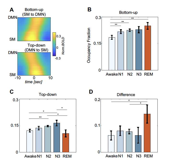
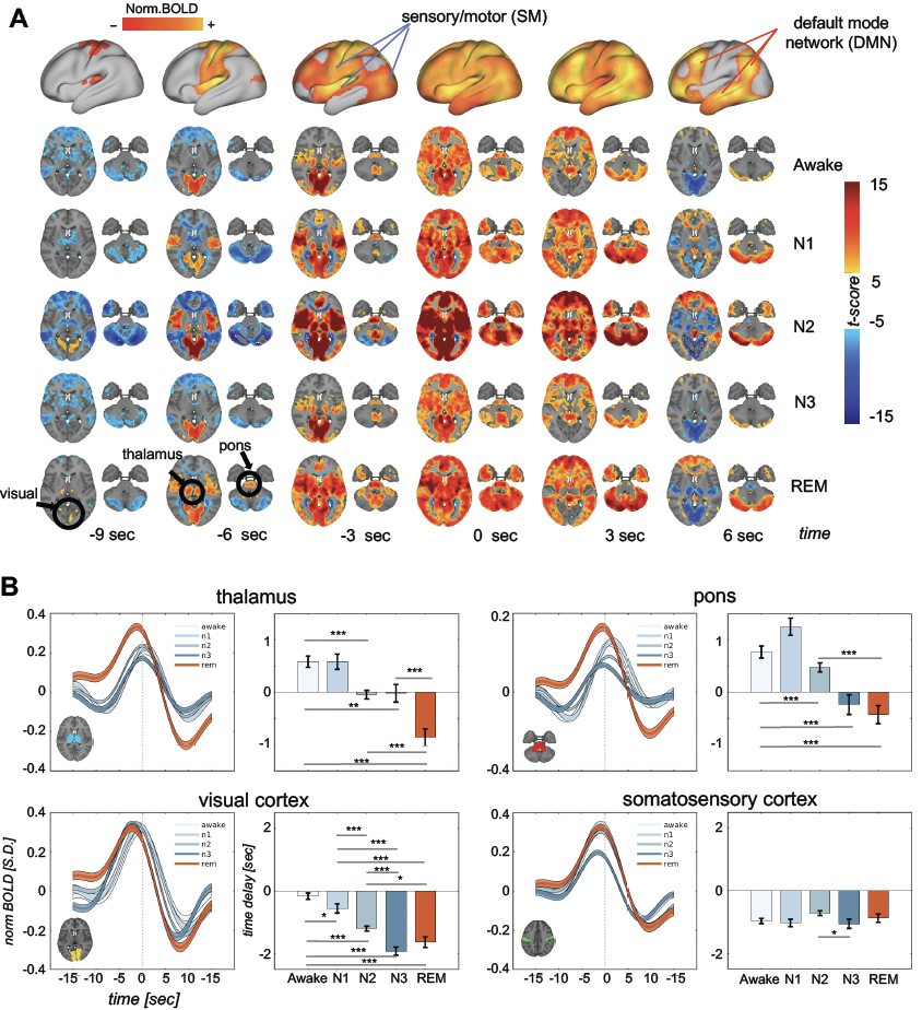
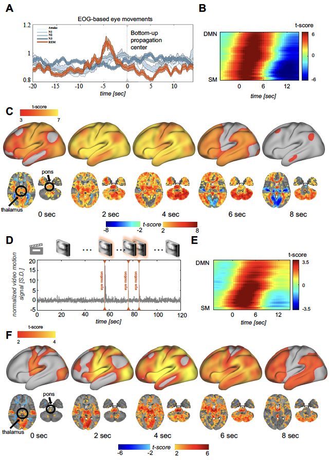
Bottom-up propagations gradually increased from wakefulness to deeper NREM sleep stages (N1 to N3), peaking during REM sleep (Fig.1B). Top-down propagations followed a similar trend during wakefulness and NREM stages but exhibited an opposite change and reached its minimum during REM sleep (Fig.1C). Consequently, REM sleep featured the largest difference between the two types of propagations (Fig.1D). Bottom-up propagating waves during REM sleep also showed distinct dynamics in the thalamus, pons and visual cortex, regions with established involvement in ponto-geniculo-occipital (PGO) waves, but not in the control somatosensory areas (Fig.2A and 2B). Their phase in the bottom-up propagations (i.e., relative time delay to the propagation center) was significantly advanced to earlier time as compared to other conditions (Fig.2B). Bottom-up propagations during REM sleep were also associated with eye movements estimated by EOG variation (Fig.3A). Consistent with this, fMRI changes at EOG-based eye movements showed gradually increased time delays along the cortical hierarchical gradient described by a principal gradient (PG) direction (Fig.3B), manifested as a bottom-up propagation on the cortical surface (Fig.3C), and is associated with early thalamic and pontine (Fig.3C). In addition, fMRI changes surrounding isolated eye movements identified through video recordings displayed similar dynamics of bottom-up propagating waves (Fig.3D-3F).discussion
Here we demonstrated significant modulations of the cross-hierarchy propagating waves across sleep stages, with REM sleep standing out as the stage characterized by notably dominant bottom-up propagations. Importantly, these bottom-up propagating waves during REM sleep exhibited features distinct from other conditions, with the PGO-related regions (i.e., the thalamus, pons, visual cortex) co-activating at the earliest phase. In addition, these waves during REM sleep were also associated with eye movements. REM sleep is known to serve vital brain functions, particularly related to learning and memory14–17. The realization of these functions has been linked to PGO waves and eye-movement-associated dreaming11,18. Our study links these previously known REM signatures to infra-slow brain dynamics, and more importantly, unveils the intricate organization of these infra-slow dynamics, in particular its cross-hierarchy organization that appears to align well with its potential role in learning and memory.conclusion
Cross-hierarchy propagating waves of fMRI activity are significantly modulated across sleep stages. REM sleep is characterized by notably dominant bottom-up propagations, which appear linked to PGO waves and eye movements.Acknowledgements
This research was supported by the Brain Initiative award (1RF1MH123247-01) and the NIH R01 award (1R01NS113889-01A1), and NIH Intramural Research Project ZNS003027.
References
1. Abbas, A. et al. Quasi-periodic patterns contribute to functional connectivity in the brain. Neuroimage 191, 193–204 (2019).
2. Mitra, A., Snyder, A. Z., Blazey, T. & Raichle, M. E. Lag threads organize the brain’s intrinsic activity. Proceedings of the National Academy of Sciences 112, E2235–E2244 (2015).
3. Mitra, A., Snyder, A. Z., Constantino, J. N. & Raichle, M. E. The Lag Structure of Intrinsic Activity is Focally Altered in High Functioning Adults with Autism. Cerebral Cortex 27, bhv294 (2017).
4. Mitra, A., Snyder, A. Z., Tagliazucchi, E., Laufs, H. & Raichle, M. E. Propagated infra-slow intrinsic brain activity reorganizes across wake and slow wave sleep. Elife 4, e10781 (2015).
5. Mitra, A. et al. Spontaneous Infra-slow Brain Activity Has Unique Spatiotemporal Dynamics and Laminar Structure. Neuron 98, 297-305.e6 (2018).
6. Gu, Y. et al. Brain Activity Fluctuations Propagate as Waves Traversing the Cortical Hierarchy. Cerebral Cortex 31, 3986–4005 (2021).
7. Yousefi, B., Shin, J., Schumacher, E. H. & Keilholz, S. D. Quasi-periodic patterns of intrinsic brain activity in individuals and their relationship to global signal. Neuroimage 167, 297–308 (2018).
8. Thompson, G. J., Pan, W.-J., Magnuson, M. E., Jaeger, D. & Keilholz, S. D. Quasi-periodic patterns (QPP): Large-scale dynamics in resting state fMRI that correlate with local infraslow electrical activity. Neuroimage 84, 1018–1031 (2014).
9. Mitra, A. et al. Human cortical–hippocampal dialogue in wake and slow-wave sleep. Proceedings of the National Academy of Sciences 113, E6868–E6876 (2016).
10. Raut, R. V et al. Global waves synchronize the brain’s functional systems with fluctuating arousal. Sci Adv 7, eabf2709 (2023).
11. Hobson, J. A., Hong, C. C.-H. & Friston, K. J. Virtual reality and consciousness inference in dreaming. Front Psychol 5, (2014).
12. Moehlman, T. M. et al. All-night functional magnetic resonance imaging sleep studies. J Neurosci Methods 316, 83–98 (2019).
13. Syeda, A. et al. Facemap: a framework for modeling neural activity based on orofacial tracking. bioRxiv 2022.11.03.515121 (2022) doi:10.1101/2022.11.03.515121.
14. Datta, S. Avoidance Task Training Potentiates Phasic Pontine-Wave Density in the Rat: A Mechanism for Sleep-Dependent Plasticity. The Journal of Neuroscience 20, 8607 (2000).
15. Datta, S., Mavanji, V., Ulloor, J. & Patterson, E. H. Activation of Phasic Pontine-Wave Generator Prevents Rapid Eye Movement Sleep Deprivation-Induced Learning Impairment in the Rat: A Mechanism for Sleep-Dependent Plasticity. The Journal of Neuroscience 24, 1416 (2004).
16. Nielsen, T. A. A review of mentation in REM and NREM sleep: “Covert” REM sleep as a possible reconciliation of two opposing models. Behavioral and Brain Sciences 23, 851–866 (2000).
17. Carlyle T Smith, M. R. N. R. S. N. Posttraining increases in REM sleep intensity implicate REM sleep in memory processing and provide a biological marker of learning potential. Learning and Memory 11, 741–9 (2004).
18. Gott, J. A., Liley, D. T. J. & Hobson, J. A. Towards a Functional Understanding of PGO Waves. Front Hum Neurosci 11, (2017).
Figures