4966
Alterations in Spatial Working Memory and Brain Activity following 24h of Acute Sleep Deprivation in Healthy Men: A Resting-State fMRI Study1Department of Magnetic Resonance, Lanzhou University Second Hospital, Lanzhou, China, lanzhou, China, 2School of Psychology, Northwest Normal University, Lanzhou, China, lanzhou, China
Synopsis
Keywords: fMRI Analysis, fMRI (resting state)
Motivation: The precise neural mechanisms through which SD induces spatial working memory impairment are currently the subject of ongoing investigation.
Goal(s): The purpose is to investigate the causes of spatial working memory deficits in subjects following SD.
Approach: we employed a combination of ROCFT and rs-fMRI.
Results: We observed elevated ALFF and ReHo in the Precuneus_L, ParaHippocampal_R, Postcentral_R, and Temporal_Mid_L regions, as well as reduced ReHo in the bilateral Frontal_Sup regions. Furthermore, we identified a negative correlation between the ReHo of Temporal_Mid_L and the duration of the 30min delayed recall, suggesting that these changes may contribute to the impaired spatial working memory following SD.
Impact: The alterations in ALFF and ReHo after SD point to potential underlying mechanism for spatial working memory impairment. These findings offer promising avenues for future research aimed at elucidating the intricate neural mechanisms responsible for SD-induced spatial working memory deficits.
Introduction
An increasing body of evidence concerning acute sleep deprivation (SD) indicates the presence of atypical brain activity in multiple regions, observed across various paradigms and imaging modalities, resulting in cognitive impairment[1]. The Rey-Osterrieth Complex Figure Test (ROCFT) is the predominant assessment tool for evaluating visuospatial structural and visual memory capabilities, making it a versatile choice for memory-related research involving patients of various age groups afflicted by cognitive disorders stemming from a range of pathologies. In preclinical studies, a substantial body of research has demonstrated that the spatial working memory of rats becomes impaired after varying durations of sleep deprivation[2].In this investigation, we examined alterations in the spatial working memory and brain activity of healthy adults following 24h of SD using resting-state functional magnetic resonance imaging (rs-fMRI). Additionally, we conducted an analysis to elucidate the correlation between spatial working memory task performance and brain activity.Methods
The current study was carried out with the approval of the Ethics Committee of Lanzhou University Second Hospital, and the study protocol is illustrated in Figure 1.3T MRI scanner with 48 channel head coil(GE Premier,US) were used to acquire the images. 33 adult male undergraduate were recruited in this forward-looking study. In this study, rs-fMRI and High-resolution T1-weighted images were collected.The collected parameters of rs-fMRI are as follows:[TR]=2000 ms,[TE]=30ms,voxel size=3×3×3 mm3,Matrix ==90×90,slices=60,volume=180,time=6min;The collected parameters of T1 are as follows:[TR]=6.7 ms,[TE]=2.7ms,Matrix ==224×224,slices=196,time=6min. The rs-fMRI and T1 images were preprocessed using DPABI software to extract two parameters, namely, the Amplitude of Low-Frequency Fluctuation (ALFF) and Regional Homogeneity (ReHo). SPSS 25.0 was used for statistical analysis, and paired t test and Pearson correlation analysis were performed on rs-fMRI and ROCFT data. A significance level of P< 0.05 was applied to determine statistical significance.Results
Compared to resting wake (RW), the duration of time spent on copying and immediate recall of Rey-Osterrieth complex shapes was significantly longer (P<0.05) after 24 hours of SD. However, no significant difference was observed in the 30-minute delayed recall. Additionally, scores were significantly lower (P<0.05) for both immediate recall and 30-minute delayed recall, while no significant difference was found in the copying scores (Figure 1).Compared to RW, the ALFF of the Precuneus_L, ParaHippocampal_R, and Postcentral_R regions exhibited a significant increase following SD, while the ReHo values of Postcentral_R and Temporal_Mid_L also significantly increased. Conversely, the ReHo values of Frontal_Sup_R and Frontal_Sup_L regions significantly decreased (P<0.001, FDR-corrected, cluster level voxels ≥100). Further details are provided in Figures 2 and 3. A Pearson correlation analysis was conducted to examine the relationship between the ALFF and ReHo values in each brain region following SD and the ROCFT index. The analysis revealed a negative correlation between the ReHo of Temporal_Mid_L and the 30min delayed recall time after SD (r = -0.375, P = 0.031).Discussion
Sleep not only primes the brain for the encoding of new memories but also affords the opportunity to consolidate and integrate information, thereby enhancing the durability of memories.Memory deficits following SD have been documented in numerous pertinent studies[3,4],Nevertheless, the precise neural mechanisms underlying spatial working memory impairment remain the subject of ongoing investigation.In the current investigation, we observed a decrease in ReHo values of the bilateral Frontal_Sup region after 24 h of SD, a finding consistent with the outcomes of prior researches on working memory[5].Nonetheless, the significance of the bilateral Frontal_Sup as a potentially vital brain region for spatial working memory is worth noting, and this may explain the observed elevation in ALFF and ReHo values in the Precuneus_L, ParaHippocampal_R, Postcentral_R, and Temporal_Mid_L regions[6],and aditional functional brain regions need to be engaged to compensate for the impairment in the bilateral Frontal_Sup.The ReHo of Temporal_Mid_L following SD exhibited a negative correlation with the duration of the 30-minute delayed recall, indicating that the heightened activity in Temporal_Mid_L as a result of SD may offer a compensatory mechanism for mitigating the impairment in spatial working memory among individual subjects.Conclusion
It is widely recognized that humans spend approximately one-third of their lifespan in sleep, and both acute and chronic sleep deprivation can result in cognitive function impairments, including memory, alertness, and attention, as well as an elevated risk of depression and neurodegenerative diseases.In this study, we observed impaired spatial working memory in subjects following 24h of SD. We noted an increase in ALFF and ReHo values in the Precuneus_L, ParaHippocampal_R, Postcentral_R, and Temporal_Mid_L brain regions, along with a reduction in bilateral Frontal_Sup ReHo. These findings suggested that alterations in brain activity within these regions after SD may represent a potential neural mechanism contributing to the impairment of spatial working memory.Acknowledgements
We thank the subjects whose participation enabled this work. We thank the GE scientists for their assistance.References
1. Zheng S, Feng S, Yao H, et al. Altered functional connectivity after acute sleep deprivation reveals potential locations for noninvasive brain stimulation techniques. Sleep Med. 2023;110:212-219.
2. Hagewoud R, Havekes R, Novati A, Keijser JN, Van der Zee EA, Meerlo P (2010a) Sleep deprivation impairs spatial working memory and reduces hippocampal AMPA receptor phosphorylation. J Sleep Res 19:280–288.
3. Chengyang L, Daqing H, Jianlin Q, et al. Short-term memory deficits correlate with hippocampal-thalamic functional connectivity alterations following acute sleep restriction. Brain Imaging Behav. 2017;11(4):954-963.
4. Poh JH, Chee MWL. Degradation of cortical representations during encoding following sleep deprivation. Neuroimage. 2017;153:131-138.
5. Guo Z, Jiang Z, Jiang B, McClure MA, Mu Q. High-Frequency Repetitive Transcranial Magnetic Stimulation Could Improve Impaired Working Memory Induced by Sleep Deprivation. Neural Plast. 2019;2019:7030286.
6. Dai C, Zhang Y, Cai X, et al. Effects of Sleep Deprivation on Working Memory: Change in Functional Connectivity Between the Dorsal Attention, Default Mode, and Fronto-Parietal Networks. Front Hum Neurosci. 2020;14:360.
Figures
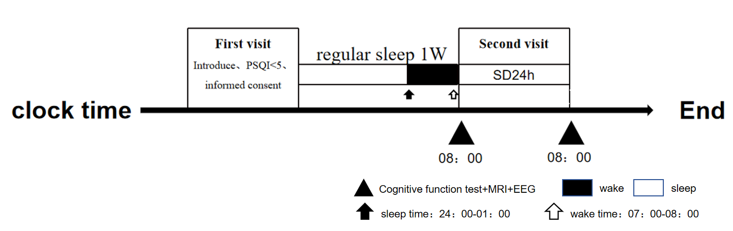
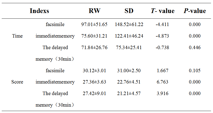
Table 1: ROCFT data in the RW state and after 24 h SD- The participants exhibited significantly extended durations in copying and immediate recall scores following 24h SD, with no significant differences observed in 30-minute delayed recall. In contrast, both immediate recall and 30min delayed recall scores were significantly lower, while no significant differences were noted in the scores for copying.
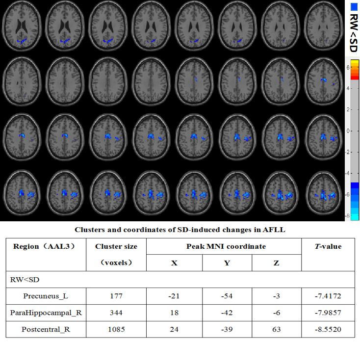
Figure 2: Whole-brain ALFF changes in RW and TSD states-Following SD, the ALFF values of the Precuneus_L, ParaHippocampal_R, and Postcentral_R regions exhibited a significant increase according to the ALFF metrics-SD (P<0.001, FDR-corrected, cluster level voxels ≥100). The color red represents ALFF values in which RW>SD, while the color blue indicates ALFF values in which RW<SD.
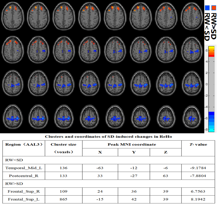
Figure 3:Whole-brain ReHo changes in RW and TSD states-Following SD, ReHo values in the Postcentral_R and Temporal_Mid_L regions showed a significant increase, while those in the Frontal_Sup_R and Frontal_Sup_L regions exhibited a significant decrease (P<0.001, FDR-corrected, cluster level voxels ≥100). The color red represents ReHo values with RW>SD, while the color blue indicates ReHo values with RW<SD.
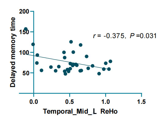
Figure 4: The correlation between the Temporal_Mid_L ReHo and the delayed memory time-A Pearson correlation analysis revealed a negative correlation between the ReHo value of Temporal_Mid_L after sleep deprivation (SD) and the 30-minute delayed recall time.