4964
Functional connectome phenotype of multiple cerebrovascular disease markers and its interaction with plasma p-tau181 on downstream outcomes1Centre for Sleep and Cognition & Centre for Translational Magnetic Resonance Research, Yong Loo Lin School of Medicine, National University of Singapore, Singapore, Singapore, 2Saw Swee Hock School of Public Health, National University of Singapore and National University Health System, Singapore, Singapore, 3Memory Ageing & Cognition Centre, National University Health System, Singapore, Singapore, 4Department of Pharmacology, Yong Loo Lin School of Medicine, National University of Singapore, Singapore, Singapore, 5St Luke’s Hospital, Singapore, Singapore, 6Raffles Neuroscience Centre, Raffles Hospital, Singapore, Singapore, 7Integrative Sciences and Engineering Programme (ISEP), National University of Singapore, Singapore, Singapore, 8Department of Electrical and Computer Engineering, National University of Singapore, Singapore, Singapore
Synopsis
Keywords: Functional Connectivity, Alzheimer's Disease, Cerebrovascular disease
Motivation: Cerebrovascular disease (CeVD) is assessed by several MRI markers, but their impact on brain functional connectivity (FC) remains unclear.
Goal(s): To examine how multiple CeVD markers influence FC, and how CeVD-related FC changes interact with Alzheimer’s disease pathology to influence downstream outcomes.
Approach: We studied multivariate associations between four CeVD markers and whole-brain FC in 529 participants, and how this CeVD-related FC phenotype interacted with plasma p-tau181 to influence longitudinal brain atrophy and cognitive decline.
Results: We identified a FC phenotype linked to high CeVD burden across all markers. This phenotype and p-tau181 contributed additively, but not synergistically, to atrophy and cognitive decline.
Impact: Using a multivariate approach, our study demonstrated that CeVD exerted widespread, non-MRI marker-specific effects on the whole-brain functional connectome. Further, we showed that AD and CeVD have additive but not synergistic effects on neurodegeneration and cognitive changes over time.
Introduction
Cerebrovascular disease (CeVD) is a major cause of cognitive impairment in older adults1, and frequently co-occurs with Alzheimer’s disease (AD)2. Despite the focal nature of its lesions, CeVD is associated with a wide range of clinical symptoms and thus has global effects3. In this regard, fMRI-derived functional connectivity (FC) measures have been instrumental in understanding how CeVD leads to diverse symptoms via its effects on the functional connectome3,4. Disrupted FC across large-scale distributed networks has been reported in individuals with CeVD3,4, and further linked to poorer cognition5,6. To date, studies have typically quantified CeVD burden using a single MRI marker. However, CeVD is assessed by several MRI markers that are heterogeneous in aetiology and disease severity3. As such, CeVD burden may not be fully reflected by a single marker. Our study thus sought to examine the effects of multiple CeVD MRI markers on whole-brain FC using a multivariate approach. Given the global effects of CeVD3, we hypothesize that CeVD would be linked to widespread FC alterations in a non-marker-specific manner. Further, we investigated how such CeVD-related FC changes interacted with plasma p-tau181, an AD marker, to influence longitudinal changes in neurodegeneration and cognition/behaviour. Converging evidence suggests additive, rather than synergistic effects of AD and CeVD on cognitive decline7-11. Accordingly, we hypothesized that expression of the CeVD-related FC phenotype and p-tau181 would have additive, but not synergistic effects on cognition/behaviour and grey matter volumes.Methods
Cross-sectional task-free fMRI, CeVD (four MRI markers: age-related white matter changes score (measuring white matter hyperintensity), number of lacunes, cerebral microbleeds and cortical microinfarcts) and plasma p-tau181 data, and longitudinal T1-weighted and cognitive data from 529 participants across the dementia spectrum12 were analyzed. Individual FC matrices were first computed for 114 cortical13 and 30 subcortical14,15 regions. FC and CeVD residuals were then obtained by regressing out age, sex, ethnicity, education, handedness, and total intracranial volume (TIV). To derive the CeVD-related FC phenotype, multivariate associations between FC and CeVD residuals were examined using partial least squares correlation (PLSC). Brain scores, indicating the extent to which each individual expressed the CeVD-related phenotype, were then derived from the PLSC analyses. Finally, main and interactive effects of brain scores and log-transformed plasma p-tau181 on baseline and longitudinal changes in grey matter volumes and cognition/behaviour were examined using linear mixed-effects models, with time as random effect, and age, sex, ethnicity, education, handedness, diagnosis, TIV, p-tau181 and brain score as fixed effects.Results
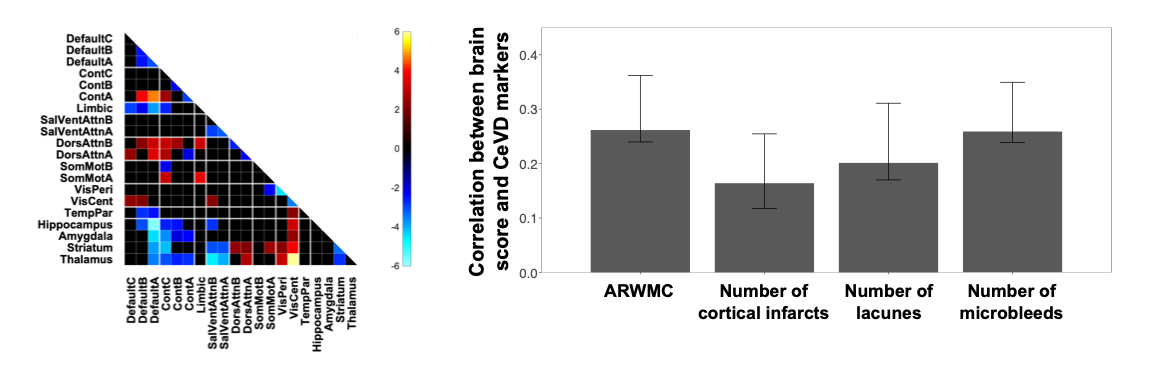
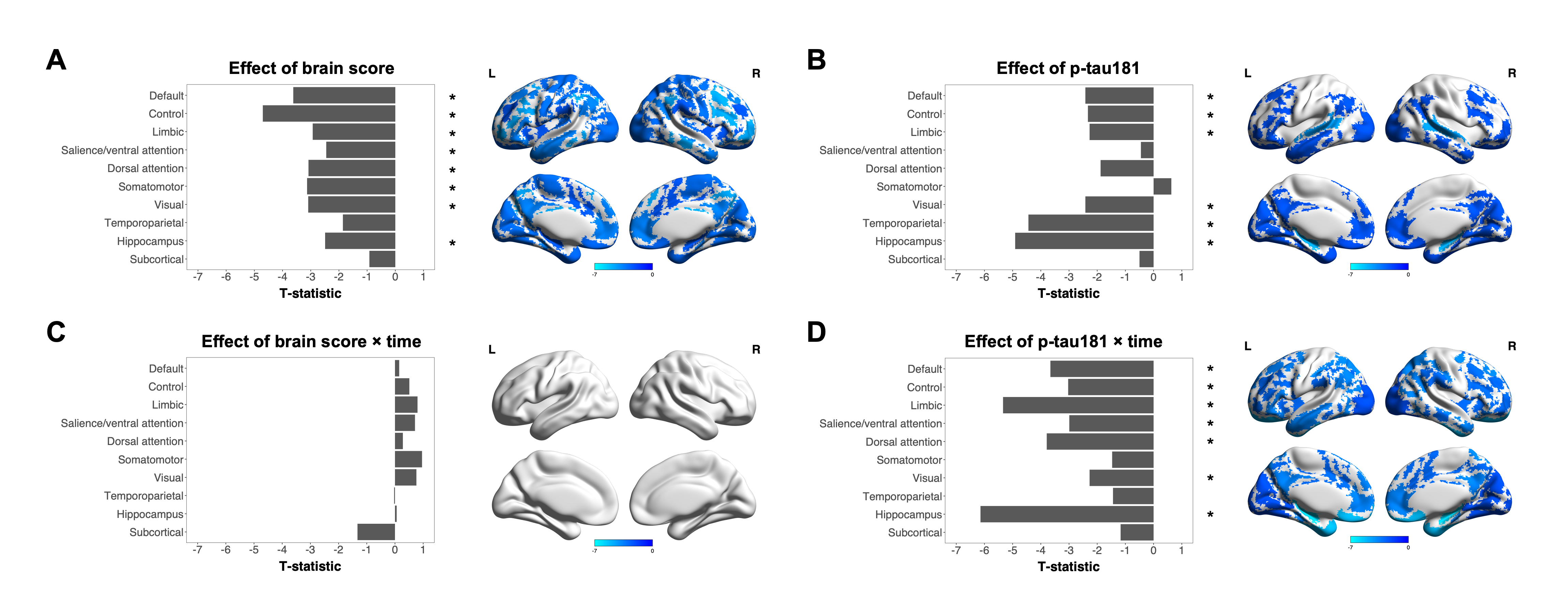
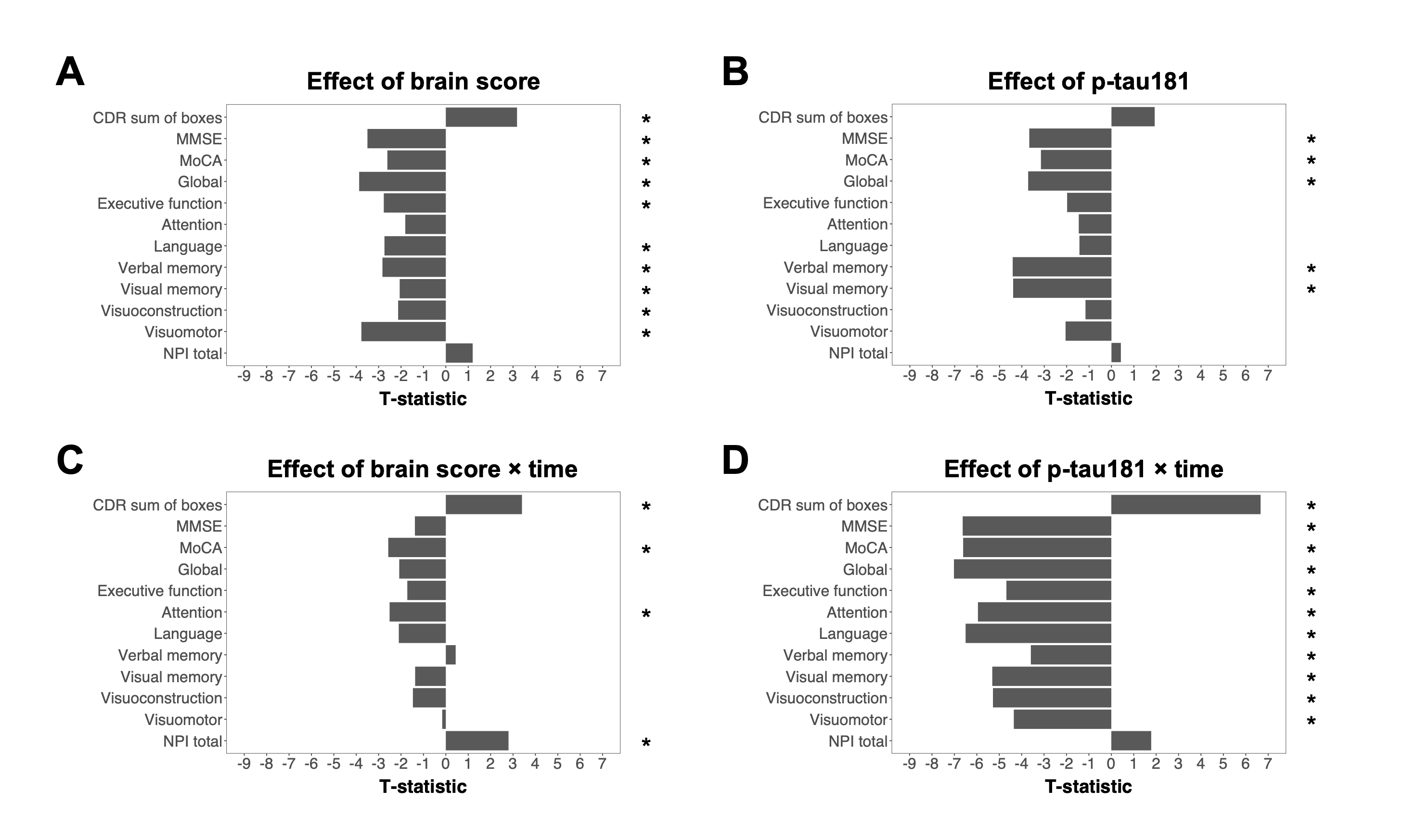
PLSC analyses revealed a significant latent variable that explained 76.6% of the covariance between FC and CeVD markers (Figure 1). This phenotype was linked to high burden across all CeVD markers, and characterized by lower within-network and higher between-network cortical FC, as well as lower subcortical FC to associative networks but higher subcortical FC to sensorimotor networks. Further, we demonstrated additive, but not synergistic effects, of the CeVD-related FC phenotype and p-tau181 on grey matter volumes and cognition/behaviour. Brain scores had widespread effects on baseline grey matter volumes and cognition, but only influenced longitudinal changes in global cognition and neuropsychiatric symptoms. By comparison, p-tau181 showed circumscribed effects on baseline cognitive performance of AD-related domains (e.g., memory) and grey matter volumes of AD-related regions, but widespread effects on atrophy and cognition over time (Figures 2-3).Discussion
We identified a phenotype that was linked to high CeVD burden across all markers and widespread FC changes, supporting studies showing widespread, global effects of CeVD on FC3,4. Importantly, we demonstrated that expression of the CeVD-related FC phenotype and p-tau181 showed additive, but not synergistic effects on both baseline and longitudinal changes in grey matter volumes and cognitive/behavioral performance, corroborating previous studies showing differential effects of AD and CeVD on cognitive function7,11, cortical thickness7, and FC9,10. Specifically, the effects of CeVD-related phenotype on neurodegeneration and cognition were widespread at baseline but limited over time, which is consistent with the global, but heterogeneous effects of CeVD on clinical symptoms3. Conversely, the effects of p-tau181 were limited to AD-related cognitive domains and brain regions at baseline, but widespread across multiple brain networks and cognitive domains over time, which mirrors AD symptom progression from early memory impairments to more widespread neurodegeneration and cognitive deficits at later stages16,17.Conclusion
Using a multivariate approach, we found widespread, non-MRI marker-specific effects of CeVD on FC. Further, we demonstrated that expression of this phenotype and plasma p-tau181 had additive, but not synergistic effects on baseline and longitudinal changes in neurodegeneration and cognition/behaviour. Collectively, our findings provide insight on the effects of CeVD on functional networks in older adults, which may be a therapeutic target.Acknowledgements
This study was supported by the National Medical Research Council Singapore Translational Research Investigator Award (MOH-000707-00) (C. L.-H. Chen), National Medical Research Council NMRC/CIRG/1485/2018 (C. L.-H. Chen), National Medical Research Council Centre Grant - NMRC/CG/M009/2017_NUH/NUHS (C. L.-H. Chen) and National Medical Research Council Senior Clinician Scientist Award 2017-2022 (NMRC/CSA-SI/0007/2016) (C. L.-H. Chen). This work was also supported by the Singapore National Medical Research Council (NMRC/OFLCG19May-0035, NMRC/CIRG/1485/2018, NMRC/MOH-00707-01, NMRC/OFLCG19May-0035) (J. H. Zhou), RIE2020 AME Programmatic Fund from A*STAR, Singapore (No. A20G8b0102) (J. H. Zhou) and Yong Loo Lin School of Medicine Research Core Funding, National University of Singapore, Singapore (J. H. Zhou).References
- Fitzpatrick, A. L. et al. Incidence and prevalence of dementia in the Cardiovascular Health Study. J Am Geriatr Soc. 52, 195-204, doi:10.1111/j.1532-5415.2004.52058.x (2004).
- Arvanitakis, Z., Capuano, A. W., Leurgans, S. E., Bennett, D. A. & Schneider, J. A. Relation of cerebral vessel disease to Alzheimer's disease dementia and cognitive function in elderly people: a cross-sectional study. The Lancet Neurology. 15, 934-943, doi:10.1016/s1474-4422(16)30029-1 (2016).
- Ter Telgte, A. et al. Cerebral small vessel disease: from a focal to a global perspective. Nat Rev Neurol. 14, 387-398, doi:10.1038/s41582-018-0014-y (2018).
- Schulz, M., Malherbe, C., Cheng, B., Thomalla, G. & Schlemm, E. Functional connectivity changes in cerebral small vessel disease - a systematic review of the resting-state MRI literature. BMC Med. 19, 103, doi:10.1186/s12916-021-01962-1 (2021).
- Zhou, X. et al. Aberrant Functional Connectivity and Structural Atrophy in Subcortical Vascular Cognitive Impairment: Relationship with Cognitive Impairments. Frontiers in Aging Neuroscience. 8, 14, doi:10.3389/fnagi.2016.00014 (2016).
- Ding, J. R. et al. Altered connectivity patterns among resting state networks in patients with ischemic white matter lesions. Brain imaging and behavior. 12, 1239-1250, doi:10.1007/s11682-017-9793-9 (2018).
- Kim, H. J. et al. Relative impact of amyloid-beta, lacunes, and downstream imaging markers on cognitive trajectories. Brain : a journal of neurology. 139, 2516-2527, doi:10.1093/brain/aww148 (2016).
- Toledo, J. B. et al. Contribution of cerebrovascular disease in autopsy confirmed neurodegenerative disease cases in the National Alzheimer's Coordinating Centre. Brain : a journal of neurology. 136, 2697-2706, doi:10.1093/brain/awt188 (2013).
- Chong, J. S. X. et al. Amyloid and cerebrovascular burden divergently influence brain functional network changes over time. Neurology. 93, e1514-e1525, doi:10.1212/wnl.0000000000008315 (2019).
- Chong, J. S. X. et al. Influence of cerebrovascular disease on brain networks in prodromal and clinical Alzheimer's disease. Brain : a journal of neurology. 140, 3012-3022, doi:10.1093/brain/awx224 (2017).
- Vemuri, P. et al. Vascular and amyloid pathologies are independent predictors of cognitive decline in normal elderly. Brain : a journal of neurology. 138, 761-771, doi:10.1093/brain/awu393 (2015).
- Hilal, S. et al. Markers of cardiac dysfunction in cognitive impairment and dementia. Medicine (Baltimore). 94, e297, doi:10.1097/MD.0000000000000297 (2015).
- Yeo, B. T. et al. The organization of the human cerebral cortex estimated by intrinsic functional connectivity. Journal of neurophysiology. 106, 1125-1165, doi:10.1152/jn.00338.2011 (2011).
- Choi, E. Y., Yeo, B. T. & Buckner, R. L. The organization of the human striatum estimated by intrinsic functional connectivity. Journal of neurophysiology. 108, 2242-2263, doi:10.1152/jn.00270.2012 (2012).
- Tzourio-Mazoyer, N. et al. Automated anatomical labeling of activations in SPM using a macroscopic anatomical parcellation of the MNI MRI single-subject brain. Neuroimage. 15, 273-289, doi:10.1006/nimg.2001.0978 (2002).
- Weintraub, S., Wicklund, A. H. & Salmon, D. P. The neuropsychological profile of Alzheimer disease. Cold Spring Harb Perspect Med. 2, a006171, doi:10.1101/cshperspect.a006171 (2012).
- Scahill, R. I., Schott, J. M., Stevens, J. M., Rossor, M. N. & Fox, N. C. Mapping the evolution of regional atrophy in Alzheimer's disease: unbiased analysis of fluid-registered serial MRI. Proceedings of the National Academy of Sciences of the United States of America. 99, 4703-4707, doi:10.1073/pnas.052587399 (2002).
Figures