4963
Elucidation of whole-brain network in operant training using functional connectivity and immediate gene expression1Human Informatics and Interaction Research Institute, National Institute of Advanced Industrial Science and Technology, Tsukuba, Japan, 2Neural Computation Unit, Okinawa Institute of Science and Technology Graduate University, Okinawa, Japan, 3Health and Medical Research Institute, National Institute of Advanced Industrial Science and Technology, Tsukuba, Japan, 4University of Tsukuba, Tsukuba, Japan
Synopsis
Keywords: Functional Connectivity, Neuroscience
Motivation: It is unclear how and what cellular-level changes cause changes in the whole-brain network in operant training.
Goal(s): To investigate the functional network changes and underlying cellular processes involved in operant learning.
Approach: We utilized resting-state functional magnetic resonance imaging (rsfMRI) and whole-brain immunohistochemical analysis of early growth response 1 (EGR1) in mice during the early and late stages of training.
Results: Increased functional connectivity and EGR1 regional correlations were observed between the limbic and thalamus or auditory cortex early, and between motor and somatosensory cortex and striatum in the late stage.
Impact: Our study is an initial effort to create a new experimental approach that combines rsfMRI and immunohistochemistry to connect large-scale and small-scale mechanisms of learning.
Introduction
Operant conditioning, the process of acquiring new skills, is used for training animals to learn certain behaviors by associating new actions, such as lever pulling, button pressing, and nose pokes, with rewards. The process involves specific regions of the brain, such as the amygdala, basal ganglia, frontal cortex, and lateral septum, which are responsible for fear learning, reward prediction, decision-making, and social playing. Electrophysiological experiments have shown enhanced activity and interactions in these regions, but most studies have focused on individual regions with implanted electrodes1,2, leaving the whole brain network unclear. To address this, we hypothesized that immunohistochemical analyses for EGR1, the protein product of Egr1, and resting-state functional connectivity (FC) could be used to reveal when, where, and how learning changes connectivity at the cellular (EGR1 expression) and macro (resting-state FC) level3,4. We used histological analysis of an immediate early gene and rsfMRI to investigate spatiotemporal functional network shifts in resting-state FC and neural activity before and after early-stage and late-stage training.Method
The study used 64 adult male C57/BL6N mice that were aged 7 weeks old. The Ethics Committee (approval number: 2017-178, 2020-0362) approved all procedures. The six groups of mice were observed to study the temporal and spatial changes in the whole-brain FC and cellular-level responses with operant conditioning (Fig. 1). Mice were trained to press two different buttons in order, left and then right. If they pressed the buttons in the correct sequence, they were given sucrose water. However, if they made a mistake, they were punished with a flash of light and had to wait 2 seconds before trying again. The rsfMRI was obtained using a Bruker BioSpec 117/11 11.7 Tesla MRI scanner for 10 min (repetition time = 2000 ms, echo time = 15 ms, 300 scans). All mice were initially anesthetized with 4% isoflurane, with 2% isoflurane during set-up on the animal bed, and 1.2% isoflurane with air for rsfMRI. We calculated the fMRI signals in 142 regions, excluding the cerebellum, which was divided based on the Allen Brain Atlas, and estimated the functional connectivity between these regions using CONN. Two-hundred and sixteen sections, excluding the olfactory bulb and cerebellum, were selected for histological analysis of EGR1-immunopositive cells. Each brain region that had significant resting-state FC was captured with a digital camera mounted to a microscope. We counted the number of EGR1-immunopositive cells on each side of the hemisphere within the targeted region with ilastik and ImageJ Fiji.Results
After three days of training, resting-state functional connectivity (rsFC) increased between the limbic areas and the thalamus or auditory cortex (Fig. 2a). After 21 days of training, the increase in rsFC primarily occurred between the motor cortex, the somatosensory cortex, and the striatum (Fig. 3a). We also tested for inter-regional correlation in the numbers of EGR-1-positive cells. During the early stage, Pearson's r in the training group was significantly higher than that in the no-training group between the auditory cortex and the amygdala, between motor cortices, and between motor and primary somatosensory cortices (p < 0.05). However, r between the auditory and primary somatosensory cortices and the auditory cortex and the striatum was significantly lower in the training group (Fig. 2b). During the later stage, r was found to be significantly higher in the training group as compared to the no-training group between the primary somatosensory cortex and the striatum, the primary somatosensory cortex and the motor cortex, and the primary somatosensory cortex and the auditory cortex (Fig. 3b, p < 0.05).Discussion
The study reveals how the combination of resting-state fMRI and immunohistochemistry can uncover the cellular-level changes underlying whole-brain dynamics. It also demonstrates the spatiotemporal shift of network dynamics during different phases of training. During the early stages of training, subcortical networks of the limbic system related to reward were dominant, whereas during the late stages of training, cortical networks related to consolidation and reconsolidation formed stronger connections. This study successfully visualized the temporal shifts in brain regions involved in behavioral learning and demonstrated their plasticity for the first time by combining MRI and histological analysis. It is the first step towards establishing an experimental system that combines both MRI and whole-brain immunohistochemical analysis.Abbreviation
Lateral septal nucleus ventral (LSv), lateral septal nucleus dorsal (LSd), central nucleus of the amygdala (CEA), dorsal auditory cortex (AUDd), striatum (Str), primary somatosensory upper limb (S1ul), secondary somatosensory (S2), primary motor (M1), and secondary motor (M2).Acknowledgements
This study was funded by the Japan Society for the Promotion of Science grants KAKANHI 21K19463 and KAKANHI 20H04236 (KK) and Japan Science and Technology Agency grant FORESTO JPMJFR206G (KK).References
1. Hamel L, Cavdaroglu B, Yeates D, et al. Cortico-Striatal Control over Adaptive Goal-Directed Responding Elicited by Cues Signaling Sucrose Reward or Punishment. J Neurosci. 2022:42:3811-3822.
2. Zimmermann KS, Yamin JA, Rainnie DG, et al. Connections of the Mouse Orbitofrontal Cortex and Regulation of Goal-Directed Action Selection by Brain-Derived Neurotrophic Factor. Biol Psychiatry 2017:81:366-377.
3. Sampaio-Baptista C, Filippini N, Stagg CJ, et al. Changes in functional connectivity and GABA levels with long-term motor learning. Neuroimage 2015:106:15-20.
4. Veyrac A, Besnard A, Caboche J, et al. The transcription factor Zif268/Egr1, brain plasticity, and memory. Prog Mol Biol Transl Sci 2014:122:89-129.
Figures
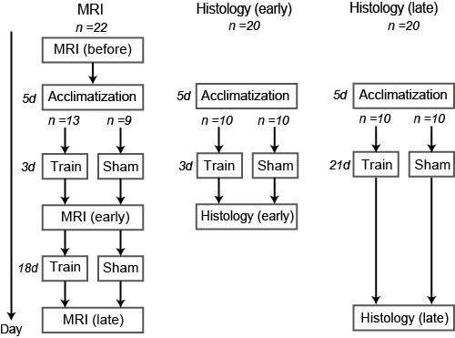
Figure 1 A schematic diagram of the experimental design.
62 mice were acclimated for 5 days, followed by MRI scans on 22 mice, with 13 receiving 21 days of behavioral training (Train) and 9 serving as a control group (No-train). Immunohistochemistry was performed on 40 mice, divided into groups that had 21 days or 3 days of behavioral training, or no training for the same duration.
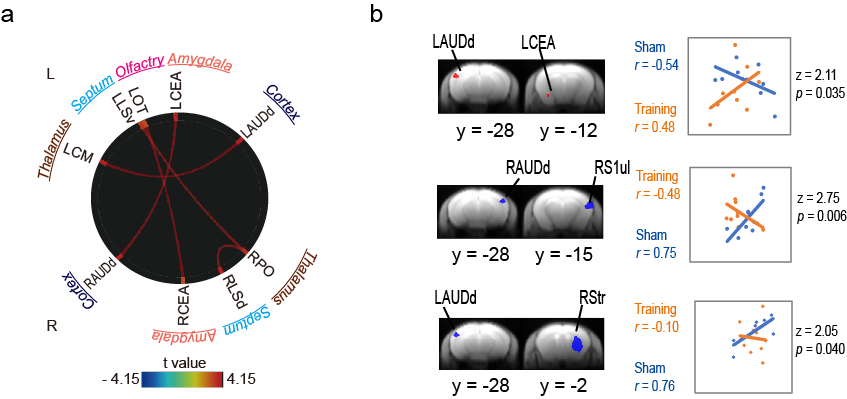
(a) Short-term (early) training enhanced resting-state functional connectivity in the auditory cortex and subcortical regions (thalamus, amygdala, septum, and olfactory), (b) In EGR-1, significant differences in correlations of LAUDd-LCEA, RM1-LM1, RM1-LS1ul, RAUDd-RS1ul, and LAUDd-RStr between no-training and training after 3 days (p < 0.05).
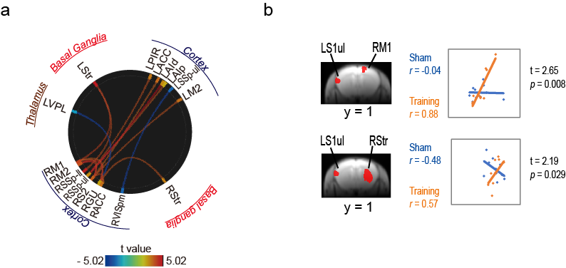
(a) Long-term (late) training enhanced resting-state functional connectivity in the cortex (motor, somatosensory, anterior cingulate, and gustatory) and striatum. Long-term training, by contrast, decreased resting-state functional connectivity between the thalamus and gustatory cortex and visual and somatosensory cortex. (b) In EGR1, significant differences in correlations of LS1ul-RM1, LS1ul-RStr, LS1ul-RM2, and LS1ul-LAUDd between no-training and training after 21 days (p < 0.05).