4962
Fast and accurate 31P B1-mapping at high magnetic fields with short TRs.1Animal imaging and technology core, CIBM Center for Biomedical Imaging, École Polytechnique Fédérale de Lausanne (EPFL), Lausanne, Switzerland, 2Laboratory for Functional and Metabolic Imaging, École Polytechnique Fédérale de Lausanne (EPFL), Lausanne, Switzerland, 3Animal imaging and technology core, CIBM Center for Biomedical Imaging, École polytechnique fédérale de Lausanne (EPFL), Lausanne, Switzerland, 4Laboratory for Functional and Metabolic Imaging, Ecole Polytechnique Fédérale de Lausanne (EPFL), Lausanne, Switzerland, 5Department of Radiology, Case Western Reserve University, Cleveland, Cleveland, OH, United States
Synopsis
Keywords: RF Pulse Design & Fields, RF Pulse Design & Fields, DAM, 31P, B1 mapping, fast B1, MRSI, X-Nuclei
Motivation: Fast and accurate B1 mapping for 31P MRI is challenging due to the low sensitivity of 31P nucleus and its long T1 relaxation times.
Goal(s): This study aims to develop a fast B1 mapping approach for 31P at 7T.
Approach: A look-up table approach was adopted to enable B1 mapping at short TR. Fast spiral encoding together with weighted averaging was implemented with a GRE sequence to further accelerate the acquisition.
Results: B1 mapping was validated in phantom and in vivo in human calf muscle and brain within a scan time of 10min.
Impact: The presented fast and accurate B1 mapping correction method is particularly suited for moderate to short repetition times, showing promise for future applications in rapid X-nuclei imaging.
Introduction
Proper knowledge about if the nominal flip angle is reached, is essential for many MRI experiments. B1 mapping is used to estimate the RF transmit field inhomogeneity. Various B1 mapping sequences, such as sa2rage [1], have been developed designed for 1H nuclei. However, their direct translation to X-nuclei MRI is challenging due to differences in sensitivity, relaxation properties, and chemical shifts. Double angle method (DAM) [2,3,4] is a common B1 mapping method, it involves acquisition of two images, one with angle α1 and the other with α2=2α1. The achieved flip angle is estimated by αe=acos(Sα2/ (2*Sα1)). The downside of this method is that it needs to be ensured that the system is fully relaxed. Thus, the repetition time TR needs to be 3-5 times of T1. Especially for 31P, this results in long acquisition times or errors when using reduced TRs. Therefore, this study aims to introduce a correction method for short TR DAM using a lookup table approach for fast and accurate B1 mapping. GRE with a stack of spiral GRE and frequency selective pulse enables an efficient 31P B1-mapping approach at high magnetic fields in vivo. We show the phantom validation and preliminary in vivo results from human calf and human brain at 7T.Methods
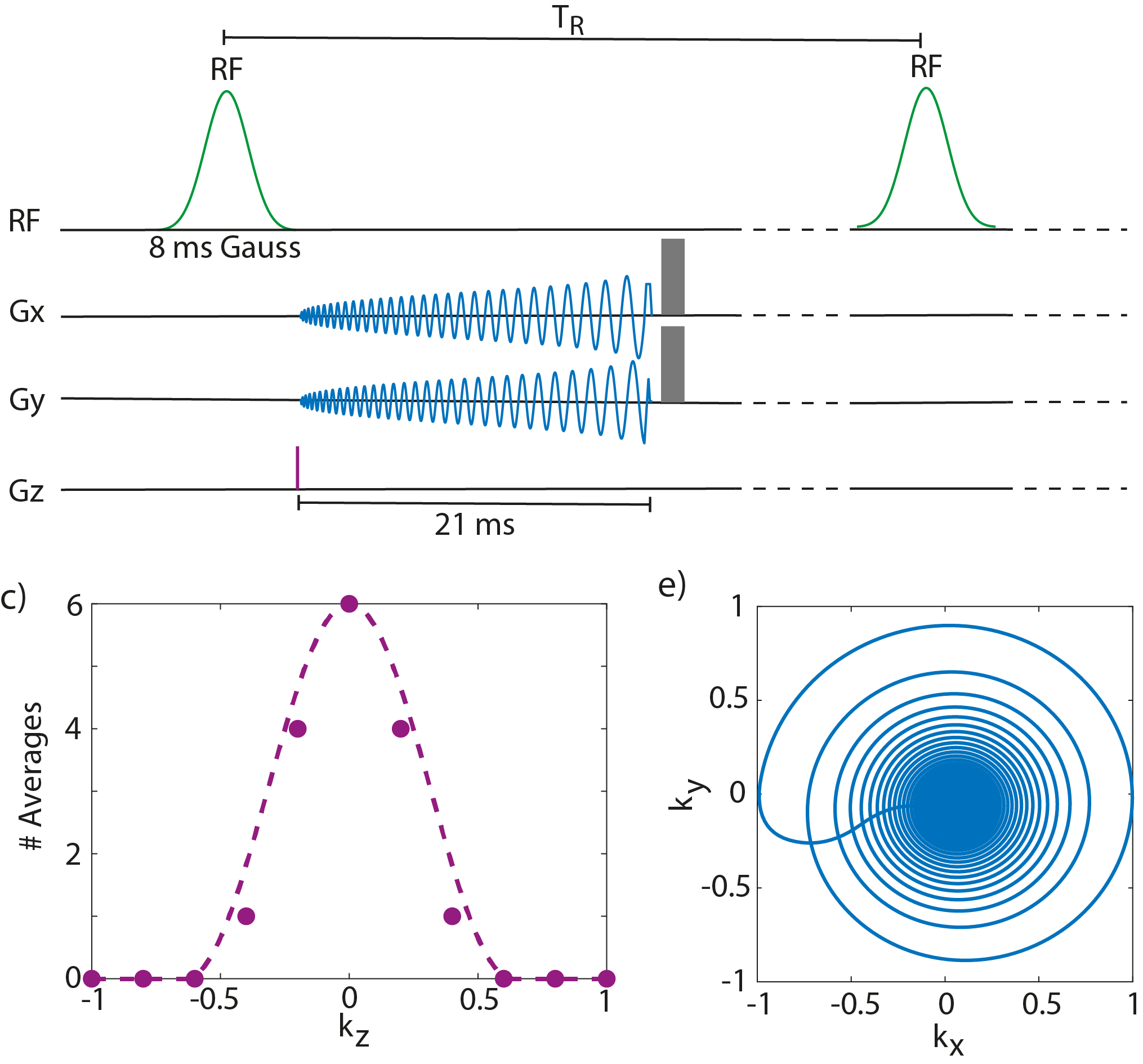
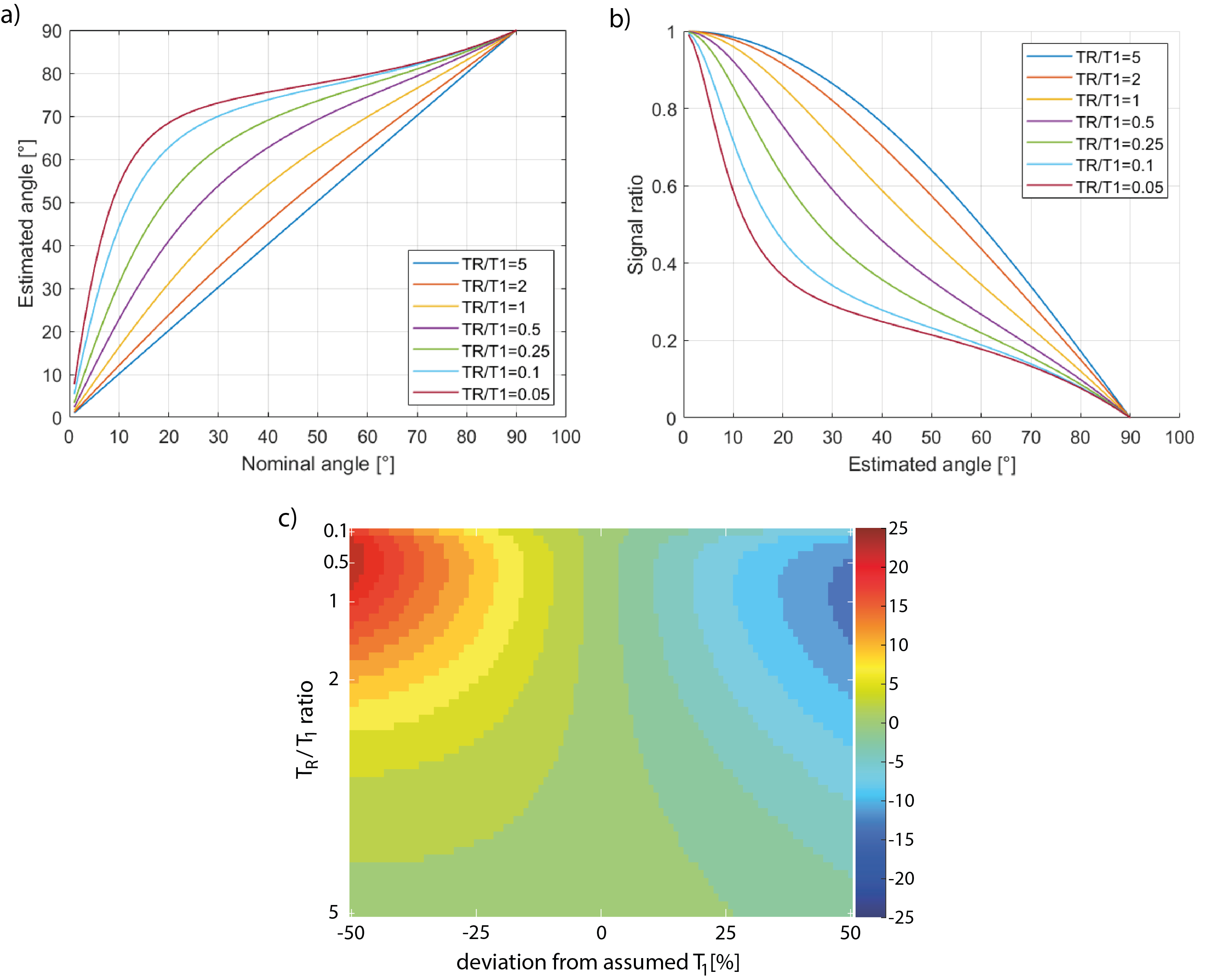
The look up table approach is displayed in Figure 2b. The look-up table is generated by calculating the signal ratios of two GRE signals with doubled flip angle α [5; equation (3)] for different B1 values. The T1 is therefore fixed assuming stable T1 relaxation times of PCr in vivo (human calf: 4s; human brain: 3.4s;[6,7]) and measured by inversion recovery experiments in vitro (50mM Pi phantom; T1=7.2s). All MR experiments were carried out on a 7T/68 cm MR scanner (Siemens Medical Solutions, Erlangen, Germany). The sequence used to acquire the 31P-GRE images is displayed in Figure 1. In-vivo data was measured on a single subject (male, 28 years old) using a 31P/1H birdcage 1Ch Rx/Tx coil for the calf muscle (TR=1.8s, α1=45°, 10 averages, 9:36 min, Vref=350V) and a 31P 32Ch-Rx/1Ch-Tx with 1H birdcage 1Ch Rx/Tx coil for the brain (TR=1.7s, α1=45°, 10 averages, 9:00 min, Vref=350V). The matrix size acquired in all experiments was 32 x 32 x 11 in a FOV of 230 x 230 x 220 mm3.Results/Discussion
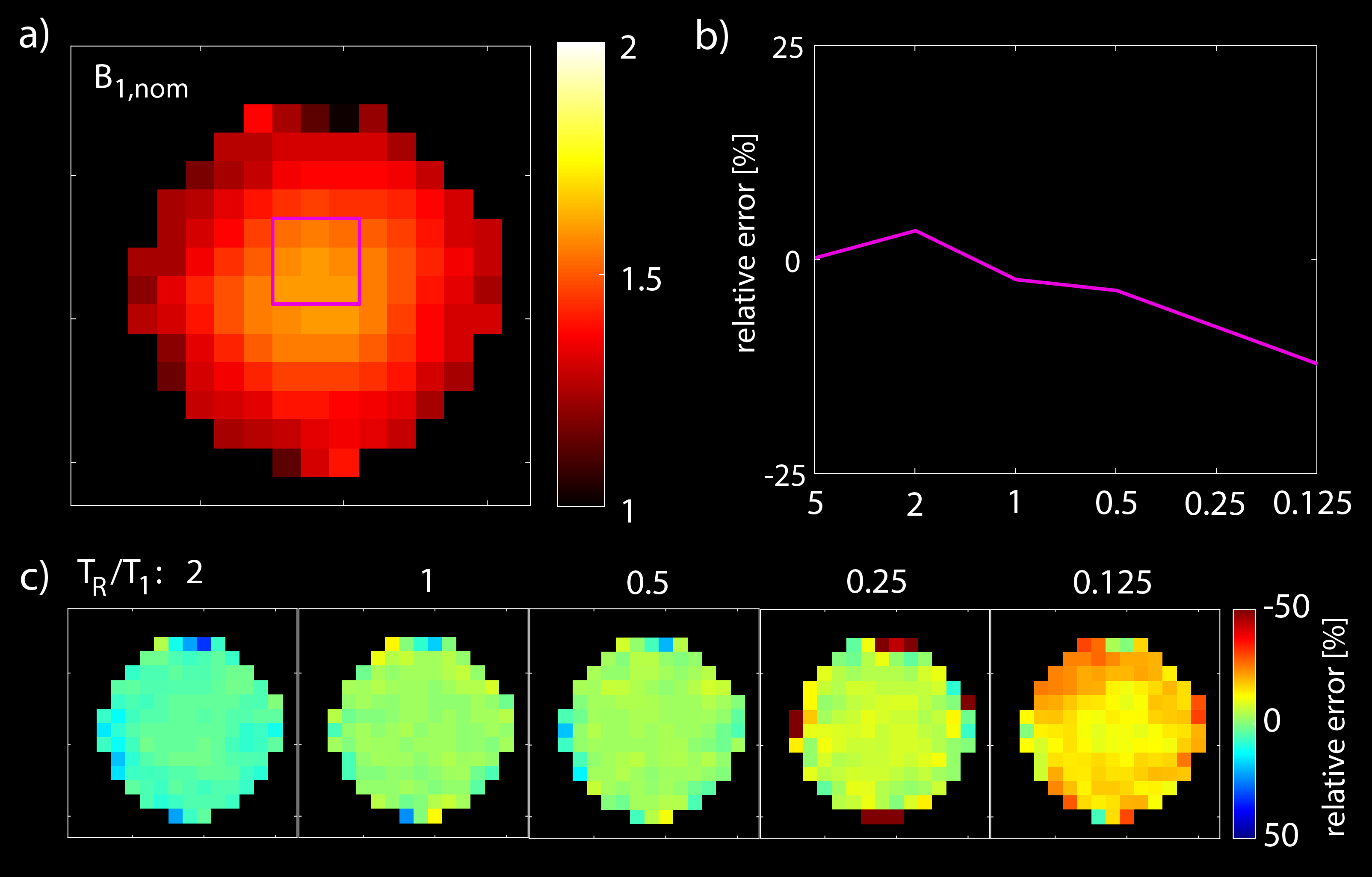
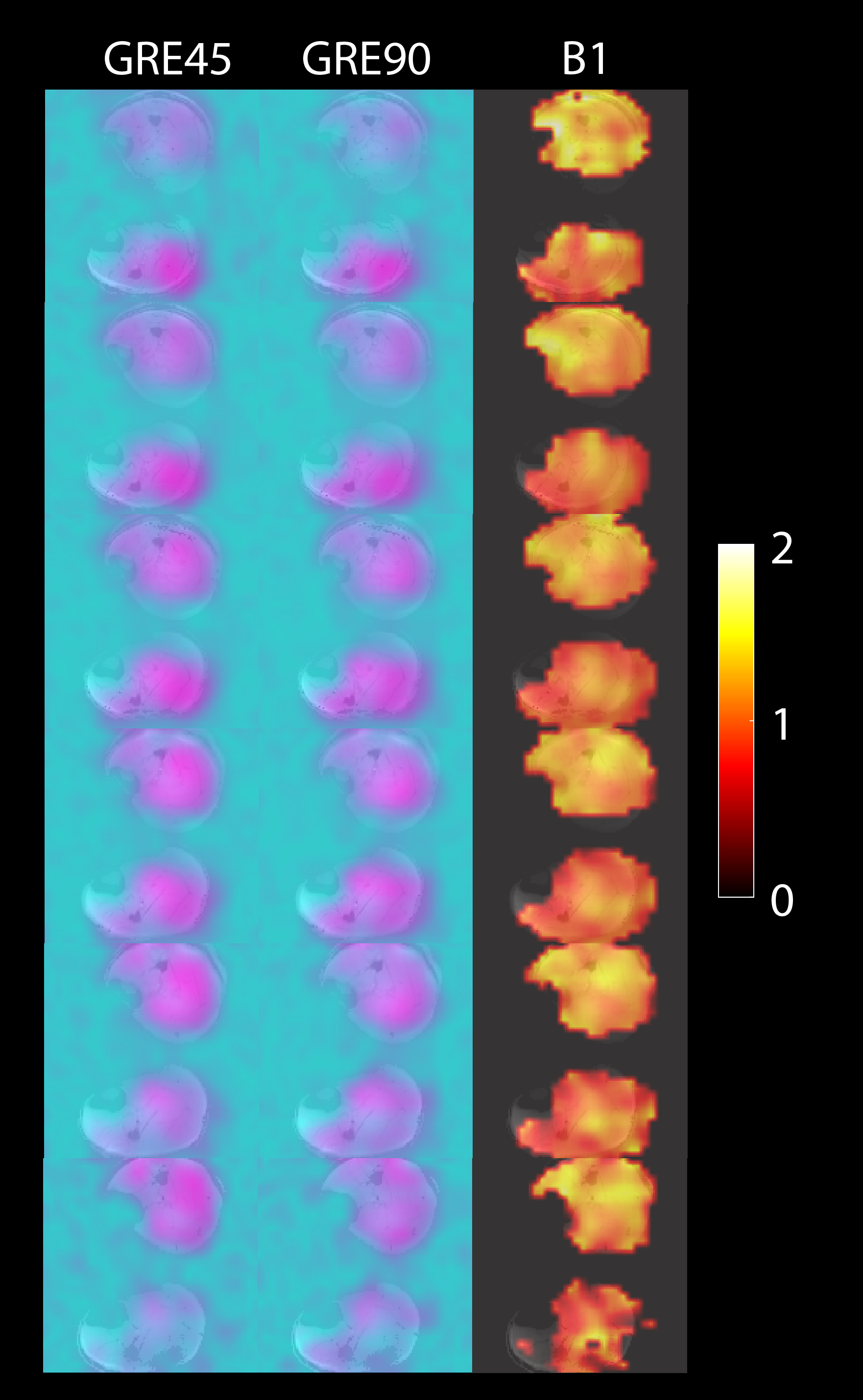
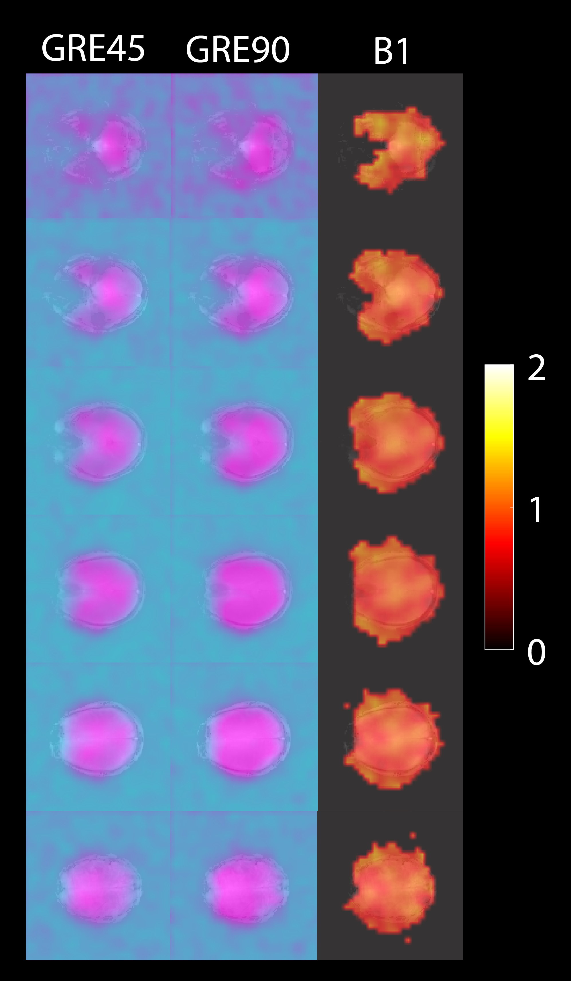
This study is investigating the effect of a wrongly assumed T1 value. The smaller the TR/T1, the more accurate the T1 needs to be assumed. However, the estimation bias does not exceed 15% as the T1 is assumed correctly in a 25% range around the ground truth. Figure 3 validates the method in vitro. For smaller TR/T1 ratio, the more underestimation is observed. The estimation error reaches about 14% for a TR/T1 ratio of 0.125. Figure 4 and 5 show the preliminary results of the short TR DAM in vivo in human calf muscle and brain, achieved in a scan time below 10 min. Both measurements are showing rather homogeneous B1 maps, which is expected for a birdcage transmit coil.Conclusion
The novel correction method introduced in this study demonstrated its effectiveness, particularly for moderate to short repetition times, showing promise for future applications in rapid X-nuclei imaging. Enhancements of this approach can be achieved by refining the quantification steps to create a more detailed lookup table.Acknowledgements
This work was supported by the Swiss National Science Foundation (grants n° 320030_189064). We acknowledge the CIBM Center for Biomedical Imaging for providing expertise and resources to conduct this study.References
[1] Eggenschwiler F, Kober T, Magill AW, Gruetter R, Marques JP. SA2RAGE: a new sequence for fast B1+ -mapping. Magn Reson Med. 2012 Jun;67(6):1609-19. doi: 10.1002/mrm.23145. Epub 2011 Aug 29. PMID: 22135168.
[2] Insko EK, Bolinger L. Mapping of the radiofrequency field. J Magn Reson A 1993;103:82-5.
[3] Cunningham CH, Pauly JM, Nayak KS. Saturated double-angle method for rapid B1+mapping. Magn Reson Med 2006;55:1326-33.
[4] Dowell NG, Tofts PS. Fast, accurate, and precise mapping of the RF field in vivo using the 180 degrees signal null. Magn Reson Med 2007;58:622-30.
[5] Ishimori Y, Shimanuki T, Kobayashi T, Monma M. Fast B1 mapping based on double-angle method with T1 correction using standard pulse sequence. J Med Phys 2022;47:93-8.
[6] Bogner, Wolfgang, et al. "Assessment of 31P relaxation times in the human calf muscle: a comparison between 3 T and 7 T in vivo." Magnetic Resonance in Medicine: An Official Journal of the International Society for Magnetic Resonance in Medicine 62.3 (2009): 574-582.
[7] Lei, Hao, et al. "In vivo 31P magnetic resonance spectroscopy of human brain at 7 T: an initial experience." Magnetic Resonance in Medicine: An Official Journal of the International Society for Magnetic Resonance in Medicine 49.2 (2003): 199-205.
Figures