4959
A Novel 30-Channel Ultra-flexible Phased-Array for High-Resolution and Parallel Imaging Optimized MR Hand/Wrist/Brachial Plexus Imaging at 3T1MR ENGINEERING, GE HEALTHCARE, Aurora, OH, United States, 2MR Research, GE HEALTHCARE, Waukesha, WI, United States, 3Hospital for Special Surgery, New York, NY, United States
Synopsis
Keywords: RF Arrays & Systems, RF Arrays & Systems
Motivation: Conventional hand/wrist coils have larger loop sizes and limited number of elements, resulting in in-sufficient SNR and acceleration capability for imaging small anatomic structures.
Goal(s): To develop a small flexible coil for hand/wrist, which improves spatial resolution, SNR and acceleration with flexibility for brachial plexus imaging as well when combined with other coils.
Approach: A 30-Channel parallel imaging optimized coil was built with the small flexible dual loop, high impedance internal cable baluns, exceptionally low noise preamplifiers and highly flexible mechanical materials.
Results: This study shows higher SNR and improved acceleration capabilities for results for this novel ultra-flexible coil compared to conventional coils.
Impact: This novel coil design provides comfortable fitting, high-resolution and higher acceleration for in-vivo 3T hand/wrist/unilateral brachial plexus imaging.
INTRODUCTION
The hand and wrist regions comprise small structures and have non-convex anatomic shapes, which create challenges for routine coil designs. Currently, most commercially available hand/wrist coils are rigid and may not adequately conform to either small or large appendages. Flexible multi-purpose coils are valuable in imaging larger regions of the upper extremity, such as the shoulder and elbow joints, but provide inadequate SNR and acceleration capabilities for imaging small anatomic structures due to relatively large loop sizes and a limited density of elements (for a given, small field-of-view (FOV)). To increase flexibility, SNR, and parallel imaging capabilities, a 30-channel ultra-flexible, high-resolution, parallel imaging optimized phased-array coil for hand/wrist MRI was developed. A prototype of the flexible 30-channel phased-array coil was constructed using flexible fabric and miniaturized electronics modules. The prototype coil was evaluated on phantoms and human volunteers under an IRB-approved protocol.METHODS
A range of hand/wrist habiti were evaluated to ensure the coil accommodated hand/wrist sizes the 95th-percentile range of males in the United States. A novel ultra-flexible, high-resolution, and parallel imaging optimized 30-channel phased-array hand/wrist coil was developed using a small dual loop configuration, miniaturized feed boards and flexible materials [3, 4]. The small dual loops (OD: 5.7cm) were made from 0.136 mm-thick highly flexible copper PCBs with no lumped components for proton imaging at 3T (127.73 MHz) (Fig.1 (a), (b) and (c)). This small dual loop provided a higher Qunloaded/Qloaded ratio and SNR compared to a single loop, and the loaded loop impedance was also significantly higher allowing for the use of a higher inductor value in the lattice balun for impedance matching resulting in higher Tx/Rx decoupling impedance and lower component temperature. Full coverage of the hand/wrist (SI 24.5cm by RL 28.2cm) was achieved by 30 elements located on the flexible fabric. The flexible materials and loops provide a light-weight flexible conformal coverage (Fig. 1 (e)). Nearest neighboring elements were decoupled by overlapping and next nearest neighbor and distant elements were decoupled by preamplifiers that provide exceptionally low noise and are tolerant of a wide range of loop loading conditions. The coil layout (6 tiles (RL) by 5 tiles (SI)) was optimized for parallel imaging. Specifically, the coil layout was proposed to support a 1D acceleration factor of up to 5(R=5) with parallel imaging and a Multi-Band acceleration factor of 5(SI) for a 24cm FOV (Fig. 1 (a)). The dual loops were situated between two thermoplastic coated thin fabrics(1mm). A medical grade foil was used as the coil cover sleeve for biocompatibility and water sealing. The SNR and max g-factors for R=2, 3, 4 were measured in phantoms. The coil was developed and evaluated on a 3T GE Signa Premier scanner (GE Healthcare, Aurora, Ohio USA). A total of 18 subjects, including healthy volunteers and patients indicated for hand/wrist examinations, were imaged.RESULTS
The 30-Channel hand/wrist prototype coil provided higher resolution and highly accelerated images with phantoms compared to two conventional coils due to the optimized loop size/layout and proximity to the targeted anatomy. As shown in Figure 2 (a), (b) and (c), the 30ch prototype coil has low noise correlation. The 30-Channel prototype shows ~19% and ~18% higher SNR than conventional coil #1 and conventional coil #2 respectively at 5cm in the loading shell/ silicon oil phantom comparison shown in Figure 3 (a) and (b). Table 1 shows the maximum g-factor at 2, 3 and 4 for 1D acceleration in the axial plane. The 30-Channel hand/wrist prototype coil shows superior acceleration performance at higher accelerations compared to conventional coil2. High-resolution in-vivo imaging was performed using the optimized 30-Channel hand/wrist prototype coil. Figure 5 (a), (b) and (c) show the coronal proton density images with DL 50% (R=4), 8 cm FOV, 1.5 mm slice thickness using the 30-Channel hand/wrist prototype coil (c) compared to two conventional coils (a and b). The 30-Channel hand/wrist prototype coil shows superior SNR and acceleration performance at higher accelerations compared to two conventional coils. Figure 5 (d) shows an image example from a 3D Oblique Coronal STIR-FSE acquisition of the brachial plexus.DISCUSSION
The 30-Channel hand/wrist prototype coil provides ultra-flexibility, comfort, high-resolution and accelerated imaging with highly flexible dual loop conductors, miniaturized electronics, exceptionally low noise preamplifiers, and light-weight materials. The hand/wrist/brachial plexus regions can be imaged with a small FOV while still maintaining high spatial resolution. The 30-Channel hand/wrist prototype coil provided better SNR and improved acceleration compared to conventional coils for human volunteers.CONCLUSION
This novel coil design provides a comfortably fitting, high-resolution and higher acceleration for in-vivo 3T hand/wrist/unilateral brachial plexus imaging.Acknowledgements
We also thank Saban Kurucay and Dan Chirayath from GE Healthcare for their continued support.
References
1. Sneag DB., et al., High-resolution Brachial Plexus Imaging using 3-D Short Tau Inversion Recovery (CUBESTIR) with IV Gadolinium for Vascular Suppression. Proceedings ISMRM 2019.
2. Chhabra A., et al., High-resolution 3T MR neurography of the brachial plexus and its branches, with emphasis on 3D imaging. AJNR Am J Neuroradiology 2013 Mar; 34:486-97.
3. Rossman P., et al., Characterization of a new ultra-flexible low-profile RF receive coil technology. Proceedings ISMRM 2017, 763.
4. Vasanawala S.S., et al., Development and Clinical implementation of very light weight and highly flexible Air technology arrays. Proceedings ISMRM 2017, 755.
5. Roemer P.B., et al., The NMR phased array. MRM, 1990; 16(2):192-225.
6. Pressman K.P., et al., SENSE: sensitivity encoding for fast MRI. MRM, 1999;42:952–962.
Figures
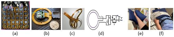
Fig. 1. (a) 30-Channel phased array layout for hand/wrist MRI (b) Dual loop with the miniaturized feedboard assembly (c) flexible loop PCB (d) Coil module schematic including the preamplifier, decoupler, matching, and baluns (e) Hand/Wrist imaging setup (f) Brachial Plexus imaging setup
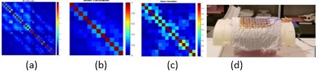
Fig. 2. Noise correlation matrices (a) 30-Channel hand/wrist prototype coil (b) Conventional coil #1 (c) Conventional coil #2 (d) Coil SNR test setup with loading shell and silicon oil brick phantom
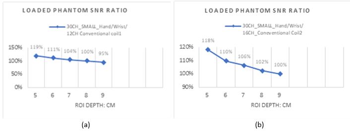
Fig. 3. Loaded shell with silicon oil brick phantom Sagittal SNR comparison results using sum of square reconstruction method (a) 30-Channel hand/wrist prototype coil vs. Conventional coil #1 (~19% higher SNR at 5cm depth) (b) 30-Channel hand/wrist prototype coil vs. Conventional coil #2 (~18% higher SNR at 5cm depth)
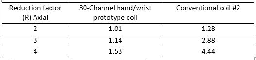
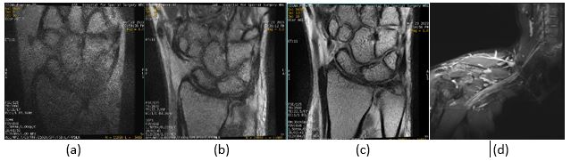
Fig. 5. Coronal proton density images with DL 50% (R=4), 8 cm FOV, 1.5 mm slice thickness using (a) 20-Channel medium conventional coil. (b) 16-Channel small conventional coil #2 (c) 30-Channel hand/wrist prototype coil. 30-Channel hand/wrist prototype coil shows superior acceleration performance at higher accelerations compared to both conventional coils. (d) Brachial Plexus Imaging with 3D Oblique Coronal STIR-FSE with 3D DL.