4956
Optimizing RF Coil Design to Minimize RF Interference in MR-Guided Microwave Liver Ablations at 1.5T1Neuroscience Research Institute, Gachon University, Incheon, Korea, Republic of, 2Department of Radiology, Mayo Clinic, Rochester, MN, United States, 3Department of Biomedical Engineering, Gachon University, Incheon, Korea, Republic of
Synopsis
Keywords: System Imperfections, Interventional Devices
Motivation: Clinical MRI-guided microwave ablations often suffer from noise due to electromagnetic interference (EMI) between ablation probes and imaging coils. Instead of modifying the clinical microwave ablation system that could compromise FDA-approval, we explore alternative coil designs to mitigate the noise.
Goal(s): Minimize EMI between the microwave ablation probes and imaging coils during clinical MR-guided microwave ablations.
Approach: We applied a band-pass filter, studying the impact of coil size, and investigating efficient coil combinations to reduce interference.
Results: A band-pass filter led to a 89% improvement in field uniformity. Also employing smaller coils can mitigate the ripple effects induced by the ablation probe.
Impact: Our study addresses the critical need to reduce electromagnetic interference (EMI) between microwave ablation probes and imaging coils during clinical MR-guided microwave ablations at 1.5T. Motivated by the interference challenges in current clinical setups, we develop methods to mitigate EMI.
Introduction
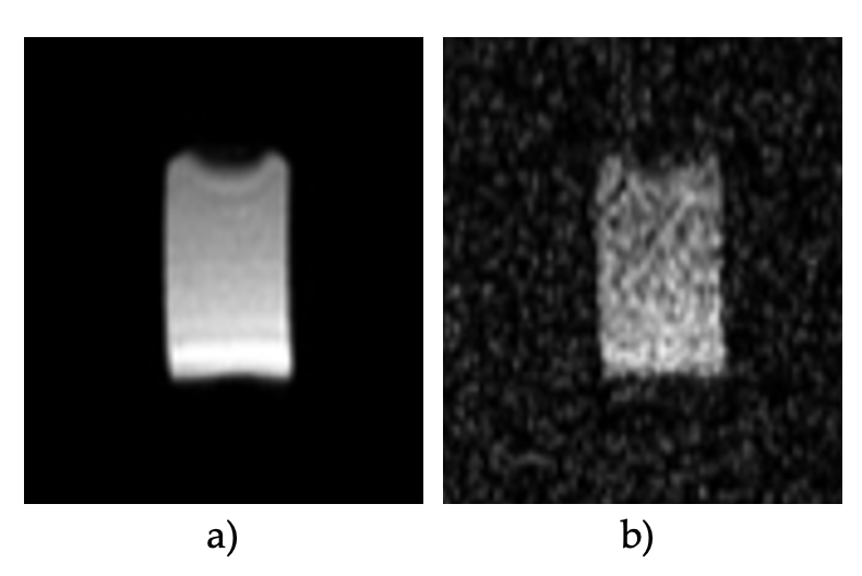
Magnetic resonance imaging (MRI) has shown as an important imaging modality to assist clinical intervention procedures, such as cancer treatments by using radio-frequency (RF) ablation [1]. RF ablation consists on destroying cancer cells by increasing the temperature to more than 60°C. One way to deliver the heat at the precise location, is the use of RF heating probes, which consist of a thin needle that is inserted on the body to the target tissue. These probes, while operating at high frequency and power, often introduce electromagnetic interference (EMI) in clinical settings. This interference can lead to reduced field intensity, loss of uniformity, and consequently, poor signal-to-noise ratio (SNR) or signal artifacts, as shown in Figure 1, for a phantom image without and with the RF ablation system turn on. Our work explores methods to mitigate EMI between RF ablation probes and imaging coils.Methods
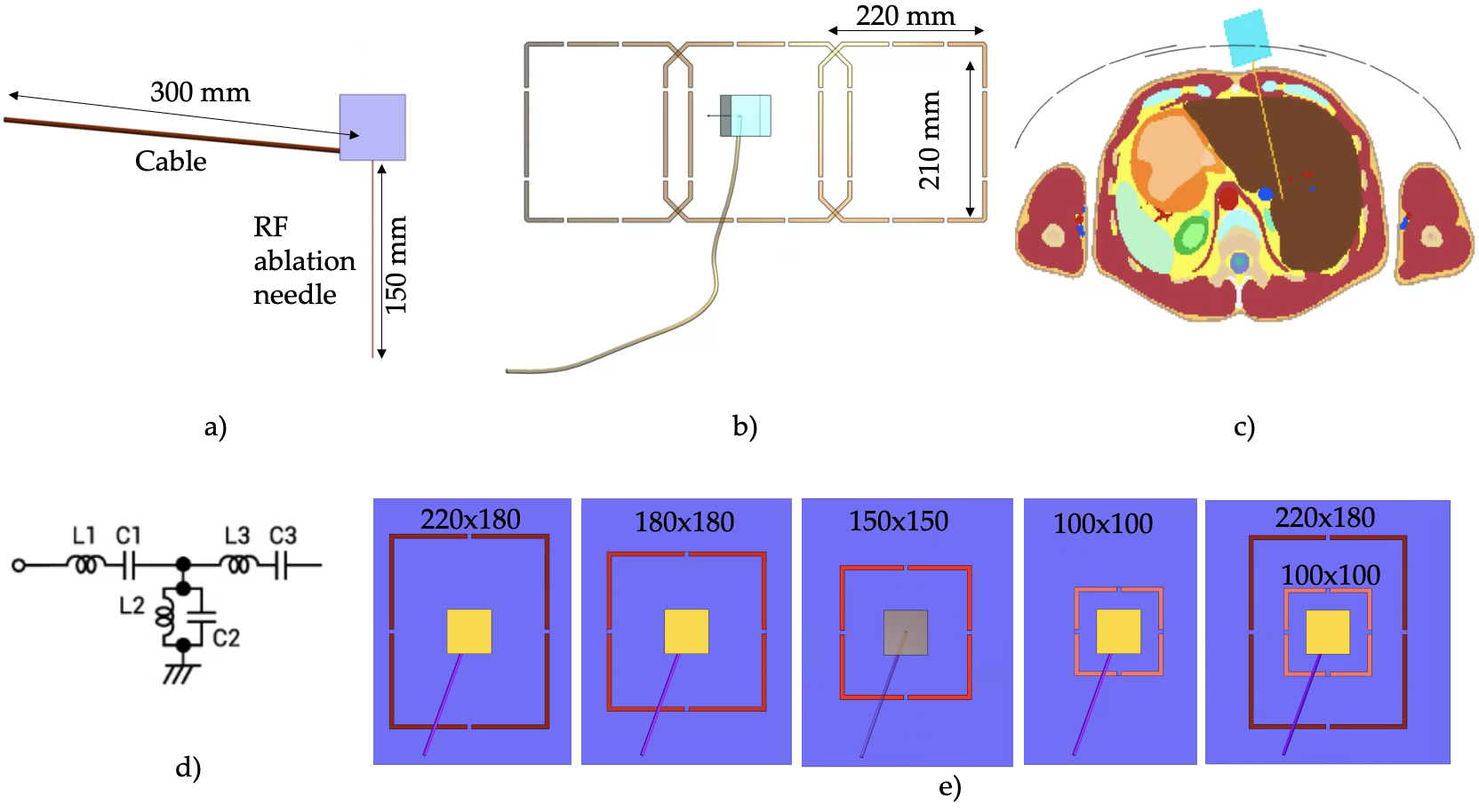
To explore methods for mitigating EMI, electromagnetic simulations were performed using a commercial software (Sim4life, Zurich, Switzerland). Two analyses are presented, the first analysis consist of including a band-pass filter to the RF ablation probe and assess its effectiveness when used in conjunction with a 3-ch loop array imaging coil focused on the liver area. The second analysis assumes that the RF ablation cannot be modified and only the RF coil can be optimized to reduce the interference between them.For the simulation, the design of the ablation probe is based on the one from the microwave system (MedWaves Inc., San Diego, CA) that is used clinically for MR-guided ablations (Fig. 2a). It consists of a needle of 0.5mm radius and 150 mm length. The microwave ablation probe was excited with a harmonic waveform of 900MHz and input power of 30W. The imaging coils comprise an array of three channels, each coil had dimensions of 220 by 210 mm (Fig. 2b). The coils were slightly curved to surround the shape of the human model (Fig. 2c). The coils were excited with a central frequency of 64MHz and a bandwidth of 1GHz. A human model was used [2], with the electrical properties to the corresponding frequency [3].In the first analysis, a bandpass filter of 3rd order based on LC network was applied to the RF ablation probe, as shown in Fig. 2d. The cut frequency was selected to 750 MHz.The second analysis involved modifying the coil size to reduce the interference. Figure 2e shows the various coil dimensions used in these analyses, including 220x180, 180x180, 150x150, 100x100 mm, and the combination of coils with two different sizes, such as 220x180 and 100x100mm. For this analysis we target a rectangular phantom measuring 280x250x300 mm, representing muscle tissue.For quantitative assessment, B1 field uniformity was determined using the coefficient of variation (CV) computed as the standard deviation scaled by the mean value.Results
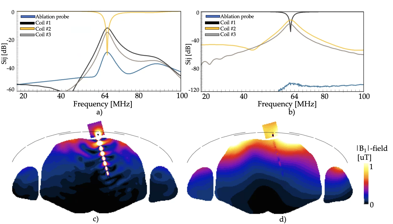
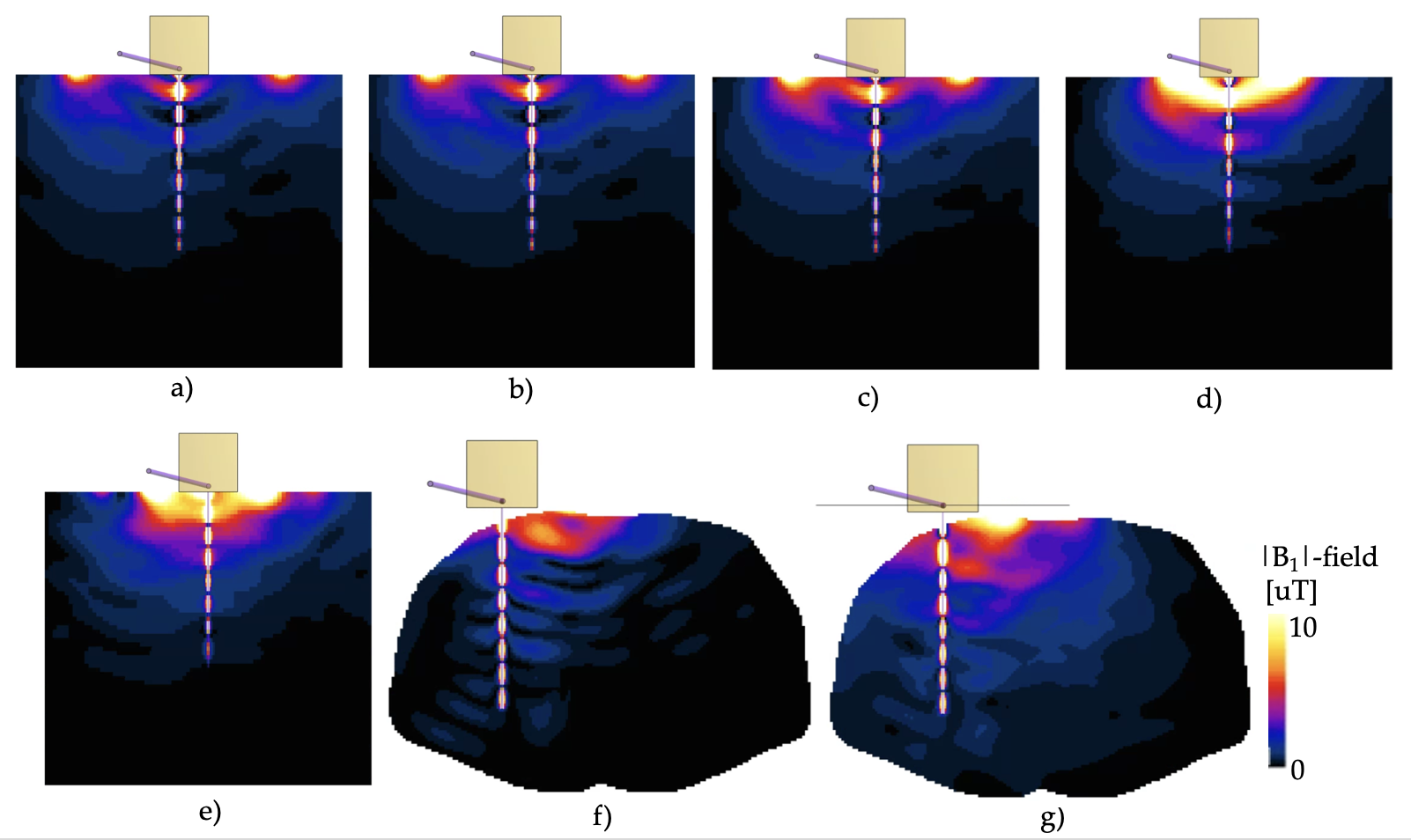
When a band-pass filter is applied to the RF ablation probe, the coupling changed from -30dB to -100dB (Fig. 3). Without the filter, the B1 field exhibits ripples along the y-axis, while with the filter applied, the B1 field becomes uniform with no noticeable distortion. The CV values before and after applying the filter were 1.37 and 0.14, respectively, indicating the improvement of 89 % in field uniformity.Figure 4 displays the computed B1 fields for each of the coils in Fig. 2e. The CV values for the B1 fields in Fig 4a-d were 2.5, 2.3, 2.1 and 2.0, respectively. It is noticeable that using a smaller coil size resulted in a more uniform B1 field and reduced interference from the microwave ablation probe. Furthermore, the combination of coils with two difference coil sizes yielded a mean and CV of 1.50 and 2.0, respectively, which contributed to improving field uniformity (Fig. 4e). These effect was also consistent in the human model simulation (Figs. 4f & 4g), resulting in a CV of 1.36 and 0.92 with and without the coil combinations, respectively.Conclusion
This work demonstrates that the RF ablation probe with a band-pass filter can reduce the interference between coils and RF ablation probe. This study indicates that the size of the coil can also be a useful method to reduce the interference.Acknowledgements
This research was supported by the K-Brain Project of the National Research Foundation (NRF) funded by the Korean government (MSIT) (No. RS-2023-00264160)References
1. Kilbride, B.F., 2022. MRI-guided endovascular intervention: current methods and future potential. Expert Review of Medical Devices, 19(10), pp.763-778.
2. Christ, 2009. The Virtual Family—development of surface-based anatomical models of two adults and two children for dosimetric simulations. Physics in Medicine & Biology, 55(2), p.N23.
3. https://itis.swiss/virtual-population/tissue-properties/database/
Figures