4955
Development and dynamic in vivo evaluation of a multi-channel stretchable self-tuning coil array1Weill Cornell Medicine, New York, NY, United States, 2Singapore University of Technology and Design, Singapore, Singapore, 3GE Healthcare, Aurora, OH, United States, 4Hospital for Special Surgery, New York, NY, United States
Synopsis
Keywords: RF Arrays & Systems, RF Arrays & Systems, liquid metal, stretchable RF coils
Motivation: Motivated by the limitations of traditional RF receive coils, this study aims to demonstrate in vivo imaging using a conformal and stretchable, self-tuning liquid metal coil array.
Goal(s): This study’s goal was to demonstrate improved signal-to-noise ratio (SNR) with the stretchable array compared to commercial coils.
Approach: We designed and fabricated a one-dimensional 6-channel stretchable coil array and tested it in vitro and in healthy volunteers using standard knee imaging sequences.
Results: In vitro and in vivo experiments demonstrated an SNR improvement of 4.7x over a dedicated commercial knee coil.
Impact: Our self-tuning stretchable coil array allows for maximized SNR and improved image quality due to its conformal fit and minimized distance from the target anatomy. This concept could also allow for dynamic imaging, leading to enhanced, clinically relevant, MRI applications
Introduction
Stretchable liquid metal-based RF receive coils offer enhanced sensitivity, adaptability to anatomical variations, and potential for dynamic imaging1-4. However, conductor stretching alters the resonance frequency, diminishing the signal-to-noise ratio (SNR) benefits of a closely fitting receive coil. To overcome this, a smart coil design with a self-tuning interdigital capacitor geometry was introduced5 and validated in a single coil element5 and a 2-channel array6. This study presents the fabrication and implementation of a one-dimensional self-tuning 6-channel stretchable receive array and demonstrates its performance in vitro and in vivo in the knee.Methods
Array fabrication: Coil elements were fabricated using direct ink writing (DIW)6. DragonSkinTM 30 silicone (Smooth-On) was spin-coated on a glass panel at 700 rpm for 40 seconds. Microchannel walls were printed on a DIW printer (SHOTmini200ΩX, Musashi, Japan), utilizing fast-curing silicone sealant (SpeedSeal) as the liquid ink. Liquid metal (GaIn) was injected into these microchannels. Copper wires were embedded at the terminals and connected to a printed circuit board containing tuning, matching, detuning, and preamplifier circuitry. Coil elements were then configured into a 1x6 array and bonded using fast-curing silicone adhesive (SIL-poxy by Smooth-On), forming a cylindrical array with a diameter of 125mm.In vitro imaging: Imaging was performed on a 3T MRI system (MR750, GE Healthcare). For in vitro experiments, the coil was loaded with a standard homogeneous cylindrical phantom (diameter=125mm, length=150mm). A T1-weighted (T1w) FSE sequence from a standard knee protocol was acquired (TR=400ms, TE=8.5ms, NEX=1, slice thickness=3mm, ETL=3, FOV=160x160mm2, resolution 0.3x0.3mm2).
In vivo imaging: For in vivo experiments, informed consent was obtained from two healthy volunteers under a locally approved IRB protocol. Two volunteers (male and female) were scanned using a proton-density weighted (PDw) sequence with axial slices using (a) the proposed 6-channel coil array and (b) a standard 8-channel knee coil array (GE Healthcare) (TR=537ms, TE=9ms, NEX=1, ETL=16, slice thickness=3, FOV=160x160mm2, resolution 0.3x0.3mm2). To demonstrate feasibility of imaging in different positions, we scanned one volunteer using a sagittal T1w sequence with the knee (a) fully extended and (b) flexed (approximately 30 degrees); note that this is not possible with the commercial coil.
Results
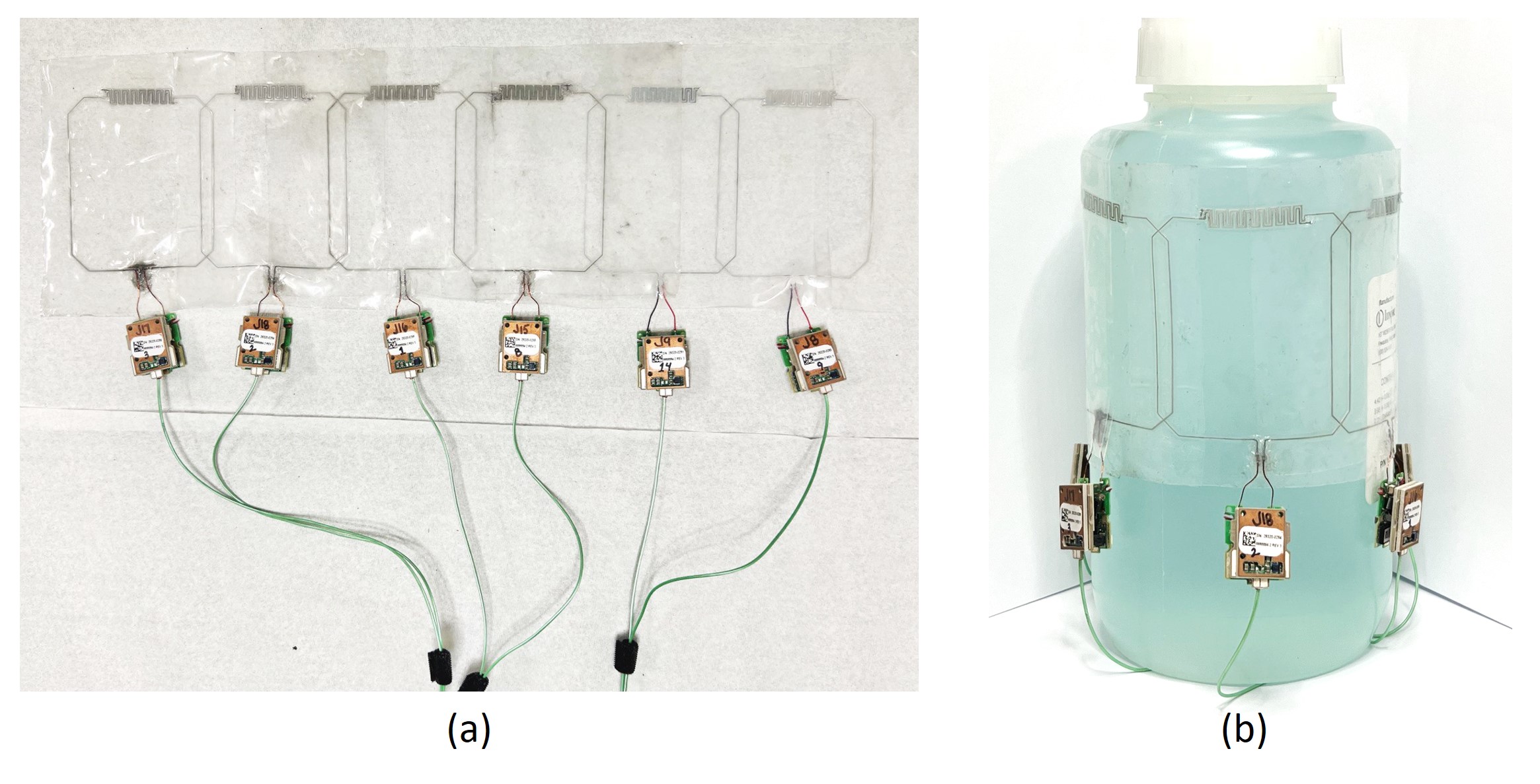
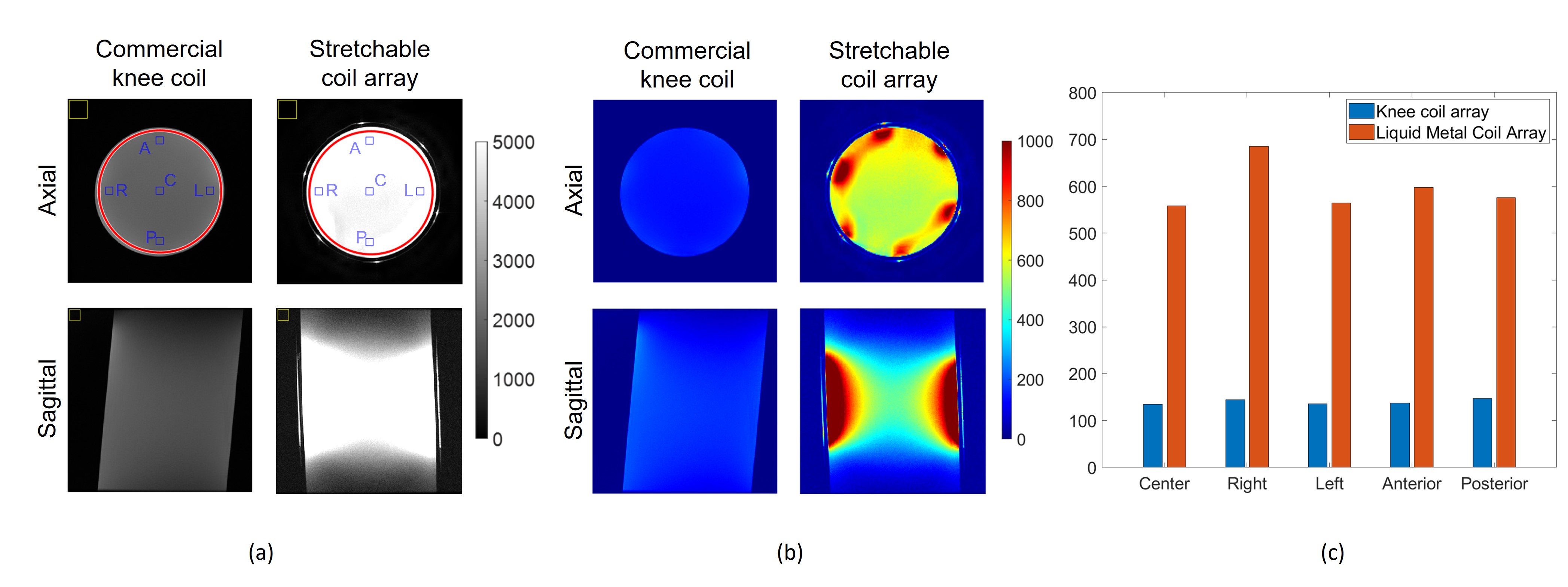
Figure 1 shows the fabricated 6-channel stretchable coil array (a) on a flat surface and (b) wrapped around a standard homogeneous phantom.Figure 2 shows (a) signal and (b) SNR images for central axial and sagittal slices within a standard homogeneous cylindrical phantom obtained with the commercial and stretchable arrays. On the axial slices in Figure 2(a), red circular lines indicate the region of interest (ROI) covering the entire cylinder, blue squares mark smaller ROIs at central (C), right (R), left (L), anterior (A), and posterior (P) locations, and yellow squares indicate the position where background noise was evaluated. On average, the stretchable coil improved SNR within the entire axial slice by 4.4 times, and by 3.9-4.7 times within smaller ROIs (Figure 2(c)).
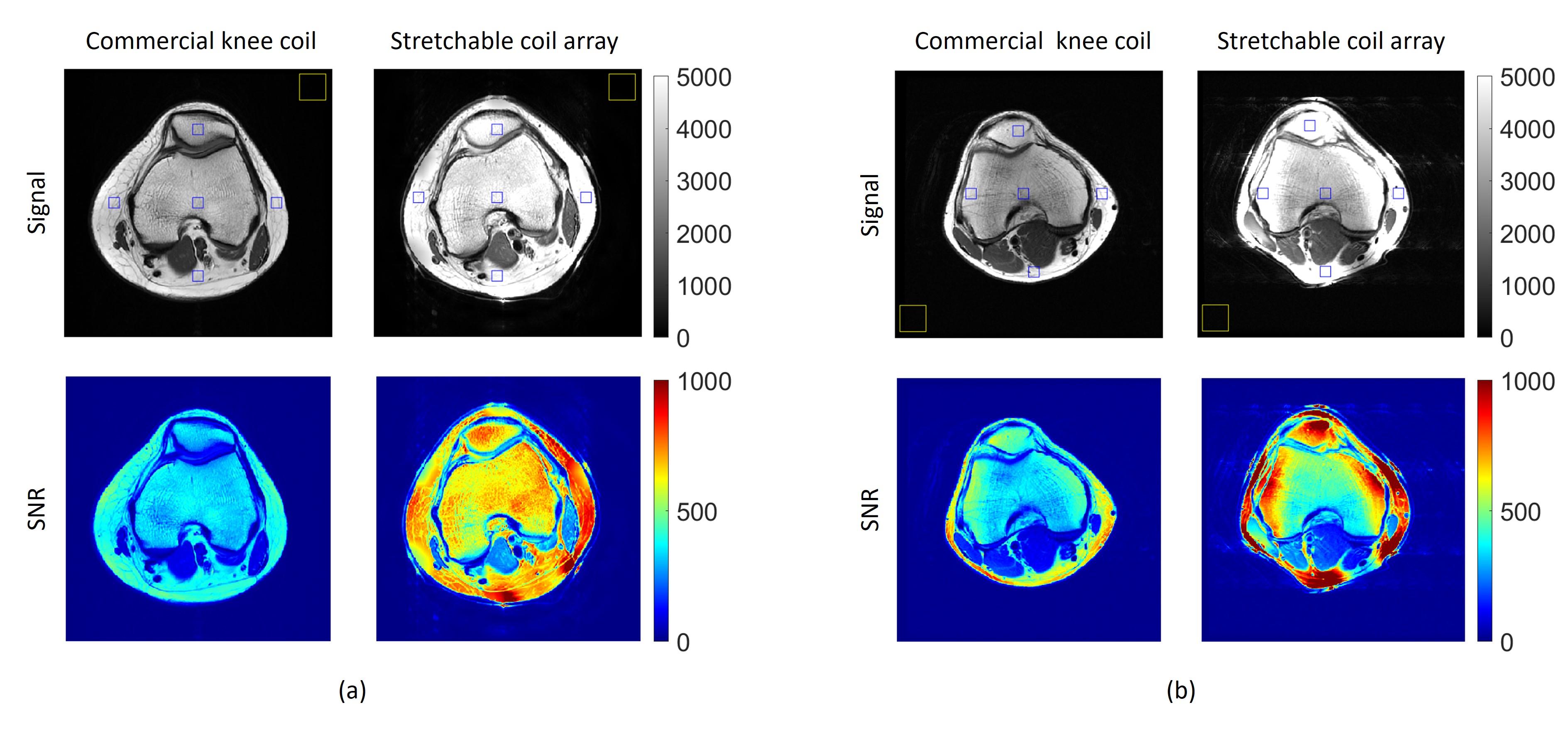
Figure 3 shows central axial PDw knee images from two volunteers using commercial and stretchable arrays. The measured average SNR improvement is 1.5-2.1 times in volunteer #1 and 1.3-4 times in volunteer #2.
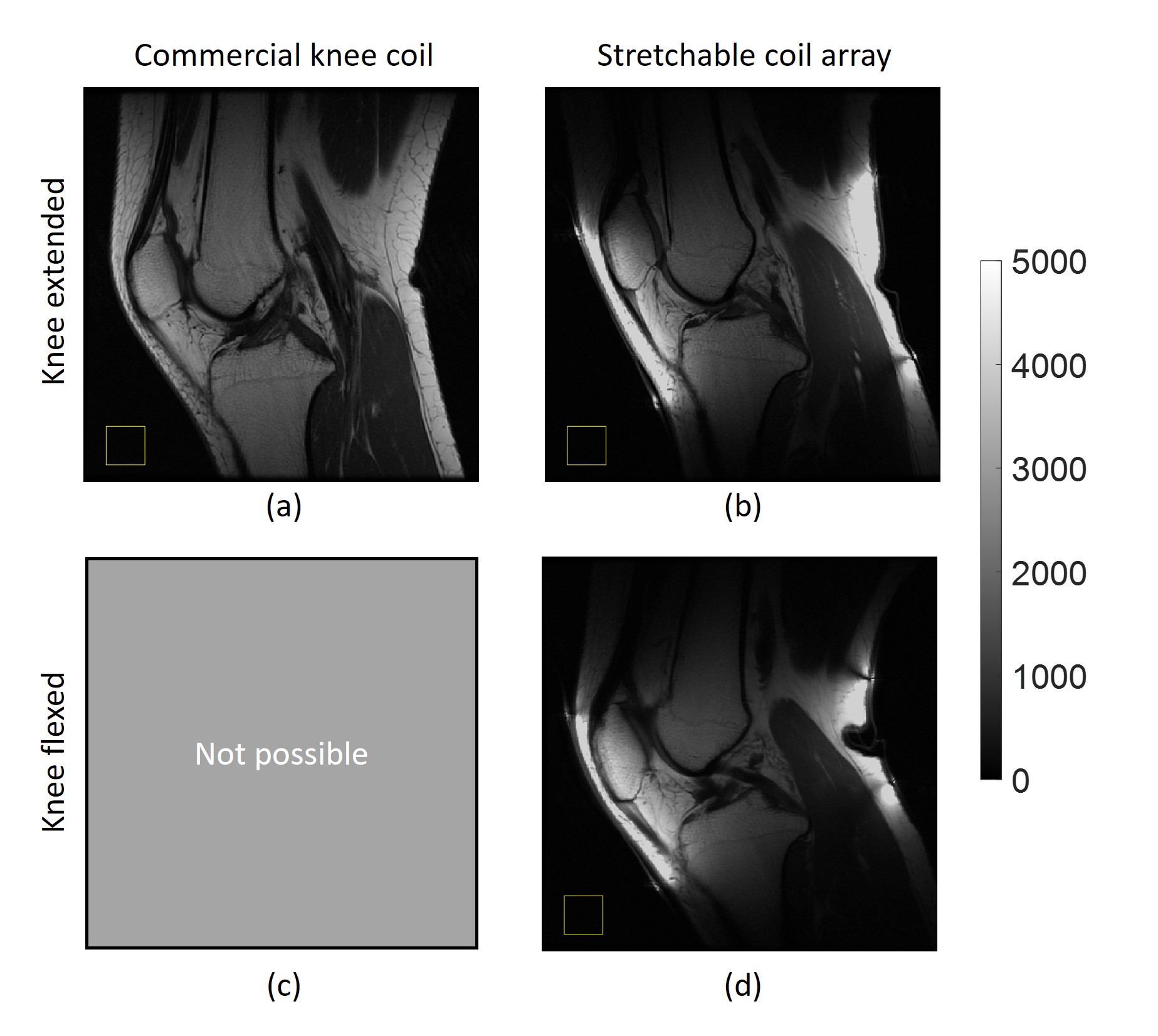
Figure 4 shows central sagittal T1w knee images (volunteer #1) using the commercial and stretchable arrays. The stretchable coil provides more signal due to its tight fit when compared to the rigid commercial coil. Figure 4(a) shows the image acquired with the rigid coil, limiting knee flexion (Figure 4(c)). Conversely, the stretchable coil array allows imaging in the extended (b) and flexed (d) positions.
Discussion
Study results showed a significant SNR improvement using the stretchable coil array both in vitro and in vivo. In vitro, SNR was increased by up to 4.7 times. In vivo, central axial and sagittal slices of knees from two volunteers demonstrated an average SNR improvement between 1.3-4 times, further supporting the efficacy of the stretchable array. Slight signal inhomogeneity introduced with the stretchable coil may require implementation of correction algorithms. The demonstrated ability to image the knee in two different positions underscores the adaptability and potential clinical relevance of the coil. Further research and clinical validation may extend the applicability of stretchable coil arrays to a broader range of imaging scenarios, paving the way for advancements in dynamic and flexible radiofrequency coil designs for MR applications.Conclusion
This study demonstrates a stretchable self-tuning 6-channel coil array and shows performance in an in vivo application of the knee. In vitro and in vivo experiments demonstrate SNR improvements of more than 4 times compared to a commercial knee coil. Furthermore, dynamic imaging was investigated using the stretchable coil array, which is not feasible with rigid commercial coils.Acknowledgements
This work was supported by NIH R01 EB031820 and GE Healthcare.References
1. Varga M, Mehmann A, Marjanovic J, et al. Adsorbed eutectic GaIn structures on a neoprene foam for stretchable MRI coils. Advanced Materials. 2017;29(44):1703744.
2. Mehmann A, Vogt C, Varga M, et al. Automatic resonance frequency retuning of stretchable liquid metal receive coil for magnetic resonance imaging. IEEE transactions on medical imaging. 2018;38(6):1420-1426.
3. Mehmann A, Varga M, Vogt C, et al. On the bending and stretching of liquid metal receive coils for magnetic resonance imaging. IEEE Transactions on Biomedical Engineering. 2018;66(6):1542-1548.
4. Port A, Luechinger R, Albisetti L, et al. Detector clothes for MRI: A wearable array receiver based on liquid metal in elastic tubes. Scientific Reports. 2020;10(1):1-10.
5. Motovilova E, Tan ET, Taracila V, et al. Stretchable self-tuning MRI receive coils based on liquid metal technology (LiquiTune). Scientific reports. 2021;11(1):1-10.
6. Motovilova E, Ching T, Vincent J, et al. Dual-Channel Stretchable, Self-Tuning, Liquid Metal Coils and Their Fabrication Techniques. Sensors. 2023;
Figures